당뇨 처방에 근거한 생약재의 α-Glucosidase 활성 저해 효과 및 이를 활용한 미백 소재 평가법
김미진․임경란․윤경섭 (주)사임당화장품 기술연구소
Effect of Medicinal Herb Prepared through Traditional Antidiabetic Prescription on α-Glucosidase Activity and Evaluation Method for
Anti-Melanogenesis Agents Using α-Glucosidase Activity
Mi Jin Kim, Kyung Ran Im, and Kyung-Sup Yoon R&D Center, Saimdang Cosmetics Co., Ltd.
ABSTRACT For the purpose of investigating the in vitro antidiabetic activity of a medicinal herb and herb mixture extracts prepared through traditional antidiabetic prescription, this study examined α-glucosidase inhibitory activity.
Tyrosinase, a type Ⅰ membrane glycoprotein, is synthesized and glycosylated in the endoplasmic reticulum (ER) and Golgi. The enzyme is subsequently transported to melanosomes, where it participates in melanogenesis. Previous studies showed that disruption of early ER N-glycan processing by an α-glucosidase inhibitor suppresses tyrosinase enzymatic activity and melanogenesis. According to the results, most oriental medicinal herbal extracts were stronger than acarbose and N-butyldeoxynojirimycin, known as an α-glucosidase inhibitor. Interestingly, ethyl acetate layer of enzyme hydro- lyzed Cheongsimyeonjaeum had an inhibitory effect on melanin synthesis in B16F1 cells, although it did not inhibit tyrosinase activity directly. Together, α-glucosidase inhibition activity could be used to evaluate anti-melanogenesis, although cross-checking with melanin inhibitory assay is recommended.
Key words: α-glucosidase inhibition activity, skin whitening, traditional antidiabetic prescription, herb
Received 11 March 2015; Accepted 5 June 2015
Corresponding author: Kyung-Sup Yoon, R&D Center, Saimdang Cosmetics Co., Ltd., Cheongju, Chungbuk 363-886, Korea E-mail: [email protected], Phone: +82-43-218-4203
서 론
최근 서구화된 식생활 습관과 운동 부족 등으로 인한 비만 인구의 증가로 당뇨병 환자가 매년 10% 이상 증가하고 있는 추세이다(1). 고열량 식사가 늘어나면서 당뇨, 고지혈증 등 대사증후군 질환의 발병률이 증가하고 있으며, 이들 대사증 후군을 식품의 섭취를 통하여 개선하고자 하는 노력이 지속 적으로 이루어지고 있다. 당뇨병의 치료 방법에는 식이요법, 운동요법과 함께 약물요법이 있으며, 임상에서 사용되고 있 는 약물로는 인슐린 제제, sulfonylurea 제제, biguanide 제 제, troglitazone 제제 등이 있지만 저혈당, 유산증 등의 치 명적 부작용이 보고되고 있다(2). 또 다른 혈당관리 방법은 섭취한 식이 중 탄수화물의 소화와 흡수를 지연시켜 식후 혈당 및 혈중 인슐린의 상승을 저하시킴으로써 당뇨병의 치 료 효과를 가져오는 것이 있으며, 이러한 물질로는 식이섬유 (3,4)와 장내소화효소인 α-glucosidase에 대한 억제제(5) 가 있다.
α-Glucosidases는 소장 내에서 탄수화물로부터 α-glu- cose를 방출하는 α-glucosidic bond의 분해를 촉진하여 포 도당을 흡수하는 데 관여하는 생체 내 필수 효소이다. 또한 당지질과 당단백질의 합성과 같은 다양한 대사 경로에도 관 여하므로 이를 조절함으로써 대사 질환의 치료 가능성을 기 대할 수 있다. α-Glucosidase inhibitor는 제2형 당뇨병의 치료에 이용되고 있으며, Actinoplanes strain SE50에서 분리한 acarbose는 강력한 α-glucosidase inhibitor로 식 후 혈당을 감소시키고 인슐린의 분비를 촉진시키는 것으로 밝혀졌다(6).
멜라닌 생합성 경로에서 가장 중요한 초기 속도결정단계 에 관여하는 효소인 tyrosinase는 type Ⅰ membrane gly- coprotein으로(7) asparagine의 side chain에 있는 NH2기 에 당을 붙이는 N-linked glycosylation 과정과 serine이나 threonine의 side chain에 있는 OH기에 당을 붙이는 O- linked glycosylation 과정(8) 중 N-linked glycosylation 과정에 의해 만들어진다. 단백질의 N-linked glycosylation 은 endoplasmic reticulum(ER)에서 단백질 합성 번역과 함 께 일어난다(9). Glycan unit의 절반인 14개짜리의 당 (Glc3Man9GlcNAc2)은 ER막의 바깥인 세포질 쪽에서 만들 어지며 이후 나머지 절반은 ER 내에서 만들어져 단백질로
전이된다(10). ER에서 이런 합성 과정은 oligosaccharide 의 말단 끝에 존재하는 mannose에서 3개의 glucose를 α- glucosidase Ⅰ과 Ⅱ가 끊어줌으로써 시작된다(11). 그러므 로 α-glucosidase를 저해하게 되면 tyrosinase의 glyco- sylation이 저해되어 구조는 변형되고 불활성 형태로 멜라 노좀으로 이동하게 되어 결과적으로 melanogenesis가 억 제된다(12-14). 이와 같은 tyrosinase glycosylation 저해 제에 대한 보고들로는 Imokawa와 Mishima(15)가 B16 melanoma 세포 배양에서 glucosamine과 tunicamycin 같 은 물질들이 tyrosinase의 glycosylation 반응을 저해하여 멜라닌 합성이 완전히 억제될 수 있음을 보고하였고, Fran- chi 등(16)은 calcium D-pantetheine-S-sulfonate가 ty- rosinase의 glycosylation을 저해하여 tyrosinase의 안정 도를 저해하는 역할을 한다고 보고하였다. 그리고 Petrescu 등(12)에 의하면 ER processing enzyme인 α-glucosi- dase Ⅰ, Ⅱ의 저해제인 N-butyl-deoxynojirimycin(NB- DNJ)이 B16 melanoma 세포의 활성을 억제하며 비처리군 에 비해 2%의 멜라닌만이 합성되었다고 보고하여 ty- rosinase glycosylation 저해제에 대한 연구들이 이루어지 고 있음을 알 수 있다.
본 연구에서는 특허 및 논문 등을 통하여 당뇨병에 효과가 있는 약재 중 α-glucosidase 저해 활성을 보이는 생약재를 검색하였다. 이 중 미백 관련 특허가 없고 α-glucosidase 저 해 활성의 IC50 값이 1 mg/mL 이하이며 쉽게 구할 수 있는 귀전우(Euonymus alatus Siebold)(17), 연자육(Nelumbo nucifera Gaertner)(18), 자화지정(Viola mandshurica W.
Becker)(19), 적양(Alnus japonica Steud)(20)을 선정하였 다. 또한 오늘날의 당뇨병과 같은 소갈의 원인과 증상, 치료 를 다룬 동의보감의 소갈문에 기재된 복합처방단과 단방처 방 중 미백과 관련된 생약재를 포함하지 않으며 쉽게 구할 수 있는 마인(Cannabis Semen), 죽력(tabasheer), 맥문동 음자(麥門冬飮子), 문동음자(門冬飮子), 청심연자음(淸心蓮 子飮), 활혈윤조생진음(活血潤燥生津飮)(21)을 선정하였다.
선정한 생약재 및 복합처방단의 α-glucosidase 저해 활성 을 알아보았으며, 이 방법이 미백 소재 스크리닝을 위한 유 용한 평가법인지를 알아보았다.
재료 및 방법
실험재료
본 실험에 사용한 생약재 및 복합처방단의 약재들은 (주) 지유본초(Chuncheon, Korea)로부터 구입하여 사용하였으 며, 죽력은 죽림식품(Damyang, Korea)에서 구입하였다. 복 합처방단은 동의보감을 참고하였으며, 맥문동(Liriope pla- typhylla), 지모(Anemarrhena asphodeloides Bunge), 천 화분(Trichosanthes kirilowii), 인삼(Panax ginseng), 오미 자(Schizandrachinensis Baill.), 갈근(Pueraria lobata), 복 령(Poria cocos), 생지황(Rehmannia glutinosa), 감초
(Glycyrrhiza uralensis), 지골피(Lycii radicis Cortex), 연 자육(Nelumbo nucifera Gaertner), 황기(Astragalus mem- branaceus Bunge), 황금(Scutellaria baicalensis), 차전자 (Plantaginis Semen), 천문동(Asparagus cochinchinensis (Lour.) Merr.), 과루인(Trichosanthis Semen), 마자인 (Cannabis Semen), 당귀(Angelica gigas Nakai), 숙지황 (Rehmannia glutinosa), 생지황(Rehmanniae Radix)으로 구성되어 있다(Table 1). 추출 과정에 사용된 용매들은 시약 급을 사용하였다.
추출물의 제조
생약재 추출물의 제조를 위하여 시료 중량 10배의 75%
EtOH 수용액을 가하여 60~90°C에서 4시간 동안 환류냉각 하면서 추출하고 여과지(Whatman No. 2, Whatman GE Healthcare, Westborough, MA, USA)로 여과하였다. 복합 처방단 추출물은 Table 1의 약재 구성 및 비율로 제조하였 다. 이 추출 과정을 2회 반복하고 40°C에서 감압 농축하여 활성 측정 및 분획물 제조용 시료로 사용하였다(75% EtOH Ext.). 이 추출물에 증류수를 가하여 가용화한 후 동량의 메 틸렌클로라이드(CH2Cl2)와 에틸아세테이트(EtOAc)를 순 차적으로 첨가하여 용매분획을 실시하여 분획물을 얻었다(각 각 CH2Cl2 layer, EtOAc layer).
Pectinase, cellulase 등의 효소들은 효모, 세균 또는 일 부 고등식물 등에서 분리되며, 일반적으로 식물조직의 세포 벽이나 세포 사이를 연결해주는 펙틴을 분해하여 과실이나 야채의 조직을 연화시키고 당을 생산 및 분해하거나 생산 수율을 높이는 데 주로 사용된다(22). 이에 청심연자음의 효소처리 에틸아세테이트 분획물(hydrolyzed EtOAc lay- er)은 75% EtOH Ext.를 10% EtOH 수용액으로 용해한 뒤 당분해 효소인 Viscozyme L(Novozymes, Copenhagen, Denmark)을 처리하여 50~60°C에서 20시간 동안 반응시 킨 뒤 여과한 후 감압 농축하였다. 이 추출물에 증류수를 가하여 가용화한 후 동량의 에틸아세테이트를 첨가하고 상 층부를 농축하여 hydrolyzed EtOAc layer를 얻었다.
α-Glucosidase 활성 저해
α-Glucosidase가 p-nitrophenyl(pNP) glycoside의 glycoside 부분을 기질로 인식하여 pNP와 glycoside를 효 소반응으로 끊어주고, 여기에서 끊어져 나온 pNP의 양을 405 nm에서 흡광도를 측정하여 이것으로 α-glucosidase 활성을 간접적으로 측정하였다(23). 0.1 M phosphate buf- fer(pH 6.8)에 시료와 α-glucosidase(1 unit/mL)를 넣고 37°C에서 5분간 반응시켰다. 여기에 2 mM 4-nitrophenyl α-D-glucopyranoside를 첨가해 37°C에서 10분간 효소반 응을 시킨 후 1 M Na2CO3을 넣어 반응을 정지시키고 생성 된 pNP의 양을 microplate reader(Synergy, BioTek In- struments, Inc., Winooski, VT, USA)로 405 nm에서 흡광 도를 측정하였다.
Table 1. Composition of traditional prescriptions for diabetes
Prescriptions Composition Dose (g)
Maekmundongeumja
Liriope platyphylla
Anemarrhena asphodeloides Bunge Trichosanthes kirilowii
Panax ginseng
Schizandra chinensis Baill.
Pueraria lobata Poria cocos
Rehmannia glutinosa Glycyrrhiza uralensis
8 4 4 4 44 4 4 4
Mundongeumja
Liriope platyphylla Schizandra chinensis Baill.
Panax ginseng Lycii radicis Cortex Poria cocos
Glycyrrhiza uralensis
8 4 4 4 44
Cheongsimyeonjaeum
Nelumbo nucifera Gaertner Poria cocos
Panax ginseng
Astragalus membranaceus Bunge Scutellaria baicalensis
Plantaginis Semen Liriope platyphylla Lycii radicis Cortex Glycyrrhiza uralensis
8 4 4 4 2.8 2.8 2.8 2.82.8
Hwalhyeolyunjosaengjineum
Asparagus cochinchinensis (Lour.) Merr.
Liriope platyphylla Schizandra chinensis Baill.
Trichosanthis Semen Cannabis Semen Angelica gigas Nakai Rehmannia glutinosa Rehmanniae Radix Trichosanthes kirilowii Glycyrrhiza uralensis
4 4 4 4 4 4 4 44 4
세포주 및 세포 배양
세포주는 마우스 흑색종 세포주인 B16F1 melanoma cell 을 ATCC(American Type Culture Collection, Manassas, VA, USA)에서 분양받아 사용하였으며, 세포 배양에 사용된 배지(Dulbecco's modified Eagle's medium; DMEM)는 10% fetal bovine serum(HyClone Lab., Logan, UT, USA), 1% antibiotic antimycotic(100 U/mL penicillin and 50 μg/mL streptomycin, Invitrogen, Carlsbad, CA, USA)을 혼합한 배지를 사용하여 37°C, 5% CO2 incubator에서 배양 하고 70~80%의 cell culture confluency를 보일 때 실험을 실시하였다.
세포 생존율 시험
본 실험에서 B16F1 melanoma cells에 대한 시료의 처리 농도를 결정하기 위해 MTT[3-(4,5-dimethythiazol-2- yl)-2,5-diphenytetrazolium bromide, Sigma-Aldrich Co., St. Louis, MO, USA] assay를 Mosmann(24)의 방법을 변 형하여 실시하였다. 이 분석법은 노란색의 수용성 기질인 MTT를 진청색의 비수용성 formazan으로 변환시키는 살아
있는 세포의 mitochondria dehydrogenase의 능력을 이용 한 방법이다. 생성된 formazan의 양은 살아있는 세포 수에 비례한다.
B16 melanoma cells을 5×10³ cells/well 농도로 96- well plate에 분주하여 37°C, 5% CO2 조건 하에서 24시간 동안 배양하였다. 배양 후 배양액을 제거하고 시료를 농도별 로 배지에 희석하여 교체한 후 최종 200 nM α-MSH(α- melanocyte stimulating hormone)가 되도록 첨가하여 48 시간 동안 더 배양하였다. 배양 후 MTT assay를 통하여 세포 생존율을 확인하였다.
멜라닌 양 측정
멜라닌 양 측정은 Oka(25)의 방법을 변형하여 사용하였 다. B16F1 melanoma cells을 6-well plate에 1×10⁵ cells/well이 되게 준비한 후 24시간 동안 37°C, CO2 항온 기에서 배양하였다. 배양액을 제거하고 시료를 농도별로 배 지에 희석하여 교체한 후 최종 200 nM α-MSH가 되도록 첨가하여 48시간 동안 더 배양하였다. 대조군은 α-MSH만 첨가한 것으로 하였으며 양성 대조군으로는 알부틴을 사용
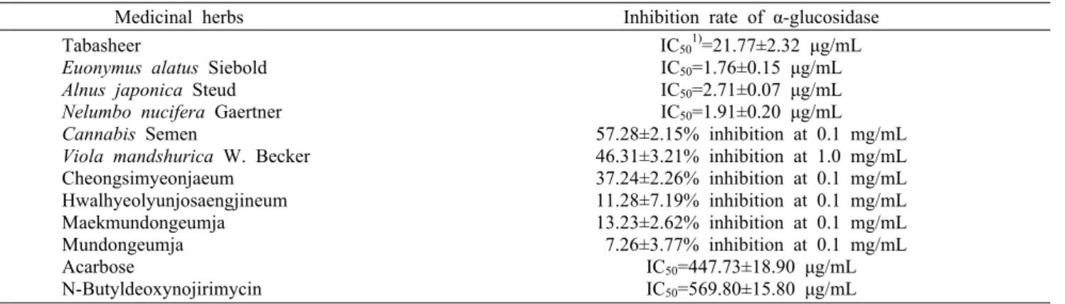
Table 2. Inhibitory effects of 75% EtOH extracted medicinal herbs on α-glucosidase
Medicinal herbs Inhibition rate of α-glucosidase Tabasheer
Euonymus alatus Siebold Alnus japonica Steud Nelumbo nucifera Gaertner Cannabis Semen
Viola mandshurica W. Becker Cheongsimyeonjaeum
Hwalhyeolyunjosaengjineum Maekmundongeumja Mundongeumja Acarbose
N-Butyldeoxynojirimycin
IC501)=21.77±2.32 μg/mL IC50=1.76±0.15 μg/mL IC50=2.71±0.07 μg/mL IC50=1.91±0.20 μg/mL 57.28±2.15% inhibition at 0.1 mg/mL 46.31±3.21% inhibition at 1.0 mg/mL 37.24±2.26% inhibition at 0.1 mg/mL 11.28±7.19% inhibition at 0.1 mg/mL 13.23±2.62% inhibition at 0.1 mg/mL 7.26±3.77% inhibition at 0.1 mg/mL
IC50=447.73±18.90 μg/mL IC50=569.80±15.80 μg/mL
1)IC50: concentration of the sample required for 50% the activity to be inhibited.
하였다. 배양 후 배양액을 제거하고 PBS(phosphate buf- fered saline)로 세척한 후 10% DMSO(dimethyl sulf- oxide)가 함유된 1 N NaOH를 첨가한 후 50°C 항온조에서 세포 내 멜라닌을 용해시켰다. 이 액을 microplate reader 를 이용하여 490 nm에서 흡광도를 측정하였으며 총 단백질 량으로 보정하였다.
Tyrosinase 저해 활성 측정
Tyrosinase에 대한 활성은 각 농도별 시료 0.9 mL, 0.1 M 인산완충액(pH 6.8) 1.0 mL, 1.5 mM L-tyrosine 용액 1.0 mL를 넣은 후, 37°C에서 10분간 반응시킨 뒤 mush- room tyrosinase(1,500 units/mL) 0.1 mL를 첨가하여 37
°C에서 10분간 반응시킨 후 microplate reader를 사용하여 475 nm에서 흡광도를 측정하였다.
DOPA(3,4-dihydroxyphenylalanine) oxidation 저해 활 성 측정
DOPA oxidation 저해 활성은 각 농도별 시료 100 μL, 0.1 M 인산완충액(pH 7.0) 800 μL, 5 mM L-DOPA 용액 50 μL를 넣은 후 37°C에서 3분간 반응시킨다. Mushroom tyrosinase(1,500 units/mL) 50 μL를 첨가하여 37°C에서 1분간 반응시킨 후 ice에서 반응을 종결시킨 다음 microplate reader를 이용하여 490 nm에서 흡광도를 측정하였다.
결과 및 고찰
α-Glucosidase 활성 저해 효과
Tyrosinase는 다양한 조절 경로 중 생체 내 당단백질 합 성과정인 glycosylation 과정을 통해 만들어진다. 이 과정 중에 문제가 생겨 tyrosinase의 당 부분에 이상이 생기면 tyrosinase는 세포 내 멜라닌 생합성 장소인 melanosome 으로 이동하지 못하거나 이동하더라도 tyrosinase의 활성 을 나타내지 못하게 되고 따라서 멜라닌이 생성되지 않는다.
α-Glucosidase는 glycosylation 과정에 관여하는 많은 효 소 중 하나로 α-glucosidase 활성 억제를 통해 미백 소재를
스크리닝 하고자 하였다.
한의학과 민간에서 당뇨의 개선 및 치료 효과가 우수하다 고 알려진 복합 처방 및 생약재들을 75% EtOH 수용액으로 추출하고 그 추출물들의 α-glucosidase 저해 활성을 평가 하였다. 그 결과 죽력, 귀전우, 적양, 연자육이 100 μg/mL 농도에서 90% 이상으로 뛰어난 활성 저해 효과를 나타내었 다. 이는 α-glucosidase 활성 저해제로 알려져 있는 acar- bose(약 29% 저해)와 N-butyldeoxynojirimycin(약 19%
저해)보다 탁월한 효과를 나타낸 것이다. 다음으로 마인은 57.28±2.15% 저해 효과를 보였으며, α-glucosidase 활성 저해 효과가 알려진 자화지정은 2.71±0.21%로 위의 측정 농도에서는 활성 저해 효과가 거의 나타나지 않았다. 복합처 방단인 청심연자음은 37.24±2.26%의 저해 효과를 보였으 며, 활혈윤조생진음(11.28±7.19% 저해), 맥문동음자(13.23
±2.62% 저해), 문동음자(7.26±3.77% 저해)의 75% EtOH Ext.에서는 α-glucosidase 활성 저해 효과가 거의 나타나 지 않았다(Table 2).
Table 2의 생약 추출물 중 우수한 α-glucosidase 활성 저해 효과가 나타난 생약재 추출물에 함유되어 있는 활성물 질 분획물을 얻고자 75% EtOH Ext.를 제조하고 이를 에틸 아세테이트, 메틸렌클로라이드로 분획하였다. 그 결과 자화 지정은 75% EtOH Ext.에 비해 EtOAc layer에서 α-gluco- sidase 활성을 50% 저해하는 농도인 IC50 값이 13.34±2.88 μg/mL로 향상되었으며, 복합처방단인 청심연자음은 EtOAc layer, CH2Cl2 layer, hydrolyzed EtOAc layer에서 IC50 값 이 각각 4.46±1.05, 37.24±2.26, 2.04±0.33 μg/mL로 효 과가 향상되었다. 귀전우, 적양, 연자육은 EtOAc layer에서 IC50 값이 각각 0.99±0.06, 1.76±0.20, 1.57±0.23 μg/mL 로 75% EtOH Ext.에 비해 유사하거나 약간 향상된 결과를 나타내었다. 반면 귀전우, 적양, 연자육의 CH2Cl2 layer에서 IC50 값이 각각 46.20±4.96, 26.64±0.99, 12.43±1.33 μg/
mL로 75% EtOH Ext.에 비해 활성 저해 효과가 감소하였 다. 그러나 α-glucosidase 저해제로 알려진 acarbose 및 N-butyl-deoxynojirimycin에 비해서는 월등한 효과를 보 였다(Table 3).
Table 3. Inhibitory effects of solvent fractions from medicinal herbs on α-glucosidase
Medicinal herbs
Inhibition rate of α-glucosidase (IC501), μg/mL) CH2Cl2 layer EtOAc layer Acarbose
Euonymus alatus Siebold
Alnus japonica Steud Nelumbo nucifera
Gaertner Viola mandshurica
W. Becker Cheongsimyeonjaeum Hydrolyzed
Cheongsimyeonjaeum
447.73 46.20±4.96 26.64±0.99 12.43±1.33
- 37.24±2.26
-
±18.90 0.99±0.06 1.76±0.20 1.57±0.23 13.34±2.88 4.46±1.05 2.04±0.33
1)IC50: concentration of the sample required for 50% the activity to be inhibited.
Table 4. Inhibitory effects of medicinal herb extracts on tyrosinase (at 1 mg/mL) Medicinal herbs Inhibition rate of tyrosinase Inhibition rate of DOPA oxidation
Arbutin (at 0.5 mg/mL) 31.45±1.52% -
Euonymus alatus Siebold
75% EtOH Ext.
CH2Cl2 layer EtOAc layer
- 21.75±2.25%
-
28.60±2.17%
48.15±3.27%
28.40±1.24%
Alnus japonica Steud
75% EtOH Ext.
CH2Cl2 layer EtOAc layer
15.50±1.35%
12.98±2.25%
33.85±3.06%
37.04±4.90%
30.86±6.08%
37.04±0.62%
Nelumbo nucifera Gaertner
75% EtOH Ext.
CH2Cl2 layer EtOAc layer
2.17±0.28%
7.34±2.14%
13.06±2.27%
17.90±1.63%
10.49±0.62%
52.47±3.86%
Viola mandshurica W. Becker
75% EtOH Ext.
CH2Cl2 layer EtOAc layer
-
-
-
-
-
-
Cheongsimyeonjaeum
75% EtOH Ext.
CH2Cl2 layer EtOAc layer
Hydrolyzed-EtOAc layer
9.92±2.35%
IC50=0.21±0.00 mg/mL 43.02±1.57%
36.73±0.50%
21.81±1.89%
IC50=0.93±0.09 mg/mL 59.26±4.82%
-
0 20 40 60 80 100 120
0 μg/mL 0 μg/mL 20 μg/mL 50 μg/mL 100 μg/mL 200 nM α-MSH
Melanin (%) .
**
**
**
*
Fig. 1. Inhibitory effect of EtOAc layer of viscozyme hydro- lyzed 75% EtOH extract of Cheongsimyeonjaeum on melanin synthesis in B16F1 cells. The cells were treated with Cheongsi- myeonjaeum at indicated concentration for 48 h. The absorbance was measured at 490 nm and corresponding total protein content was used to normalize absorbance. The results were expressed as the mean±SD from the three independent experiments. *P<0.05 and **P<0.01 vs. control group treated with α-MSH.
멜라닌 생성 저해 효과
앞선 연구에 따르면 mushroom tyrosinase 활성 저해 효 과는 없으면서 α-glucosidase 활성 저해 효과가 우수한 연 자육 추출물이 tyrosinase의 N-glycosylation 과정을 저해 함으로써 멜라닌 합성을 저해한다는 보고가 있으며, α-glu- cosidase 활성 저해 효과가 우수한 적양 추출물의 멜라닌 생성 저해 효과도 연구된 바 있다(26,27). 이에 본 연구에서 는 연자육을 포함하며 α-glucosidase 활성 저해 효과를 나 타낸 청심연자음 추출물의 멜라닌 생성 저해 효과에 대해 알아보았다.
MTT assay를 통하여 청심연자음 hydrolyzed EtOAc layer는 100 μg/mL 농도까지 세포 생존에 영향을 주지 않 는 것을 확인하였다(data 미첨부). Fig. 1에서와 같이 청심 연자음 hydrolyzed EtOAc layer는 20, 50, 100 μg/mL 농 도에서 각각 26.13±1.78, 32.17±12.71, 49.13±3.92%의
멜라닌 생성 저해 효과를 나타내었다. 이러한 결과로 보아 청심연자음 hydrolyzed EtOAc layer의 α-glucosidase 활 성 저해 효과가 멜라닌 생성에 영향을 준 것으로 생각된다.
Tyrosinase 활성 및 DOPA oxidation 저해 효과 본 연구의 생약 추출물들에서 in vitro 미백효능 스크리닝 을 위한 평가법으로 널리 이용되는 tyrosinase 활성 저해 및 DOPA 자동산화 억제 효과가 있는지 알아보았다. Tyrosi- nase는 멜라닌 생합성 경로에서 가장 중요한 초기 속도결정 단계에 관여하는 효소이며, DOPA oxidation은 멜라닌 합성 과정에서 tyrosine이 DOPA를 거쳐 DOPAquinone으로 전 환되고, DOPAquinone으로부터 자동산화 반응과 효소 반응 으로 DOPAchrome을 거쳐 멜라닌이 생성되는 데 관여하는
단계이다(28).
α-Glucosidase 활성 효과가 양호하였던 생약재 추출물 들의 tyrosinase 활성 측정 결과는 Table 4에 나타내었다.
비교한 결과 arbutin은 500 μg/mL 농도에서 31.45%의 저 해 효과가 나타났으며, 귀전우 및 자화지정 추출물에서는 저해 활성이 나타나지 않았다. 그 외 추출물에서는 arbutin 과 유사하거나 낮은 저해 효과를 나타내었으며, 청심연자음 의 CH2Cl2 layer에서는 IC50=0.21±0.00 mg/mL로 양호한 저해 효과가 나타났으나 청심연자음 hydrolyzed EtOAc layer에서는 tyrosinase 활성 저해 효과가 나타나지 않았 다. DOPA 자동산화 저해 활성 측정 결과 자화지정 추출물 에서는 효과가 나타나지 않았으며, 그 외의 추출물군에서는 유사한 저해 효과가 나타났다. 이러한 결과로 볼 때 청심연 자음 hydrolyzed EtOAc layer는 tyrosinase 활성 억제제 가 아닌 tyrosinase의 glycosylation inhibitor로 작용함으 로써 멜라닌 생성 저해 효과를 나타낸 것으로 사료되며, 이 러한 작용 기전을 확인하기 위해서는 청심연자음 hydro- lyzed EtOAc layer에서의 tyrosinase N-glycosylation 저 해 효과에 대해 알아봐야 할 것이다.
요 약
본 연구에서는 선정한 생약재 및 복합처방단의 α-glucosi- dase 저해 활성을 알아보았으며, 이 방법이 미백 소재 스크 리닝을 위한 유용한 평가법인지를 알아보았다. 한의학과 민 간에서 당뇨의 개선 및 치료 효과가 우수하다고 알려진 생약 재 및 처방 중 죽력, 귀전우, 적양, 연자육, 마인, 청심연자음 의 α-glucosidase 활성 저해 효과는 식후 혈당조절제인 acarbose와 비교하여 볼 때 우수한 효과를 나타내었다. 미 백 효과가 알려진 연자육을 함유한 청심연자음 hydrolyzed EtOAc layer는 100 μg/mL 농도에서 약 50% 멜라닌 생성 저해 효과를 보였다. 또한 청심연자음 hydrolyzed EtOAc layer는 α-glucosidase 활성 저해 효과가 우수하였으나 mushroom tyrosinase 활성 저해 효과는 나타나지 않았다.
이로써 청심연자음 hydrolyzed EtOAc layer는 α-gluco- sidase 활성을 저해시켜 tyrosinase의 glycosylation을 저 해함으로써 멜라닌 생성 억제 효과가 나타나는 것으로 생각 된다. 이상의 결과로 볼 때 α-glucosidase 활성 억제 효과가 있으면서 당뇨병에 효과가 있는 생약재들은 N-linked gly- coprotein인 tyrosinase의 glycosylation을 저해하여 ty- rosinase의 세포 내 이동이나 활성을 억제함으로써 멜라닌 생성을 억제할 것으로 사료되며, 본 연구에서 선정된 생약재 들은 당뇨병 치료를 위한 목적뿐만 아니라 화장품에서 새로 운 미백 소재로서의 활용가치가 있을 것으로 판단된다. 또한 미백에 효과가 있는 소재 스크리닝을 위해 현재 널리 사용되 고 있는 mushroom tyrosinase 활성 저해 효과와 다른 접근 방법으로써 α-glucosidase 활성 측정 방법도 하나의 평가 법으로 유용할 것으로 생각된다.
감사의 글
본 연구는 2013년 중소기업기술혁신개발사업(과제번호 SA112778)의 연구비 일부에 의하여 이루어진 것으로 지원 에 감사드립니다.
REFERENCES
1. Xu ML, Wang L, Xu GF, Wang MH. 2011. Antidiabetes and angiotensin converting enzyme inhibitory activity of Sonchus asper (L) Hill extract. Kor J Pharmacogn 42: 61- 67.
2. Kim TW, Kwon YB, Lee JH, Yang IS, Youm JK, Lee HS, Moon JY. 1996. A study on the antibodiabetic effect of mulberry fruits. Korean J Seric Sci 38: 100-107.
3. Torsdottir I, Alpsten M, Andersson H, Einarsson S. 1989.
Dietary guar gum effects on postprandial blood glucose, in- sulin and hydroxyproline in humans. J Nutr 119: 1925-1931.
4. Blackburn NA, Redfern JS, Jarjis H, Holgate AM, Hanning I, Scarpello JH, Johnson IT, Read NW. 1984. The mecha- nism of action of guar gum in improving glucose tolerance in man. Clin Sci (Lond) 66: 329-336.
5. Puls W, Krause HP, Müller L, Schutt H, Sitt R, Thomas G. 1984. Inhibitors of the rate of carbohydrate and lipid absorption by the intestine. Int J Obes 8: 181-190.
6. Puls W, Keup U, Krause HP, Thomas G, Hoffmeister F. 1997.
Glucosidase inhibition. A new approach to the treatment of diabetes, obesity, and hyperlipoproteinaemia. Naturwissen- schaften 64: 536-537.
7. Branza-Nichita N, Negroiu G, Petrescu AJ, Garman EF, Platt FM, Wormald MR, Dwek RA, Petrescu SM. 2000.
Mutations at critical N-glycosylation sites reduce tyrosinase activity by altering folding and quality control. J Biol Chem 275: 8169-8175.
8. Goochee CF. 1992. Bioprocess factors affecting glycoprotein oligosaccharide structure. Dev Biol Stand 76: 95-104.
9. Petrescu AJ, Butters TD, Reinkensmeier G, Petrescu S, Platt FM, Dwek RA, Wormald MR. 1997. The solution NMR structure of glucosylated N-glycans involved in the early stages of glycoprotein biosynthesis and folding. EMBO J 16: 4302-4310.
10. Kornfeld R, Kornfeld S. 1985. Assembly of asparagine- linked oligosaccharides. Annu Rev Biochem 54: 631-664.
11. Winchester B, Fleet GW. 1992. Amino-sugar glycosidase inhibitors: versatile tools for glycobiologists. Glycobiology 2: 199-210.
12. Petrescu SM, Petrescu AJ, Titu HN, Dwek RA, Platt FM.
1997. Inhibition of N-glycan processing in B16 melanoma cells results in inactivation of tyrosinase but does not pre- vent its transport to the melanosome. J Biol Chem 272:
15796-15803.
13. Branza-Nichita N, Petrescu AJ, Dwek RA, Wormald MR, Platt FM, Petrescu SM. 1999. Tyrosinase folding and copper loading in vivo: a crucial role for calnexin and alpha-gluco- sidase Ⅱ. Biochem Biophys Res Commun 261: 720-725.
14. Wang Y, Androlewicz MJ. 2000. Oligosaccharide trimming plays a role in the endoplasmic reticulum-associated degra- dation of tyrosinase. Biochem Biophys Res Commun 271:
22-27.
15. Imokawa G, Mishima Y. 1984. Functional analysis of ty- rosinase isozymes of cultured malignant melanoma cells during the recovery period following interrupted melano-
genesis induced by glycosylation inhibitors. J Invest Derma- tol 83: 196-201.
16. Franchi J, Coutadeur MC, Marteau C, Mersel M, Kupferberg A. 2000. Depigmenting effects of calcium D-pantetheine-S- sulfonate on human melanocytes. Pigment Cell Res 13: 165- 171.
17. Kim TJ, Song HJ, So JN, Lee JH. 1996. Preventive effect of Euonymus alatus extract on experimental diabetes. Kore- an J Immunol 18: 591-600.
18. Mani SS, Subramanian IP, Pillai SS, Muthusamy K. 2010.
Evaluation of hypoglycemic activity of inorganic constitu- ents in Nelumbo nucifera seeds on streptozotocin-induced diabetes in rats. Biol Trace Elem Res 138: 226-237.
19. Lee BB, Park SR, Han CS, Han DY, Park EJ, Park HR, Lee SC. 2008. Antioxidant activity and inhibition activity against α-amylase and α-glucosidase of Viola mandshurica extracts. J Korean Soc Food Sci Nutr 37: 405-409.
20. Kwak TH, Joo JH, Park MG. 2005. Active extracts from natural plants showing anti-obesity and anti-diabetes. Korea Patent 523,443.
21. Heo J. 1610. Japbyeongpyeon. In Donguibogam. 3rd ed.
Yeogang, Seoul, Korea. p 1998-2023.
22. Park MK, Kim CH. 2009. Extraction of polyphenols from apple peel using cellulase and pectinase and estimation of
antioxidant activity. J Korean Soc Food Sci Nutr 38: 535- 540.
23. Angelov A, Putyrski M, Liebl W. 2006. Molecular and bio- chemical characterization of α-glucosidase and α-mannosi- dase and their clustered genes from the thermoacidophilic archaeon Picrophilus torridus. J Bacteriol 188: 7123-7131.
24. Mosmann T. 1983. Rapid colorimetric assay for cellular growth and survival: application to proliferation and cyto- toxicity assays. J Immunol Methods 65: 55-63.
25. Oka M, Ichihashi M, Chakraborty AK. 1996. Enhanced ex- pression of protein kinase C subspecies in melanogenic compartments in B16 melanoma cells by UVB or MSH.
J Invest Dermatol 106: 377-378.
26. Lee JY, Im KR, Jung TK, Yoon KS. 2013. The inhibitory effects of Alnus Japonica Steud. extract on melanogenesis.
J Soc Cosmet Scientists Korea 39: 159-166.
27. Lee JY, Im KR, Jung TK, Yoon KS. 2013. The inhibitory effects of Nelumbo nucifera Gaertner extract on melano- genesis. KSBB J 28: 137-145.
28. Alaluf S, Heath A, Carter N, Atkins D, Mahalingam H, Barrett K, Kolb R, Smit N. 2001. Variation in melanin con- tent and composition in type Ⅴ and Ⅵ photoexposed and photoprotected human skin: the dominant role of DHI.
Pigment Cell Res 14: 337-347.