Yeungnam Univ J Med 2015;32(1):1-7
간암 줄기세포의 기원
은 종 렬
서남대학교 의과대학 명지병원 내과학교실
Cellular origin of liver cancer stem cells
Jong Ryeol Eun
Department of Internal Medicine, Myongji Hospital, Seonam University College of Medicine, Goyang, Korea
Over several decades, a hierarchical cancer stem cell (CSC) model has been established in development of solid can- cers, including hepatocellular carcinoma (HCC). In terms of this concept, HCCs originate from liver CSCs. Clinically HCCs show a wide range of manifestations from slow growth to very aggressive metastasis. One of the reasons may be that liver CSCs originate from different cells. This review describes the basic concept of CSCs and the cellular origin of liver CSCs.Keywords: Stem cells; Liver cancer stem cells; Hepatocellular carcinoma
Received: May 19, 2015, Revised: June 3, 2015, Accepted: June 5, 2015
Corresponding Author: Jong Ryeol Eun, Department of Internal Medicine, Myongji Hospital, Seonam University College of Medicine, 55 Hwasu-ro 14beon-gil, Deogyang-gu, Goyang 412-826, Korea
Tel: +82-31-810-5114, Fax: +82-31-969-0500 E-mail: [email protected]
서 론
간세포암(hepatocellular carcinoma, HCC)은 원발 간암의 85-90%를 차지한다[1]. 현재 암 발생은 암 줄기세포를 기점으 로 시작되는 계층모델(hierarchical cancer stem cell model)이 널리 받아들여지고 있다[2]. 계층모델 개념에 따르면 간세포암 은 간암 줄기세포를 기점으로 발생한다. 실제 임상에서 간세포 암은 매우 다양한 발현 양상을 보인다. 잘 분화된 간세포암이 있는 반면, 분화도가 낮은 것이 있다. 또 진행이 느린 간세포암 이 있는 반면, 어떤 경우는 매우 빠르게 진행하고 심한 전이 를 일으키는 경우도 있다. 암세포의 모양과 표지자(surface markers) 발현이 담관세포암종과 분명하게 구분되는 경우도 있지만, 간세포암과 담관세포암(cholangiocarcinoma, CC)이 섞여 있거나 면역화학염색에서 두 세포 표지자를 모두 발현하 는 경우도 있다(combined or intermediate HCC-CC type) [3].
이처럼 간세포암종이 다양한 형태와 임상소견을 보이는 이 유는 비록 간세포암이 간암 줄기세포로부터 발생하지만 간암 줄기세포가 다양한 세포들로부터 기원하기 때문일 수 있다.
예를 들면, 잘 분화된 간세포에서 간암 줄기세포로 역분화 후 발생한 간세포암과 간 줄기세포에서 간암 줄기세포로 암 전환 된 후 발생한 간세포암은 서로 다른 표지자 발현 및 임상 양상 을 보일 것이다.
여기서는 간 재생에 관여하는 세포들, 암 발생의 기본 개념 과 함께 간암 줄기세포가 기원하는 세포들을 중심으로 기술하 고자 한다.
본 론
1. 간 재생(liver regeneration)
간세포의 정상 수명은 약 200-300일 정도이다[4,5]. 수명이 다한 간세포는 제거되고 새로운 간세포가 만들어진다. 새로운 간세포의 공급원은 기존 간세포이다. 장 세포의 증식과 탈락 에서와 마찬가지로 간에서는 문맥주변의 간세포가 노화되면 서 점차 중심정맥쪽으로 이동하여 탈락된다[6,7].
간이 손상될 때에는 다양한 세포들이 간 재생에 관여한다.
간 손상이 미미할 때에는 간세포 스스로의 재생을 통하여 간
Fig. 1. Liver homeostasis. Mature hepatocytes are the major source in repair of minor liver injury. In ongoing liver injury, such as chronic viral hepatitis, HPCs contribute to liver regeneration.
When hepatocytes or HPCs are not able to restore normal homeo- stasis in severe liver injury such as cirrhosis, BM stem cells mobilize to circulation and engraft to the liver. HPC, hepatic progenitor cell; BM, bone marrow.
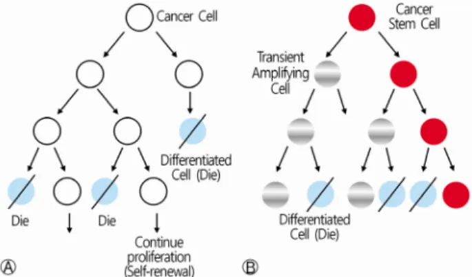
Fig. 2. Two general models of carcinogenesis. The stochastic model (A) asserts that tumor formation is the consequence of accumulation of random genetic events in any normal differen- tiated cell. All cancer cells have the same potential for growth and division, but each cell chooses at random between self-renewal and differentiation. The hierarchical cancer stem cell model (B) postulates that a single CSC gives rise to a hierarchical organiza- tion within a tumor, thus forming a heterogenous tumor. CSC, cancer stem cell.
의 항상성을 유지한다. 그러나 간 손상이 지속될 때에는 간세 포 이외에도 간 전구세포가 간 재생에 동원된다[5]. 심한 간 손상에서는 골수 세포들이 간 재생에 관여한다(Fig. 1) [8].
골수와 간은 밀접한 관련이 있다[8]. 사람의 간은 배아기때 혈액생성을 담당한다. 태아 사슴에 사람의 골수 조혈세포를 주입하면 사슴 간세포의 10% 이상에서 사람의 골수 조혈세포 가 관찰된다[9]. 또 간 섬유화 또는 간경변증에서는 골수로부 터 말초혈액으로 조혈줄기세포의 이동이 증가한다[10]. 골수 세포가 간에 생착하는 방법에 대하여 몇 가지 가설이 있는데, 세포융합(cell fusion)을 통하여 하나의 단일세포를 만드는 것 [11-14]이 우세하지만, 줄기세포의 능력(plasticity)으로 세포 융합 없이 간세포로 직접 분화[15-17]하거나, 상피-간엽 전환 (epithelial-mesenchymal transition)을 통한 교차분화(trans- differentiation) [18]도 보고되었다.
설치류에서 간을 2/3 절제하여도 10일 이내에 빠르게 복원 된다[5]. 앞서 설명한 것처럼 간세포, 간 전구세포 뿐만 아니라, 간엽줄기세포 및 조혈줄기세포 등 다양한 세포들이 간 복원에 관여한다[19]. 이때 hepatocyte growth factor (HGF), interleu- kin-6(IL-6), tumor necrosis factor (TNF)-α, transforming growth factor (TGF)-α, epidermal growth factor (EGF) 등의 신호전달 물질들이 관여한다[5]. 간 복원이 완료된 후 어떻게 종결되는 지 잘 알려져 있지는 않지만, TGF-β1같은 신호전달물질이 중요한 역할을 하는 것으로 생각된다[20].
2. 암 발생(cancer initiation)
암 발생에는 두 가지 개념이 있는데, 유전적 변이가 축적되 는 과정에서 무작위적이고도 임의적으로 종양이 발생한다는 확률 모델(stochastic model, clonal evolution)과, 암 줄기세포 를 기점으로 종양 내 세포들이 계층을 이룬다는 계층모델
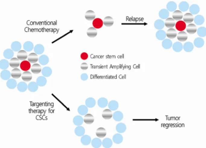
(hierarchical model)이 있다(Fig. 2) [3,21,22]. 확률 모델은 고전적 개념으로 개개의 암세포는 자기 복제 또는 분화 가운데 한가지를 선택하게 되고, 종양 내의 암세포들은 동일한 성상 을 띄게 된다. 확률 모델로는 암 치료 후의 재발, 항암치료에 대한 저항을 설명하기 어렵다. 현재는 암 줄기세포를 기점으로 암이 발생한다는 계층모델이 널리 인정되고 있다. 계층모델 에서 암 줄기세포는 세포분열 사이클의 G0 (quiescent state) 상태에 있다. 고식적 항암치료는 분화하는 암세포를 표적으 로 하므로 고식적 항암치료는 암 줄기세포를 남겨둠으로써 이후 암 재발의 원인이 된다. 암 줄기세포를 표적으로 하는 치료가 이루어져야 궁극적으로 암을 근치할 수 있게 된다 (Fig. 3) [22]. 암 줄기세포를 표적으로 하는 치료법의 개발을 위해서는 먼저 암 줄기세포의 기본 특성을 이해하여야 한다.
3. 줄기세포와 암 줄기세포(stem cell and cancer stem cell)
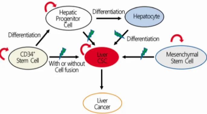
줄기세포는 자신과 동일한 세포를 무제한 만들어내는 자기 재생(self-renewal) 능력과 다양한 세포로 분화(differentiation) 하는 능력을 가진 세포를 말한다[22]. 우리 몸의 각종 장기와 조직의 재생에 있어서 줄기세포는 필수적이다. 암 줄기세포는 줄기세포의 자기 재생 및 분화 능력에 더하여 암 시작, 전이, 재발, 항암제에 대한 저항능력을 갖춘 세포를 말한다. 암 줄기 세포는 정상 줄기세포의 유전적 및 후생학적 변화에 의해 전 환되거나 분화된 세포의 역분화로 생길 수 있다(Fig. 4) [23].
Fig. 3. The therapeutic concept of conventional chemotherapy and targeting therapy for cancer stem cells. Conventional chemo- therapy kills proliferating cells but leaves behind cancer stem cells, then tumor will regrow (A). If the therapy eliminates cancer stem cells, the tumor will eventually be regressed (B).
Fig. 4. Cellular origin of CSCs. CSCs can originate from normal stem cells by genetic mutation or epigenetic alteration. CSCs can also originate from mature differentiated cells that already have tumorigenic potential by mutations. CSCs, cancer stem cells.
Fig. 5. Cell surface markers, molecular signaling and micro RNAs in liver cancer stem cells. Liver CSCs can be identified by surface markers including CD133, CD90, CD44, EpCAM, and CD34.
Molecular signaling pathways are essential to normal cellular processes such as development, growth and self-renewal. Disrup- tion or overexpression of these pathways can result in develop- ment of liver cancers. Aberrant regulation of microRNAs can also cause liver cancers. Therefore, surface antigens, signaling pathways and microRNAs could be therapeutic targets for CSCs.
CSCs, cancer stem cells.
4. 간암 발생에 관여하는 신호전달체계(molecular signaling pathway in hepatocarcinogenesis)
정상 줄기세포에서 암 줄기세포로의 전환에 관여하는 신호 전달경로로는 Wnt/β-catenin, TGF-β, Notch, Sonic Hedgehog, Bmi-1 등이 알려져 있다[24]. 이들 신호전달체계는 정상세포 및 줄기세포에서 발생, 성장, 재생(regeneration) 및 자기재생 에 필수적이다. 어떤 이유로 신호전달경로가 과발현되면 종양 이 생기게 된다(Fig. 5) [24]. 마이크로 RNA (miRNA) 역시 발 암 유전자 또는 암 억제유전자의 발현에 관여하여 암 발생에 관여한다. 간세포암에서는 miR-122, miR-26, miR-223이 암 억제에, miR-181, miR-130b, miR-221, miR-222가 암 유발에 관여하는 것으로 보고되었다(Fig. 5) [25-30]. 특히 miR-181은 epithelial cell adhesion molecule (EpCAM) 양성 간세포암에서 과발현되어 간암 줄기세포를 억제하기 위한 치료 표적으로
제시되었다[30].
5. 간 줄기세포(liver stem cells)
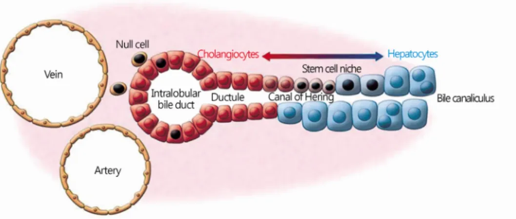
1) 간 전구세포(hepatic progenitor cells)간에 존재하는 정상 줄기세포로 가장 잘 알려진 것이 난원 세포(oval cells) 또는 간 전구세포이다. 설치류에서의 난원세 포와 비슷한 세포가 사람의 간에 존재하여 난원세포로 혼용해 서 사용하지만 사람에서는 간 전구세포라 부르는 것이 바람직 하다[31]. 간 전구세포는 canal of Hering에 존재한다(Fig. 6).
Canal of Hering은 담세관(bile canaliculi)과 소엽사이담관 (interlobular bile duct) 사이의 연결부위로 간 줄기세포의 미세 환경(niche)을 제공한다.
간 전구세포는 조혈줄기세포처럼 CD34, c-kit와 같은 표지 자를 발현하여 골수 조혈줄기세포로부터 기원한 것으로 생각 된다. 또한 간세포 표지자인 AFP, 담관세포 표지자인 CK7, CK19도 발현한다[31-33]. 간 전구세포는 스스로 재생하는 자 기재생 능력과 함께 간세포와 담관세포로 분화되는 능력을 가짐으로써 줄기세포의 성질을 가진다. 간세포암과 담관세포 암이 섞여 있는 중간형태의 암(intermediate HCC-CC)은 간 전구세포에서 기원하였을 것으로 생각된다. CK7, CK19을 발 현하는 간세포암종은 예후가 불량하다[34].
Fig. 6. Hepatic stem cell niche in the canals of Hering. Hepatic progenitor cells generate hepatocytes and cholangiocytes in canals of Hering. J Clin Invest 2013;123:1874-80.
2) 혈관주위세포(pericytes)
과거 간성상세포를 pericytes라고 부르기도 하였으나 혈관 주위세포를 명명할 때의 pericytes는 간성상세포의 pericytes와 는 다르다[35]. 간 내에 존재하는 혈관주위세포는 다른 장기의 혈관주위세포와 유사하다. 형태적으로는 모세혈관벽에서 혈관 내피세포를 지지하는 세포지만, 세포 표면에는 CD146, NG2, CD140b와 같은 간엽줄기세포의 표지자를 발현하고, CD34, CD31, CD45와 같은 혈관내피세포 또는 조혈세포 표지자를 발현하지 않는다. 다른 장기의 혈관주위세포처럼 CD146+ CD45-CD56-CD34-세포를 혈관주위세포로 정의하여 분리하 였을 때 성인 간에는 0.56%, 태아 간에는 0.45%가 존재한다 [35]. 이 혈관주위세포를 장기간 계대 배양하면 간엽줄기세포 의 모양과 성상을 띄게 되고, 지방세포, 뼈세포 및 연골세포로 분화하는 능력을 가진다. 이처럼 간의 혈관주위세포는 간엽줄 기세포의 근원으로 생각된다[35].
6. 간암 줄기세포의 분리(isolation of liver cancer stem cells)
혈액암에서 암 줄기세포의 존재가 확인된 이후 많은 고형암 에서 암 줄기세포가 분리되었다. 암 줄기세포를 증명하기 위해 서는 자기재생 능력과 분화능력 및 암 발생능력을 증명하여야 하며, 암 줄기세포 표지자를 발현해야 한다. 다른 고형암에서 발견된 것과 비슷하게 현재까지 알려진 간암 줄기세포 표지자 로는 CD133 (Prominin-1), CD90 (Thy-1), CD44, EpCAM (CD326), CD13 등이 있다[36-41]. 간암 세포주에서 CD133 의 빈도는 0-65%로 다양하였다[36]. CD133 양성 세포들은
in vitro
에서 콜로니를 형성하였고,in vivo
에서 종양 형성능력 및 방사선에 대한 저항을 보여주었다[36,37]. 그러나 암 줄기 세포가 전체 종양에서 5% 이하로 아주 적게 존재하는 점을감안할 때 CD133 단독 표지자로서 유용성은 제한적일 수 있다. Ma 등은 aldehyde dehydrogenase (ALDH)를 이용하여 CD133+ALDH+>CD133+ALDH->CD133-ALDH- 순으로 종양 형성능에 차이가 있음을 보고하였다[38]. 또 Zhu 등은 CD133+ CD44+ 세포들이 CD133+CD44- 세포들보다 강한 종양 생성능, 항암제에 대한 저항성을 보인다고 하였다[39]. CD90은 종양 의 0-2.5% 정도 존재하는 것으로 알려지는데, CD90+ 세포들 은 강한 종양 형성능을 보였다[40]. 여기에 더하여 CD90+ CD44+가 동시 발현되는 경우는 CD90+ 단독 발현 때보다 더 욱 강한 종양 형성능과 전이를 보였다[40]. EpCAM+ 세포들 역시 암 줄기세포의 특성과 함께 강한 종양 형성능을 보였다 [41].
최근 Park 등은 CD34+ 조혈줄기세포가 간 재생에 중요하다 는 사실에 착안하여 간암 발생에 있어서 CD34+ 정상 줄기세 포가 CD34+ 암 줄기세포로 전환될 수 있음을 가정하였다[42].
그들은 PLC/PRF/5 간암세포주에서 CD34+ 세포들을 분리하여 NOD/SCID/IL-2 reg 면역결핍쥐에 주입하였는데, 100개의 세 포만으로 종양이 형성되었고, 22회 연속 계대 배양에도 자기 재생과 종양 형성능을 계속 유지하였다[42,43].
7. 간암 줄기세포의 기원(cellular origin of liver cancer stem cells)
간세포암은 간암 줄기세포를 기점으로 생긴다. Holczbauer 등은 간 계통(hepatic lineage)에 있는 다양한 세포들이 간암 줄기세포로 재설정(reprogramming)될 수 있음을 증명하였다 [44]. 그 외 다른 세포들로부터도 기원할 수 있다(Fig. 7).
1) 간세포(hepatocytes)
Fig. 7. Cellular origins of liver CSCs. Liver CSCs can originate from mature hepatocytes, hepatic progenitor cells, hematopoietic stem cells, and mesenchymal stem cells. Hematopoietic stem cells can convert to liver CSCs by cell fusion with hepatocytes or Kupffer cells, or directly without fusion. CSCs, cancer stem cells.
간세포암의 17.7%가 분화된 간세포에서 기원한다고 보고 되었다[45]. 간세포암은 통상 만성염증 또는 간경변증 상태에 서 발생한다. 간경변증은 세포외 기질(extracellular matrix, ECM)의 재형성(remodeling)의 결과로 생긴다. 세포외 기질의 변화는 간세포의 HNF-1, HNF-4와 같은 간 특이 전사인자의 변화를 통하여 유전자 변이를 일으킨다[46]. 또 세포외 기질의 화학적 구성, 분자구조 및 공간 변형이 간세포내 중간 필라멘 트의 변화를 초래한다[47]. 또 역분화 과정에 중요한 상피- 간엽 전환에 TGF-β가 중요한 것으로 보고되었다[48].
최근에는 TNF-α, IL-6, TGF-β와 같은 염증 사이토카인이 간세포의 역분화에 관여한다고 보고되었다. 특히 IL-6는 간세 포암 발생에 필요한 전사인자인 STAT3를 활성화하였고, IL-6 가 높은 경우 예후가 불량하였다[49].
2) 간 전구세포(hepatic progenitor cells)
일부 간 종양세포는 중간형태, 즉 간세포와 담관세포가 섞 여 있는 형태를 보인다. 그리고 이러한 형태의 종양에는 간 전구세포가 포함되어 있다. 이러한 사실로 intermediate or combined type HCC는 간 전구세포에서 기원하였음을 설명할 수 있다.
만성간염에서 간경변증으로 진행하는 과정에서 간세포의 증식이 활발히 일어나다가 간손상이 매우 심해진 간경변증의 어느 시점에서는 간세포 증식이 급감하면서, 대신 간 전구세포 가 활성화된다. 그러므로 간 전구세포의 활성화는 심한 간경 변을 의미하고 암성 변화의 위험이 높음을 의미한다[50].
3) 골수 조혈줄기세포(hematopoietic stem cells)
CD34+ 조혈줄기세포는 간의 재생에 중요한 역할을 한다.
그러므로 CD34+ 조혈줄기세포가 CD34+ 암 줄기세포로의 전 환을 통하여 간세포암이 발생할 수 있다. Park 등은 CD34+ PLC/PRF/5 세포들이 기능적으로 간암 줄기세포임을 증명하 였다[42,43]. 이 세포들을 면역결핍쥐에 주입하였을 때 간세 포암, 담관세포암 및 혼합형태의 암을 유발하였다. CD34와 CD31, CD133, CD90, CD44, OV6, EpCAM이 동시에 양성인 경우는 간세포-담관세포의 혼합형 종양을, CD34 단독 양성인 경우에는 간세포암 만을 유발하였다. 조혈줄기세포가 어떻게 간암 줄기세포로 변환되는지에 대하여는 좀 더 연구가 필요하 지만, 세포융합을 통하여 암 줄기세포로의 전환이 가능하다.
그 이유로 세포표면의 표지자는 이전 세포의 기원을 의미하는 단서가 되는데, 쿠퍼세포(Kupffer cell)는 CD34와 31을 발현하 고 간 손상때 중요한 역할을 하는 세포이다. 그러므로 조혈줄 기세포가 쿠퍼세포와의 융합을 통하여 간암 줄기세포로 전환 되었을 수 있고[42], 간세포와의 융합을 통하여 간암 줄기세포 로 전환되었을 수도 있다[11-13]. 또 세포융합 없이 직접 간세 포로 분화할 수도 있다[15-17].
4) 간엽줄기세포(mesenchymal stem cells)
골수 이외에 우리 몸의 다양한 고형 장기에 간엽줄기세포가 존재한다. 간에는 혈관주위세포가 약 0.5% 정도로 존재한다.
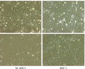
혈관주위세포가 형태적으로는 혈관을 지지하는 세포이지만, 장기간 배양하면 간엽줄기세포로 변하게 되어 간엽줄기세포 의 전구세포로 알려지고 있다[34]. 간세포암은 간세포, 간 전 구세포, 조혈줄기세포 등 다양한 세포들에서 기원하는 것으로 보고되었다. 저자와 Duan 등은 간암세포주인 SK HEP-1 세포 가 육안적으로 간엽줄기세포와 유사한데 착안하여 간엽줄기 세포와의 관련성을 조사하였다(Fig. 8) [51]. SK Hep-1 세포는 간엽줄기세포 대부분의 표지자를 발현하였고, 간엽줄기세포 의 특징인 뼈세포, 연골세포 및 지방세포로 분화하였다. 특히 놀라운 사실은 단 한 개의 세포만으로 면역억제쥐에서 암을 유발하여 강력한 종양 형성능을 보이는 암 줄기세포의 성질을 보였다. 이처럼 적은 수의 암세포로 짧은 시간에 전신 다발 전이를 일으키는 것은 육종(sarcoma)에서 보는 매우 공격적인 양상이었다. 그러므로 육종처럼 심한 진행과 전이를 보이는 간세포암은 간엽줄기세포에서 기원하였을 가능성이 있다.
결 론
계층모델 개념에 따르면 암은 암 줄기세포를 기점으로 시작 된다. 그러므로 암을 근치하기 위한 노력으로 암 줄기세포에
Fig. 8. Morphology of SK Hep-1 cells and MSC-L. Both are si- milar in appearance. SK Hep-1 cells exhibit an oncogenic mesen- chymal stem cell line with a significant metastatic capacity. MSC-L, mesenchymal stem cells of liver.
연구가 집중되고 있다. 그러나 암 줄기세포의 기원이 되는 세 포의 종류에 따라 표지자의 발현과 교란된 신호전달경로가 서로 다르다.
간세포암의 기원이 되는 세포로는 분화된 간세포, 간 전구 세포, 조혈줄기세포, 간엽줄기세포 등 다양하다. 같은 간세포 암이라 하더라도 기원하는 세포에 따라 임상 양상과 예후가 달라지는 것으로 생각된다. 간세포암 환자에게 개별적인 맞춤 치료를 하기 위해서는 간세포암의 세포기원을 확인할 필요가 있다. 아직 간세포암의 세포기원에 대한 연구는 부족하고 임상 적용은 되고 있지 않다. 간세포암 진료에서 세포기원을 확인 하는 것은 예후판정과 치료법 개발에 중요할 것으로 생각한다.