http://dx.doi.org/10.5468/ogs.2016.59.2.97 pISSN 2287-8572 · eISSN 2287-8580
Introduction
C-reactive protein (CRP) is a widely used marker which reflect inflammation and tissue damage [1]. The measurement of CRP is quick, safe, and relatively inexpensive method. The in- crease in serum CRP accompanies inflammatory diseases such as cancer, asthma, and neonatal intrapulmonary disease [2]. In obstetrics, the elevated serum level of CRP is associated with adverse pregnancy outcomes which include preeclampsia, chorioamnionitis, intrauterine growth restriction and preterm birth [1,3-6].
Ultrasound-indicated cerclage (UIC) is beneficial for prolong- ing pregnancy in women with prior preterm birth and short
Preoperative and postoperative serum C-reactive protein levels to predict the outcome of ultrasound- indicated cerclage
Hyun-Jeong Yim, Ji Eun Song, Ji-Eun Kim, Ga Hyun Son, Keun Young Lee
Department of Obstetrics and Gynecology, Kangnam Sacred Heart Hospital, Hallym University Medical Center, Hallym University College of Medicine, Seoul, Korea
Objective
To assess the role of preoperative and postoperative serum C-reactive protein (CRP) level on the prediction of pregnancy outcomes following ultrasound-indicated cerclage (UIC).
Methods
We retrospectively reviewed the medical records 44 women who underwent UIC between January 2011 and December 2011. UIC was performed between 14 and 24 weeks of gestation in women with short cervix. We divided UIC patients into two groups according to the gestational age at delivery (34 weeks) and compared the two groups. Group A was defined as patients who delivered ≥34 weeks of gestation, and group B as patients delivered <34 weeks. Correlation and receiver- operating characteristic curves were also analyzed for the prediction of preterm birth after UIC.
Results
Thirty women delivered ≥34 weeks (group A) and 14 women delivered <34 weeks (group B). Pre- and post-cerclage CRP were significantly lower in group A (pre-cerclage CRP, 1.1±1.0 vs. 11.4±6.2 mg/dL, P<0.001; post-cerclage CRP, 0.6±0.5 vs. 7.4±7.2 mg/dL, P<0.001). The mean gestational age at delivery in group A was 37.7±1.8 weeks and that in group B was 26.9±4.3 weeks (P<0.001). There were significant negative correlations between pre- and post-cerclage CRP and latency from UIC to delivery (r=-0.82, P<0.001; r=-0.70, P<0.001, respectively).
Conclusion
Both pre- and post-cerclage CRP were useful in predicting the preterm birth following UIC.
Keywords: C-reactive protein; Ultrasound-indicated cerclage
Articles published in Obstet Gynecol Sci are open-access, distributed under the terms of the Creative Commons Attribution Non-Commercial License (http://creativecommons.
org/licenses/by-nc/3.0/) which permits unrestricted non-commercial use, distribution, and reproduction in any medium, provided the original work is properly cited.
Copyright © 2016 Korean Society of Obstetrics and Gynecology Received: 2015.8.22. Revised: 2015.10.6. Accepted: 2015.10.27.
Corresponding author: Ji Eun Song
Department of Obstetrics and Gynecology, Kangnam Sacred Heart Hospital, Hallym University Medical Center, 1 Singil-ro, Yeongdeungpo- gu, Seoul 07441, Korea
Tel: +82-2-829-5151 Fax: +82-2-833-5323 E-mail: [email protected]
http://orcid.org/0000-0002-1030-0096
cervix (<25 mm) [7]. There are some reports on the outcomes of UIC [8,9]. The authors demonstrated that UIC significantly prolonged pregnancy and resulted in favorable pregnancy outcomes. However, the predicting factors for the success of UIC have not been thoroughly evaluated. There is a need to develop biomarkers predicting the outcome of UIC. In this study, we investigated the relation between the preoperative and postoperative serum CRP levels and the efficacy of UIC.
Materials and methods
This was a retrospective study of 44 women with UIC be- tween January 2011 and December 2011. All women under- went UIC at Kangnam Sacred Heart Hospital, Hallym Univer- sity Medical Center. UIC was performed between 14 and 24 weeks of gestation in singleton pregnant women with short cervix (<25 mm) and previous preterm birth. Short cervix was diagnosed by cervical sonographic screening using a standard method [10]. Women with visible bulging membrane into va- gina, multiple pregnancy, fetus with structural anomalies, fetal chromosomal anomalies, vaginal bleeding, preterm premature rupture of membranes, preterm labor pain, chorioamnionitis, or women with chronic medical diseases were excluded. Prior to the UIC, microbiological analyses were performed by vagi- nal swabs with sterile swabs. The maternal serum CRP levels were measured twice at pre- and post-cerclage day 1.
UIC was performed using McDonald technique with Mersi- lene tape (Braun, Tuttlingen, Germany) under spinal anesthe- sia. UIC was technically successful in all patients. Prophylactic antibiotics, 1.0 g intravenous cefotiam twice a day, were given at least 3 days after admission. Tocolytics were not routinely administered after UIC. Cerclage sutures were routinely re- moved at 37 weeks of gestation if patients have no complica- tions. When combined with preterm labor, premature rupture of membranes, or chorioamnionitis, cervical cerclages were removed at the discretion of maternal-fetal medicine special- ists.
Maternal demographics and pregnancy outcomes following UIC were reviewed from patients’ medical records. Patients with UIC were divided into two groups: one group is consisted of women who delivered at ≥34 weeks of gestation; the other group is consisted of women who delivered at <34 weeks of gestation. We compared the maternal demographics , preop- erative and postoperative laboratory findings, and pregnancy
outcomes between two groups.
Statistical analysis was performed with the IBM SPSS ver.
22.0 (IBM Corp., Armonk, NY, USA). Data are represented ei- ther as number and percent, or mean±standard deviation for continuous variables. Continuous variables were evaluated by Student’s t-test or Mann-Whitney U-test, where appropriate.
Chi-square analysis was used for categorical data. Correlations for continuous variables were assessed using either the Pear- son or the Spearman test, depending on normal distribution.
Receiver-operating characteristic curves were analyzed to pre- dict preterm birth after UIC.
Results
Table 1 demonstrates maternal demographics and pregnancy outcomes after UIC. The mean gestational age at cerclage was 20.5 weeks, and the mean cervical length at cerclage was 15.1 mm. Maternal serum CRP at post-cerclage was de- creased compared to pre-cerclage CRP (pre-cerclage CRP, 4.4 mg/dL vs. post-cerclage CRP, 2.8 mg/dL). However, there was no statistically significant difference between pre- and post- cerclage CRP. Pregnancy outcomes following UIC were favor- able. All 44 patients had live births and there was no neonatal death. The mean gestational age at delivery was 34.2 weeks,
Table 1. Patient characteristics and pregnancy outcome after ultrasound-indicated cerclage (n=44)
P atient characteristics &
pregnancy outcome Value
Maternal age (yr) 32.3±4.4
Body mass index (kg/m2) 17.5±3.8
Artificial abortion 16/44 (36.4)
Cervical surgery 2/44 (4.5)
GA at cerclage (wk) 19.6±3.3
CL at cerclage (mm) 15.1±4.3
CL after cerclage (mm) 26.5±6.8
Pre-cerclage CRP 4.4±6.0
Post-cerclage CRP 2.8±5.1
At delivery
GA at delivery (wk) 34.2±5.8
Birthweight (g) 2,372.5±995.9
Days from cerclage 102.5±48.2
Values are presented as mean±standard deviation or n (%).
GA, gestational age; CL, cervical length; CRP, C-reactive protein.
and the average birthweight was 2,372.5 g. The mean pro- longation days after UIC were 102.5 days.
Thirty women delivered ≥34 weeks of gestation (group A), and 14 women delivered <34 weeks of gestation (group B).
Table 2 shows the comparison of maternal demographics and pregnancy outcomes between two groups. There were no statistical differences in maternal demographics between two groups. Gestational age at cerclage, cervical length at cerclage, and cervical length at post-cerclage showed no significant differences between two groups. No significant difference in pre-cerclage labs was detected between two groups. The mean gestation age at delivery in group A was 37.7 weeks, whereas that in group B was 26.9 weeks. The major causes of preterm birth in group B were preterm premature rupture of membrane, chorioamnionitis, and preterm labor. There were significant differences in pre- and post-cerclage CRP between two groups: The mean pre-cerclage CRP in group A was 1.1 mg/dL, whereas that in group B was 11.4 mg/dL (P<0.001);
The average post-cerclage CRP in group A was 0.6 mg/dL, whereas that in group B was 7.4 mg/dL (P<0.001). Both group showed decreased post-cerclage CRP level compared to
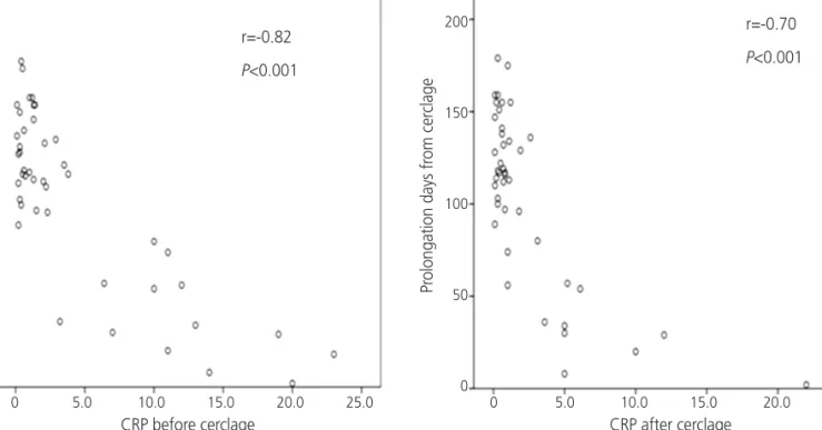
pre-cerclage CRP level. However, it was not a statistically sig- nificant decrease (P=0.07). Fig. 1 demonstrates the negative correlations between pre- and post-cerclage CRP and latency from cerclage to delivery (r=-0.82, P<0.001; r=-0.70, P<0.001, respectively). Patients with lower pre- and post-cerclage CRP showed less preterm birth following UIC. We constructed re- ceiver-operating characteristic curves of pre- and post-cerclage CRP to predict delivery <34 weeks of gestation. Area under curves of both pre- and post-cerclage CRP were significantly large (Fig. 2). Both pre- and post-cerclage CRP were useful in the prediction of preterm birth after UIC.
Discussion
The present study suggests that both of pre- and post-cerclage CRP are useful in predicting the risk of preterm birth following UIC. Women with lower pre- and post-cerclage CRP is likely to prolong pregnancy after UIC.
It is widely known that a short cervix less than 25 mm in mid-trimester is high risk for preterm birth [11]. There were Table 2. Comparison of patient characteristics and pregnancy outcome according to GA at delivery
GA at delivery <34 wk
(n=14) GA at delivery ≧34 wk
(n=30) P-value
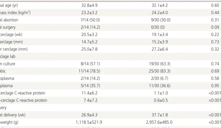
Maternal age (yr) 32.8±4.9 32.1±4.2 0.60
Body mass index (kg/m2) 23.2±3.2 24.2±4.0 0.44
Artificial abortion 7/14 (50.0) 9/30 (30.0) 0.31
Cervical surgery 2/14 (14.2) 0/30 (0) 0.09
GA at cerclage (wk) 20.5±3.2 19.1±3.4 0.22
CL at cerclage (mm) 14.7±5.2 15.2±3.9 0.73
CL after cerclage (mm) 25.0±7.8 27.2±6.4 0.32
Pre-cerclage lab
Gram culture 8/14 (57.1) 19/30 (63.3) 0.74
Aerobic 11/14 (78.5) 25/30 (83.3) 0.69
Mycoplasma 2/14 (14.2) 2/30 (6.7) 0.58
Ureaplasma 5/14 (35.7) 11/30 (36.6) 0.95
Pre-cerclage C-reactive protein 11.4±6.2 1.1±1.0 <0.001
Post-cerclage C-reactive protein 7.4±7.2 0.6±0.5 <0.001
At delivery
GA at delivery (wk) 26.9±4.3 37.7±1.8 <0.001
Birthweight (g) 1,118.5±521.9 2,957.6±485.0 <0.001
Days from cerclage 44.7±33.6 129.5±23.9 <0.001
Values are presented as mean±standard deviation or n (%).
GA, gestational age; CL, cervical length.
debates on the efficacy of UIC, but a meta-analysis demon- strated that UIC in women with prior preterm birth and short cervix (<25 mm) is beneficial in reducing preterm birth [12].
There have been some reports on the pregnancy outcomes following UIC. The authors reported that UIC resulted in fa-
vorable pregnancy outcome with high incidence of live births [13,14].
However, the predicting factors for the pregnancy outcome after UIC have not been thoroughly studied. There was a re- port on cervical mucus interleukin (IL)-8 in UIC. The increase of cervical mucus IL-8 was associated with preterm birth fol- lowing UIC [15]. There are some limitations on cervical mucus IL-8; it is not a widely used lab and obstetricians could not get the prompt result of cervical mucus IL-8. CRP is stable, inex- pensive, quick marker for inflammation. In this respect, serum CRP is more advantageous than cervical mucus IL-8.
There were two reports on serum CRP in patients with emergency cerclage [16,17]. The authors demonstrated that serum CRP predicted the outcome of emergency cerclage in women with dilated cervix and bulging membrane [16,17].
The study participants in the current study were different from those studies; The present study mainly focused on women with sonographically short cervix and prior preterm birth. We excluded women with dilated cervix and bulging membrane in this study. To our knowledge, our study is the first report to evaluate the efficacy of serum CRP in UIC.
Interestingly, there were no significant differences in pre- cerclage microbiologic labs between group A and group B in this study. It may represent that cervical shortening in women Fig. 1. Correlations between pre- and post-cerclage C-reactive protein (CRP) and latency from ultrasound-indicated cerclage (UIC) to delivery.
(A) Negative correlation between pre-cerclage CRP and prolongation days after UIC. (B) Negative correlation between post-cerclage CRP and latency from UIC to delivery.
Prolongation days from cerclage
CRP before cerclage r=-0.82 P<0.001
r=-0.70 P<0.001
0 5.0 10.0 15.0 20.0 25.0 0 5.0 10.0 15.0 20.0 25.0 CRP after cerclage
Prolongation days from cerclage
200
150
100
50
0
200
150
100
50
0
A B
Specificity
0.0 0.2 0.4 0.6 0.8 1.0
Sensitivity
Pre-cerclage CRP Post-cerclage CRP Reference line
Pre-cerclage CRP; AUC: 0.939, P<0.001 Post-cerclage CRP; AUC: 0.971, P<0.001 1.0
0.8
0.6
0.4
0.2
0.0
Fig. 2. Receiver-operating characteristic curves of pre- and post- cerclage C-reactive protein (CRP) to predict delivery <34 weeks of gestation. The areas under curve (AUCs) are demonstrated in the charts.
with previous preterm birth is affected by other inflammatory process in cervix itself. In the current study, post-cerclage CRP is decreased compared to pre-cerclage CRP. Although the dif- ference between pre- and post-cerclage CRP was not statisti- cally significant, it may suggest that UIC and perioperative an- tibiotics prevent further inflammatory process in women with short cervix. Our study showed that women with high level of pre-cerclage CRP tended to deliver more prematurely. It may be beneficial for women with high level of preoperative CRP to have cerclage after confirming decreased level of CRP with antibiotic therapy.
Although our study was limited by the small sample size and retrospective study design, the present study suggests the ef- ficacy of pre- and post-cerclage serum CRP for the prediction of preterm birth after UIC in women with short cervix and prior preterm birth. In the future prospective randomized trial is need.
Conflict of interest
No potential conflict of interest relevant to this article was reported.
References
1. Kwak DW, Cho HY, Kwon JY, Park YW, Kim YH. Useful- ness of maternal serum C-reactive protein with vaginal Ureaplasma urealyticum as a marker for prediction of im- minent preterm delivery and chorioamnionitis in patients with preterm labor or preterm premature rupture of mem- branes. J Perinat Med 2015;43:409-15.
2. Ahmed SK, Mahmood N, Malalla ZH, Alsobyani FM, Al-Kiyumi IS, Almawi WY. C-reactive protein gene vari- ants associated with recurrent pregnancy loss indepen- dent of CRP serum levels: a case-control study. Gene 2015;569:136-40.
3. Lohsoonthorn V, Qiu C, Williams MA. Maternal serum C-reactive protein concentrations in early pregnancy and subsequent risk of preterm delivery. Clin Biochem 2007;40(5-6):330-5.
4. Best LG, Saxena R, Anderson CM, Barnes MR, Hakonar- son H, Falcon G, et al. Two variants of the C-reactive pro- tein gene are associated with risk of pre-eclampsia in an
American Indian population. PLoS One 2013;8:e71231.
5. Tjoa ML, van Vugt JM, Go AT, Blankenstein MA, Oudejans CB, van Wijk IJ. Elevated C-reactive protein levels during first trimester of pregnancy are indicative of preeclampsia and intrauterine growth restriction. J Reprod Immunol 2003;59:29-37.
6. Morales E, Guerra S, Garcia-Esteban R, Guxens M, Alva- rez-Pedrerol M, Bustamante M, et al. Maternal C-reactive protein levels in pregnancy are associated with wheezing and lower respiratory tract infections in the offspring. Am J Obstet Gynecol 2011;204:164.e1-9.
7. Berghella V, Rafael TJ, Szychowski JM, Rust OA, Owen J.
Cerclage for short cervix on ultrasonography in women with singleton gestations and previous preterm birth: a meta-analysis. Obstet Gynecol 2011;117:663-71.
8. Nelson L, Dola T, Tran T, Carter M, Luu H, Dola C. Preg- nancy outcomes following placement of elective, urgent and emergent cerclage. J Matern Fetal Neonatal Med 2009;22:269-73.
9. Chan LL, Leung TW, Lo TK, Lau WL, Leung WC. Indica- tions for and pregnancy outcomes of cervical cerclage: 11- year comparison of patients undergoing history-indicated, ultrasound-indicated, or rescue cerclage. Hong Kong Med J 2015;21:310-7.
10. Berghella V, Bega G. Ultrasound evaluation of the cervix.
In: Callen PW, editors. Ultrasonography in obstetrics and gynecology. 5th ed. Philadelphia (PA): Saunders/Elsevier;
2008. p. 698-720.
11. Iams JD, Goldenberg RL, Meis PJ, Mercer BM, Moawad A, Das A, et al. The length of the cervix and the risk of spon- taneous premature delivery. National Institute of Child Health and Human Development Maternal Fetal Medicine Unit Network. N Engl J Med 1996;334:567-72.
12. Berghella V, Odibo AO, To MS, Rust OA, Althuisius SM.
Cerclage for short cervix on ultrasonography: meta- analysis of trials using individual patient-level data. Obstet Gynecol 2005;106:181-9.
13. Berghella V. Transvaginal ultrasound assessment of the cervix and prediction of spontaneous preterm birth [In- ternet]. [place unknown]: UpToDate [cited 2016 Feb 6].
Available from: http://www.uptodate.com.
14. Skupski DW, Lin SN, Reiss J, Eglinton GS. Extremely short cervix in the second trimester: bed rest or modified Shi- rodkar cerclage? J Perinat Med 2014;42:55-9.
15. Sakai M, Shiozaki A, Tabata M, Sasaki Y, Yoneda S, Arai T,
et al. Evaluation of effectiveness of prophylactic cerclage of a short cervix according to interleukin-8 in cervical mu- cus. Am J Obstet Gynecol 2006;194:14-9.
16. Kobayashi M, Ohkuchi A, Matsubara S, Izumi A, Hirashi- ma C, Suzuki M. C-reactive protein levels at pre-/post- indicated cervical cerclage predict very preterm birth. J
Perinat Med 2011;39:151-5.
17. Minakami H, Matsubara S, Izumi A, Kosuge S, Watanabe T, Iwasaki R, et al. Emergency cervical cerclage: relation be- tween its success, preoperative serum level of C-reactive protein and WBC count, and degree of cervical dilatation.
Gynecol Obstet Invest 1999;47:157-61.