건양대학교 의과대학 내과학교실 윤세희, 손지웅, 정청일, 최유진
A Case of Behcet’s Disease with Multiple Cavitary Lung Lesion
Se Hee Yoon, M.D., Ji Woong Son, M.D., Chung Il Joung, M.D., Eu Gene Choi, M.D.
Department of Internal Medicine, Konyang University College of Medicine, Daejeon, Korea
Behcet’s disease is a systemic vasculitis of an unknown etiology involving the arteries and veins of all sizes. There are reports showing that a pulmonary artery aneurysm or thromboembolism and superior vena cava thrombosis are present in 5-10% of patients with Behcet's disease and that lung parenchymal lesions are mainly airway consolidations resulting from hemorrhage or infarction. We encountered a patient with increasing pulmonary cavitary changes and localized aspergilloma. The patient was a 43-year-old man diagnosed with Behcet’s disease with a history of recurrent oro-genital ulceration and uveitis, and who was administered methotrexate, colchicines, prednisolone. During the follow up he developed progressive dyspnea upon exertion and finger clubbing. Therefore further evaluations were performed. Chest computed tomography showed more advanced consolidations and cavitations than the previous film with the previously known aspergilloma still observable. An open lung biopsy was carried out to determine the presence of malignant changes, which revealed nonspecific vasculitis. Azathioprine was added resultion in an improvement of symptoms.
(Tuberc Respir Dis 2006; 61: 65-69)
Key words: Behcet’s disease, Cavitary lung lesion, Aspergilloma.
Address for correspondence: Eugene Choi, M.D.
Department of Internal Medicine, Konyang University Gasowon-Dong, Seo-Gu Daejeon 302-718, Korea Tel: 042-600-9100,8834
Fax: 042-545-9103 E-mail: [email protected] Received : Apr. 18. 2006 Accepted : Jun. 20. 2006
서 론
베체트병은 전신적인 혈관염에 의해 재발성의 구 강 및 외음부 궤양을 주증상으로 하여 포도막염등의 안질환과 피부병변등 전신적 장기를 침범하며 다양 한 임상양상을 나타내는 질환으로 최근 자가면역기 전이 주요한 병인으로서 대두되고 있으나 아직 원인 은 잘 밝혀져 있지 않은 질환이다1). 베체트병의 기본 적인 병리학적 변화는 혈관염이며 전신 증상은 혈관 염의 정도에 따라 별현한다. 점막 궤양만이 재발하는 경우는 예후가 양호하지만 중증 장기의 침범시 환자 의 생명을 위협할 수 있다. 베체트병에서 폐침범은 5% 정도에서 발생하며 폐경색이나 출혈을 일으키며 예후가 나쁜 경우가 많다2,3). 베체트병에서 폐아스페 르길루스종이 증식할 만큼 큰 공동을 형성하며 폐를
침범한 경우는 드물며 저자들은 활동시 호흡곤란으 로 내원한 환자에서 큰 다발성 폐공동을 형성한 베체 트병 한 예를 경험하였기에 문헌고찰과 함께 보고하 는 바이다.
증 례
환 자: 43세 남자 주 소: 활동시 호흡곤란
현병력: 반복적인 구강궤양 있었고 5년전 급성 녹 내장으로 좌안을 실명하였으며 1년 6개월 전부터 활 동시 호흡곤란 있어 1년전 본원 호흡기내과에 입원하 여 흉부 전산화 단층 촬영등 검사결과 폐공동을 포함 한 폐종괴, 상대정맥협착, 폐동맥 색전증 소견 있어 세침흡입 폐생검 시행하였다. 당시 조직 검사에서 미 만성 혈관염 소견 있었으며 폐침범한 베체트병 진단 하에 methotrexate, colchicines, folic acid 복용하며 외래 추적 관찰 중이었다. 추적 관찰중 3개월 마다 촬 영한 흉부 전산화 단층 촬영에서 폐병변의 호전 보이 다가 최근 촬영한 흉부 전산화 단층 촬영상 폐공동 새로 생기며 크기 증가하는 소견 있어 폐암등 폐공동
Figure 2. At initial chest CT (A) shows multifocal consolidation with ground glass haziness and some cavitary change in subpleural area of both lung. After 16 months chest CT (B) shows newly developed cavitary lesion is at both lower lung field and the cavity of left lower lung field was more greater than before. The small mass like lesion is in the center of cavity on left lower lobe. It is suggesting aspergilloma. A: 2003/10/17 chest CT, B:
2005/01/20 chest CT
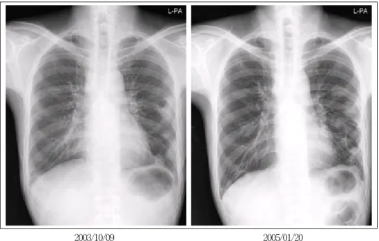
2003/10/09 2005/01/20
Figure 1. On chest radiography A, there is cavitary consolidative mass-like lesion at left upper lung field. On chest radiography B, there is more increased size of cystic lesion with air fluid level in both upper lung field and newly developed cystic lesion in right lower lung field and increased ill defined haziness in lower lung field 형성 질환 감별위해 입원하였다.
과거력 : 특이사항 없음.
가족력 : 특이사항 없음.
사회력 : 음주나 흡연은 하지 않았다.
이학적 소견 : 혈압 120/80 mmHg, 맥박 80 회/ 분, 호흡 20 회/ 분이었고 체온은 36.5℃ 였다. 만성 병색 이었고 두경부 진찰상 결막의 빈혈은 없었고, 공막의 황달 소견도 없으며, 혀와 구강점막에 궤양은 발견되
Figure 3. Histologic findings of open lung biopsy. These microscopic findings show interstitial inflammation, diffuse alveloar hemorrhage and vasculitis (A)(B)(C). and mass like lesion in the cavity reveals the septated hyphae(D) (H&E, X100,A and B) (H&E, X400, C) (GMS, X200, D).
지 않았다. 흉부진찰상 심음은 규칙적이며 심잡음은 들리지 않았다. 흉부 및 복부소견은 정상이었고 생식 기의 궤양반흔은 없었다. 입원 당시 피부 및 관절의 이상소견도 없었다.
검사실 소견: 혈색소 6.53 mmol/L, 백혈구 9.24х 109/L, 혈소판 448х109/L, 혈침속도 74 mm/hr 이었고, 혈액응고 검사상 prothrombin time 13.8초, activated partial thromboplastin time 59.2초 였다. 소변검사는 정상 소견 이었고, AST/ALT 0.26/0.20 ukat/L, 총빌 리루빈 8.5 umol/L, alkaline phosphatase 126 IU/L, 혈청 총단백/알부민 62.5/32.8 g/L, BUN 5.0 mmol/L, Cr 93 umol/L , CRP 4.58 g/L 이었다. 항핵항체 음성, 류마티스인자 음성이었다. 객담 결핵균 도말검사상 음성이었다. 심전도 검사상 특이 소견 없었다. 폐기능 검사는 노력성폐활량 2.88 L, 1초간노력성호기량 2.57
L, 1초간노력성호기량의 노력성폐활량에 대한 비는 87.5%였다.
방사선 소견: 흉부 방사선 사진상 양측 하폐야에 1 년 6개월전 처음 내원 당시와 비교 했을 때 크기가 증 가한 공동이 있었다, 우측 하폐야에 새로 생긴 공동이 있었으며 좌측 상폐야에 폐침윤 있었다. 폐 전산화 단 층 촬영에서는 상대정맥 협착과 좌폐엽간 동맥에 혈 전을 동반한 폐동맥류 관찰되었다. 좌상엽과 하엽에 공동 있으며 우하엽에 새로생긴 공동이 있었다.
(Fig.1, Fig.2)
임상경과: 내원 당시 운동시 호흡곤란 외에는 특이 증상 없었고 다발성 공동에 대한 조직검사 위해 입원 하여 복용하고 있던 면역억제제는 중단 하였다. 내원 당일 시행한 사지의 도플러 검사상 심부 정맥 혈전증 등의 특이 소견 없었다. 내원 5일째 시행한 기관지 내
시경은 특이 이상 소견 없었다. 내원 7일째 흉부외과 로 전과되어 전신 마취하에 개방적 폐생검을 시행하 였다. 흉부 전산화 단층 촬영에서 폐아스페르길루스 종이 의심되며 공동으로 보이던 좌하엽의 병변은 흉 막하의 반복적 염증이 섬유성 밴드를 형성하며 물혹 을 이루고 있었으며 좌상엽에 5х5 cm 의 공동이 있어 서 좌상후분절 절제술을 시행하였다. 환자는 기도삽 관 시행후 전신마취하에서 상대정맥 협착으로 인해 안면부종등 심해지며 상대정맥 증후군 증상 보여서 2 시간 안에 수술을 끝냈으며 수술 후 자연 회복 되었 다. 절제된 폐엽은 단면 육안 소견상 1.5х1 cm 의 노 란색의 물질을 가지고 있는 4.5х3.5х3.5 cm 크기의 낭 성 공간이 관찰되었다. 병리소견상 폐간질의 염증과 섬유화 소견이 있었으며 광범위한 혈관염을 동반한 폐출혈 양상이었다. 공동 안의 노란색 물질은 유격균 사(septated hyphea)가 관찰되었으며 진균의 조직내 침범은 없었다(Fig. 3). 따라서 조직 검사 소견으로 보아서 폐종양등 다른 폐공동 형성 질환을 배제하였 고 베체트병에 의한 폐공동 형성 폐침범을 확진 할 수 있었다. 환자는 수술 10일 후에 흉관 제거 하였고 운동시 호흡곤란은 악화나 호전등 변화 없이 퇴원하 였다. 현재 개방적 폐생검 전에 복용하였던 약에 azathioprin을 추가하여 외래 추적 진료 중이다.
고 찰
공동을 형성하는 폐병변은 염증, 종양, 선천성 질 환, 혈관염 등에 의한 것으로 나눌 수 있다. 공동을 일 으키는 염증에 인한 병은 폐농양, 괴사성폐렴, 결핵등 이 있으며 종양은 기관지폐암, 전이암이 있고 선천성 질환으로는 기관지 낭종, 선천성 낭성선 종양기형, 엽 내 기관지 폐분리증, 거대 결절성 섬유화 등이 있다.
혈관염에 의해 공동을 형성하는 질환은 Wegener granulomatosis, 패혈성 색전증, 폐경색, 혈종 등이 있 으며 베체트병에서 공동을 형성하는 경우는 드물다.
베체트병은 Hippocrates에 의해 언급된 바 있으며 1937년 터어키의 피부과 의사인 Hulusi Behcet이 구 강, 외음부 및 눈의 재발성 궤양 등을 주증상으로 하 여, 최초로 그 임상상을 기술하여 명명 하였으며4,5) 현
재는 1990년에 International Study Group for Behcet’s Disease 에서 제정한 진단기준이 가장 널리 이용되고 있으며, 반복적인 구강궤양이 필수적으로 있어야하고, 안병변, 피부병변, 외음부 병변, pathergy 검사 양성 등 4가지 중 2가지 이상을 만족할 때로 규 정되어 진다3,6,7). 본 예에서는 안병변, 구강궤양 양성 으로 베체트병을 진단 할 수 있었으며 pathergy 검사 는 음성이었고 외음부 병변은 없었다. 국내에서는 이 미 베체트병으로 진단된 환자에서의 폐혈관 및 폐침 범, 국소적 폐경색등에 대한 연구 및 몇 예의 증례보 고가 있었으나2,4,7,8) 본 증례에서처럼 다발성의 큰 폐 공동을 형성한 경우는 보고된 바가 없다.
베체트병에서 폐병변은 5-10% 환자에서 나타나며 증상으로는 객혈이 가장 흔하다. 폐병변의 형태는 폐 색전증, 폐경색, 상대 정맥 혈전, 폐동맥류 등으로 나 타나며 2년동안 30%의 사망률을 보인다. 방사선학적 소견으로 56% 환자에서 출혈과 경색으로 인한 폐침 윤이 나타나며 그외에 흉막하 폐야에 결절성 폐침윤 이 33%-83% 나타난다고 보고된다. 흉막하 폐야의 결절성 소견은 아주 드물게 공동을 형성하며 대부분 자발적으로 호전된다5). 베체트병에서 폐혈관 침범의 주된 기전은 혈관염으로 생각되며 이로 인해 동맥류 나 혈전이 생긴다고 알려져 왔다. 혈관염과 혈전이 경 색이나 국소적 또는 미만성 출혈, 국소적 무기폐를 일 으킨다. 폐경색을 일으키는 정맥 및 동맥 혈전증은 여 러가지 기전이 복합적으로 작용하는 것으로 생각되 며 혈관 내피세포에 대한 항체가 18%에서 34% 의 베 체트병 환자에서 관찰되는 것으로 보아 혈관내피세 포의 면역학적 손상이 주된 기전 중 하나로 보고 되 고 있다9-11,13). 이 항체가 직접적으로 혈관내피세포에 손상을 일으키는지 아니면 내피세포의 변화가 이차 적으로 항체생성을 유도하는지에 대하여서는 논란이 있으며 혈관염으로 인한 내피세포의 손상은 prosta- cyclin, thromboxane B2, tissue factor pathway in- hibitor 등의 생산에 이상을 초래하여 과응고 경향을 유발하는 것으로 추정된다12-14). 본 증례에서는 상대 정맥을 침범하였고 폐동맥류를 형성하며 폐동맥 색 전증을 동반하며 폐실질에서 미만성의 출혈, 경색소 견을 나타내어 베체트병의 폐침범시 나타날 수 있는
대부분의 병변을 가지고 있었다. 본 예에서 발견된 큰 공동의 형성은 혈관염에 의한 국소적 경색이 반복되 어 공동을 형성 한 것으로 추정된다. 공동에서 발견된 폐아스페르길루스종은 천식등 임상 소견이 없으며 혈청 IgE 농도 증가, 호산구혈증도 없어 Greenber- ger-Patterson등의15) allergic aspergillosis의 진단 기 준에 해당사항은 없었다. 그리고 조직침범의 소견은 보이지 않아 공동에 단순 집락을 이룬 것으로 생각된 다.
요 약
저자들은 43세 남자환자에서 반복적인 구강궤양을 가지고 있으며 안증상으로 진단한 베체트병 환자에 서 폐동맥 색전증, 폐동맥류, 상대정맥 협착등 다양한 폐혈관 침범과 함께 폐아스페르길루스종을 동반한 다발성 공동 형성을 경험하였기에 문헌고찰과 함께 보고하는 바이다.
참고문헌
1. Sakane T, Takeno M, Susuki N, Inaba G. Behcet’s disease. N Engl J Med 1999;341:1284-91.
2. Jegal YJ, Chang HK, Ryu DS, Won KS. A case of Behcet’s disease with pulmonary infarction. Korean J Med 2000;59:535-9.
3. Raz I, Okon E, Chajek-Shaul T. Pulmonary mani- festations in Behcet’s syndrome. Chest 1989;95:585-9.
4. Kim YJ, Lee SM, Ahn Y. A case of Behcet’s disese with superior vena cava syndrome. Tuberc Respir Dis 2004;56:657-63.
5. Erkan F, Gul A, Tasali E. Pulmonary manifestation
of Behcet’s disease. Thorax 2001;56:572-8.
6. Park KJ, Park SH, Kim SJ, Kim HJ, Chang J, Ahn CM, et al. Clinical manifestations of the lung involvement in Behcet’s syndrome. Tuberc Respir Dis 1996;43:763-73.
7. Kim HS, Cho JH, Yang MH, Kim HJ, Park BJ, Kim YS, et al. A case of suspected Behcet’s disease diagnosed by manifestation of pulmonary artery aneurysm. Tuberc Respir Dis 2002;52:405-10.
8. Yoo DH, Jung SS, Choi YC, Lee J, Ahn JH, Kim SY, et al. A case of Behcet’s disease with pulmonary arteritis manifestaed as multiple pulmonary nodules.
Korean J Med 1989;36:695-700.
9. Aydintug AO, Tokhoz G, D’Cruz DP, Gurler A, ervera R, Duzgun N, et al. Antibodies to endotherlial cells in patients with Behcet’s disease. Clin Immunol Im- munopathol 1993;67:157-62.
10. Haznedaroglu IC, Celik I, Buyukasik Y, Kosar A, Kirasli S, Kundar SV. Haemostasis, thrombosis, and endothelium in Behcet’s disease. Acta Haematol 1998;99:236-7.
11. Cervera R, Navarro M, Lopez-Soto A, Cid MC, Font J, Esparrza J, et al. Antibodies to endothelial cells in Behcet’s disease: cell-binding heterogeneity and asso- ciation with clinical activity. Ann Rheum Dis 1994;
53:265-7.
12. Han SW, Kang YM, Kim YW, Lee JT. Cardiovascular involvement in Behcet’s disease. Korean J Med 2003;
64:542-51.
13. Cines DB, Pollak ES, Buck CA, Loscalzo J, Zimmerman GA, McEver RP, et al. Endothelial cells in physiology and in the pathophysiology of vascular disorders. Blood 1998;91:3527-61.
14. Broze GJ Jr. Tissue factor pathway inhibitor and the current concept of blood coagulation. Blood Coagul Fibrinolysis 1995;6(Suppl):S7-13.
15. Greenberger PA, Patterson R. Diagnosis and management of allergic bronchopulmonary asper- gillosis. Ann Allergy 1986;56:444-8.