I Unusual Acute Encephalitis Involving the Thalamus: Imaging Features
7
0
0
전체 글
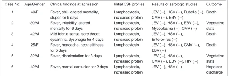
(2) Imaging Features of Unusual Acute Encephalitis. brain CT and MR imaging findings of these six patients.. MATERIALS AND METHODS Between 1997 and 1999, we retrospectively reviewed the medical records and CT and/or MR imaging findings of six patients [four men and two women aged 25 to 42 (mean, 37) years] with acute encephalitis involving the thalamus. The condition was diagnosed on the basis of clinical features and CSF examinations. The clinical features included fever, headache, disorientation, and confusion or mental alteration. In all patients, CSF examinations revealed lymphocyte-dominant pleocytosis in the range of 32 to 442 cells per microliter, elevated protein, and normal glucose. The clinical and laboratory findings are summarized in Table 1. No patient had been recently immunized or immunocompromised. In all patients, initial serologic tests for JE and HSE viruses were performed during the acute and convalescent stages (7-40 days after the onset of illness). In three, the serologic test for JE virus was performed twice, at an interval of 7 to 30 days, and in two, the serologic test for HSE virus was also performed twice, at an interval of 5 to 7 days. Serologic diagnosis of JE was based on the criteria developed by the research group for JE in Japan (i.e. a fourfold or greater rise in positive results in the haemagglutination inhibition test for the JE virus in paired sera) (9). For serological diagnosis of HSE, polymerase chain reaction (PCR) was used for the detection of herpes simplex virus DNA in CSF (10). In addition, serologic tests for a variety of pathogens were performed: cytomegalovirus in four patients, Epstein-Barr virus in four, mycoplasma in one, enterovirus in one, rubella in one, and human immunodeficiency virus in one. In all patients,. serum and CSF were also examined for ordinary bacteria, fungus and acid-fast bacili (AFB) by direct staining and culture. All the serologic examinations mentioned above were negative in all patients. Despite the fact that all patients were treated with acyclovir, clinical outcomes were very poor: death (n=3) or a vegetative state (n=3). Brain imaging studies were performed during the acute and/or convalescent stage of the illness. CT (n=6) and MR imaging (n=6) were performed 3 to 18 days and 5 to 40 days, respectively, after the onset of the condition. Four patients underwent follow-up MR imaging 11 to 45 days after onset, and one of these underwent further follow-up imaging 48 days after onset. For MR imaging, a 1.5 T imager (Magnetom; Siemens, Erlangen, Germany) was used in four cases, and a 1.0 T imager (from the same source) in two. All patients underwent spin-echo T1-weighted (repetition time [msec]/echo time [msec]: 500 800/20 25), fast spin-echo T2-weighted (2,200 2,500/60-90), and fluid-attenuated inversion recovery MRI sequences. Contrast-enhanced (IV injection of 0.1 mmol/kg of gadopentetate dimeglumine) T1-weighted spin-echo images were obtained in all patients except one (case 2). Two radiologists evaluated the imaging features, focusing on the involved area, attenuation at CT and signal intensity at MR imaging of the lesion, and whether or not enhancement was observed.. RESULTS The brain CT and MR imaging findings are summarized in Table 2. Brain CT performed 3-18 days after the onset of illness revealed diffuse brain swelling (n=2), low attenuation in both thalami (n=1), or no abnormal findings (n=3).. Table 1. Clinical and Laboratory Findings of Six Patients Case No.. Age/Gender. Clinical findings at admission. Initial CSF profiles. Results of serologic studies. 1. 40/F. 2. 39/M. 3. 42/M. 4. 25/F. 5. Lymphocytosis, increased protein Lymphocytosis, increased protein Lymphocytosis, increased protein Lymphocytosis. 32/M. Fever, chill, altered mentality, stupor for 5 days Fever, irritability, altered mentality for 6 days Mild febrile sense, sore throat dysarthria, dysphagia for 4 days Fever, headache, neck stiffness for 5 days Fever, disorientation for 3 days. 42/M. Fever, mental confusion for 2 days. JEV ( ), HSV ( ), Rubella ( ), Death CMV ( ), EBV ( ) Vegetative JEV ( ), HSV ( ), EBV ( ), state Mycoplasma ( ), CMV ( ) Death JEV ( ), HSV ( ), Enterovirus ( ) Death JEV ( ), HSV ( ), CMV ( ), EBV ( ) Vegetative JEV ( ), HSV ( ), state CMV ( ), EBV ( ), HIV ( ) Hopeless JEV ( ), HSV ( ) discharge. 6. Lymphocytosis, increased protein Lymphocytosis, increased protein. Outcome. Note. (-) = negative, CMV = cytomegalovirus, EBV = Epstein-Barr virus, HIV = human immunodeficiency virus, HSV = herpes simplex virus, JEV = Japanese encephalitis virus. Korean J Radiol 2(2), June 2001. 69.
(3) Kim et al.. Table 2. Radiological Findings of Six Patients Involved areas seen on MR images (days after the Sx onset). Case CT Findings (days No.. after Sx onset). Initial. First follow-up. 1. Normal (5). Both thalami, left hippocampus, medial area of left temporal lobe (8). 2. Normal (6). 3. Low density on both thalami (6) Diffuse brain swelling (18) Diffuse brain swelling (3). Both thalami, both hippocampi, pons, right substantia nigra, medial area of both temporal lobes (40) Both thalami, both substantia nigra, both tegmenta of the midbrain (10) Right thalamus, right insular cortex, right tempotal lobe cortex (19) Both thalami, left hippocampus medial area of left temporal lobe (6). Both thalami, both hippocampi, midbrain, Not examined pons, medulla, medial area of left temporal lobe (11) Not changed (45) Not examined. 4 5. 6. Normal (3). Both thalammi, medial area of both temporal lobes, pons, midbrain, medulla (5). In all patients, initial MR images of the brain obtained 5-40 days after onset revealed multiple patchy areas of focal abnormality. The thalamus was involved either bilaterally (n=5) or unilaterally (n=1), thalamic lesions being homogeneously hypointense on T1-weighted and hyperintense on T2-weighted images. In no patient did the signal intensities observed on initial and follow-up MR images suggest hemorrhage. Lesions were also seen in the midbrain (n=5), the medial area of the temporal lobe (n=4; 3 bilateral and 1 unilateral), the pons (n=3), both hippocampi (n=3), the insular cortex (n=2; 1 bilateral and 1 unilateral), the medulla (n=2), the lateral temporal lobe cortex (n=1), both cingulate gyri (n=1), both basal ganglia (n=1), and the cortex of the left hemisphere (n=1). Follow-up MR images obtained in four patients showed that in two (cases 1 and 5), the extent of the lesion had increased markedly. In one of these two (case 5) the lesion subsequently became smaller, and this was associated with diffuse brain atrophy and periventricular white matter change, suggesting microcystic cerebromalacia or gliosis. In the remaining two patients (cases 2 and 4), follow-up MR imaging of the lesion revealed no apparent change. Contrast-enhanced T1-weighted MR images showed no parenchymal enhancement, though diffuse corticosulcal vascular or leptomeningeal enhancement was seen in two patients (cases 3 and 5).. 70. Second follow-up. Not examined Not changed, but more definite lesion in the right temporal lobe cortex (32) Both thalami, both basal ganglia, Both insular cortices, both cingulate gyri, medial area of both temporal lobes, both hippocampi, both substantia nigra, both tegmenta of midbrain, cortex of left hemisphere (18) Not examined. Not examined Decreased size of previous lesions associated with diffuse brain atrophy and periventricular leukomalacic change (48). DISCUSSION A wide range of pathogenic organisms, the most common of which are viruses which include herpes simplex types 1 and 2, herpes zoster, arboviruses and enteroviruses, cause acute encephalitis (1, 11). In general, acute viral encephalitis causes diffuse parenchymal infiltration of inflammatory cells, and this leads to chromatolysis and pyknosis of neurons and at times extensive necrosis (1, 11). These pathologic findings are reflected by areas of low attenuation on CT, low signal intensity on T1-weighted MR images, and high signal intensity on T2-weighted MR images, depending on the degree and severity of inflammation. In our cases, although no specific viral etiology was proved in any patient, the CT and MR imaging findings appear to be similar to those of JE in terms of the frequency of involvement of the thalamus and brain stem, and -inparticular-, of the substantia nigra and pons. In JE, pathologic changes occur mainly in the gray matter and predominantly affect the diencephalon, mesencephalon, brain stem and cerebellar Purkinje cells, involving both thalami and the substantia nigra the most severely (3, 12 13). It has been reported that the CT and MR imaging findings of JE are consistent with the distribution of pathologic change. Previous articles describing these findings stated that the thalamus was usually symmetrically involved and other arKorean J Radiol 2(2), June 2001.
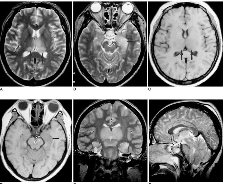
(4) Imaging Features of Unusual Acute Encephalitis. A. B. C. D E F Fig. 1. Case 1. A 40-year-old woman who presented with fever, chill and altered mentality. A-D. T2-weighted (A, B) and T1-weighted (C, D) images obtained 8 days after the onset of illness show abnormal hyperintensity and hypointensity in both thalami (large black arrows), the left hippocampus (small black arrow), and the medial area of the left temporal lobe (open white arrow). E-F. Follow-up T2-weighted images obtained 11 days after onset show marked enlargement of both thalamic lesions and the development of new lesions in the right hippocampus (white arrow) and brain stem.. eas including the basal ganglia, midbrain, pons, cerebellum, cerebral cortex and spinal cord also showed frequent involvement (3 8). Hemorrhagic changes were very often seen in the primary lesion of JE, especially in the thalamus. Kumar et al. (5) reported that in all JE patients, follow-up images obtained 10-60 days after the onset of illness showed hemorrhagic lesions in the thalamus. Kimura et al. (8) reported that HMPAO uptake in the bilateral thalami and putamina, as seen on single-photon emission CT (SPECT), increased markedly in all the four patients in whom JE was confirmed, a finding which might be useful in differentiating JE from HSE and other types of encephalitis. In our cases, the hippocampus and medial area Korean J Radiol 2(2), June 2001. of the temporal lobe were also frequently involved, though these sites were very rarely involved in JE (3 8). Therefore, in our cases, imaging findings of no hemorrhagic foci, a low rate of involvement of the basal ganglia and cerebellum, frequent involvement of the hippocampus and medial area of the temporal lobe, and the negative result of serologic tests militate against a diagnosis of JE. In our cases, lesions were also seen in the medial area of the temporal lobe, hippocampus and insular cortex, regions which are more frequently involved in HSE than in JE. HSE is known to lead to abnormal lesions in characteristic locations of the brain; the medial temporal lobe, subfrontal area, insular cortex and cingulate gyrus are preferentially 71.
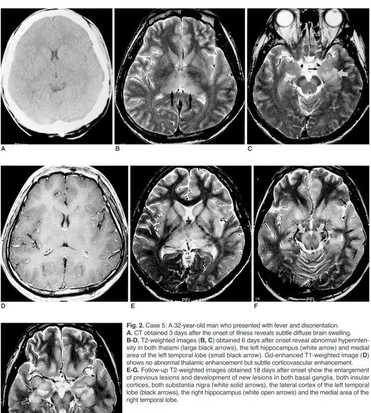
(5) Kim et al.. A. D. B. C. E. F. Fig. 2. Case 5: A 32-year-old man who presented with fever and disorientation. A. CT obtained 3 days after the onset of illness reveals subtle diffuse brain swelling. B-D. T2-weighted images (B, C) obtained 6 days after onset reveal abnormal hyperintensity in both thalami (large black arrows), the left hippocampus (white arrow) and medial area of the left temporal lobe (small black arrow). Gd-enhanced T1-weighted image (D) shows no abnormal thalamic enhancement but subtle corticovascular enhancement. E-G. Follow-up T2-weighted images obtained 18 days after onset show the enlargement of previous lesions and development of new lesions in both basal ganglia, both insular cortices, both substantia nigra (white solid arrows), the lateral cortex of the left temporal lobe (black arrows), the right hippocampus (white open arrows) and the medial area of the right temporal lobe.. G. 72. Korean J Radiol 2(2), June 2001.
(6) Imaging Features of Unusual Acute Encephalitis Fig. 2. H, I. Follow-up T2-weighted images obtained 48 days after onset show decreased size of previous lesions. Periventricular white matter abnormality around both frontal horns and diffuse brain atrophy are noticed.. H. I. involved, and rhombencephalitis involving the midbrain has sometimes been reported (2, 14 15). In these preferred areas, HSE lesions are seen as focal areas of low attenuation on CT, and as areas of low and high signal intensity, respectively, on T1-weighted and T2-weighted MR images. In addition, a parenchymal or gyral pattern of enhancement and foci of subacute hemorrhage may be observed slightly later. These imaging findings can help make a diagnosis of HSE fairly certain and prompt, though involvement of the thalamus, as in our cases, is very unusual in HSE. In our cases, dual infection with the JE and the HSE virus was a possibility. In analogous experiments in mice, JE viral antigen was localized in herpes virus-infected areas of the brain, suggesting that the JE virus gains access to the central nervous system (CNS) at sites of blood-brain barrier disruption caused by the HSE virus (12, 16), and imaging findings suggested a combination of JE and HSE. However, repetitive serologic tests failed to identify either the JE or HSE virus. Unilateral thalamic involvement was also reported in a CNS lesion associated with Epstein-Barr virus infection (17), though the good prognosis and fleeting imaging abnormality seen in this infection are different from the observed findings in our cases. The possible involvement, in our cases, of a new or different viral agent, for which serologic tests were not performed, could not be excluded. In our series, repeated serologic testing for JE involved the hemagglutination inhibition test, which in an appraisal of a recent diagnostic assay for JE showed a sensitivity of about sixty percent (18). The use of polymerase chain reaction for detecting HSE virus DNA in CSF has been reported as the most sensitive noninvasive method for early diagnosis of HSE (19), but in our cases the technique failed to demonstrate that either Korean J Radiol 2(2), June 2001. the JE or HSE virus was an etiologic agent. The mortality rate for JE has been reported as 20-50% (20), and in cases of herpes encephalitis, acyclovir effectively reduces mortality and morbidity if administered early (19). Compared with the prognosis of JE and HSE reported previously, that of our patients was very poor despite the fact that in all cases, acyclovir was injected intravenously since the onset of the condition. Yagishita et al. (21) reported acute encephalopathy with bilateral thalamotegmental involvement in infants and children, which can be a postviral or postinfectious brain disorder. There was, however, no clinical or laboratory evidence of encephalitis (no pleocytosis in the CSF). Although the involved sites in our patients were similar to those in Yagishita’s, the CSF findings and age range are completely different. The acute disseminated and immune-mediated forms of encephalomyelitis may have clinical and CSF features similar to those of our cases. The pathological findings of the first of these are, however, diffuse bilateral perivenular inflammation and demyelination, mainly involving the cerebral white matter, and for this reason - unlike in our cases, in which the gray matter was involved - MR imaging usually demonstrates bilateral abnormalities in the cerebral white matter (22). Bilateral increased signal intensity on T2-weighted images of the basal ganglia and thalamus has been reported in patients with sporadic and variant Creutzfeldt-Jakob disease (CJD) (23, 24). CJD, however, shows no leukocyte response in CSF and has clinical features different from those of our patients (25). In conclusion, the CT and MR imaging findings of acute encephalitis involving the thalamus were similar to a combination of those of JE and HSE. Further investigation, aimed at documenting the specific causative agent showing 73.
(7) Kim et al.. these imaging features, as well as histopathologic study, is needed.. References 1. Jubelt B, Miller JR. Viral infections. In Rowland LP ed. Merritt’s Textbook of Neurology, 9th ed. Philadelphia: Williams & Wilkins, 1995:142-179 2. Tien RD, Felsberg GJ, Osumi AK. Herpes virus infection of the CNS: MR findings. AJR 1993;161:167-176 3. Abe T, Kojima K, Shoji H, et al. Japanese encephalitis. J Magn Reson Imaging 1998;8:755-761 4. Shoji H, Hiraki Y, Kuwasaki N, et al. Japanese encephalitis in the Kurume region of Japan: CT and MRI findings. J Neurol 1989;236:255-259 5. Kumar S, Misra UK, Kalita J, et al. MRI in Japanese encephalitis. Neuroradiology 1997;39:180-184 6. Misra UK, Kalita J, Jain SK, Mathur A. Radiological and neurophysiological changes in Japanese encephalitis. J Neurol Neurosurg Psychiatry 1994;57:1484-1487 7. Shoji H, Murakami T, Murai I, et al. A follow-up study by CT and MRI in 3 cases of Japanese encephalitis. Neuroradiology 1990;32:215-219 8. Kimura K, Dosaka A, Hashimoto Y, et al. Single-photon emission CT findings in acute Japanese encephalitis. AJNR 1997;18:465-469 9. Ishii K. Virological and serological diagnosis of Japanese encephalitis. Adv Neurol Sci 1967;11:300-311 10. Rowley AH, Whitley RJ, Lakeman FD, Wolinsky SM. Rapid detection of herpes-simplex virus DNA in cerebrospinal fluid of patients with herpes simplex encephalitis. Lancet 1990;335:440441 11. Adams RD, Victor M, Ropper AH. Principles of Neurology, 6th ed. New York: McGraw-Hill, 1997:749-759 12. Fields BN, Knipe DM, Chanock RM, eds. Virology. New York: Raven Press, 1985:967 13. Johnson RT, Burke DS, Elwell M, et al. Japanese encephalitis: immunocytochemical studies of viral antigen and inflammatory. 74. cells in fatal cases. Ann Neurol 1985;18:567-573 14. Soo MS, Tien RD, Gray L, Andrews PI, Friedman H. Mesenrhombencephalitis: MR findings in nine patients. AJR 1993;160:1089-1093 15. Tien RD, Dillon WP. Herpes trigeminal neuritis and rhombencephalitis on Gd-DTPA-enhanced MR imaging. AJNR 1990;11:413-414 16. Hayashi K, Arita T. Experimental double infection of Japanese encephalitis virus and herpes simplex virus in mouse brain. Japan J Exp Med 1977;47:9-13 17. Tolly TL, Wells RG, Sty JR. MR features of fleeting CNS lesions associated with Epstein-Barr virus infection. J Comput Assist Tomogr 1989;13:665-668 18. Gajanana A, Samuel PP, Thenmozhi V, Rajendran R. An appraisal of some recent diagnostic assays for Japanese encephalitis. Southeast Asian J Trop Med Public Health 1996;27:673-679 19. Corey L. Herpes simplex virus. In: Fauci AS, Braunwald E, Isselbacher KJ, eds. Harrison’s Principles of Internal Medicine, 14th ed. New York: McGraw-Hill, 1998:1080-1086 20. Fauci AS, Braunwald E, Isselbacher KJ, eds. Harrison’s Principles of Internal Medicine, 14th ed. New York: McGrawHill, 1998:1137 21. Yagishita A, Nakano I, Ushioda T, Otsuki N, Hasegawa A. Acute encephalopathy with bilateral thalamotegmental involvement in infants and children: imaging and pathology findings. AJNR 1995;16:439-447 22. Osborn AG. Diagnostic Neuroradiology. St. Louis: Mosby, 1994:704-706 23. Finkenstaedt M, Szudra A, Zerr I, et al. MR imaging of Creutzfeldt-Jakob disease. Radiology 1996;199:793-798 24. Zeidler M, Sellar RJ, Collie DA, et al. The pulvinar sign on magnetic resonance imaging in variant Creutzfeldt-Jakob disease. Lancet 2000;355:1412-1418 25. Zeidler M, Stewart GE, Barraclough CR, et al. New variant Creutzfeldt-Jakob disease: neurological features and diagnostic tests. Lancet 1997;350:903-907. Korean J Radiol 2(2), June 2001.
(8)
수치

+2

관련 문서
