INTRODUCTION
Cardiovascular complications are an important cause of mor-
tality in persons with diabetes.1,2 Vascular complications in di- abetes are characterized pathologically by proliferation and migration of vascular smooth muscle cells (VSMCs), thicken- ing of intima, and limited blood vessels in atherosclerotic ves- sels.3 Proliferation of VSMCs is an important pathologic pro- cess in various cardiovascular diseases, including atheroscle- rosis, restenosis, and hypertension. Diabetes mellitus-induced early atherosclerosis is used as a model of vascular disease in animal research and cell culture studies.3 Some clinical studies have demonstrated that postprandial hyperglycemia is asso- ciated with further cardiovascular disease and is more signifi- cant than total exposure of hyperglycemia.4 In addition, glu- cose fluctuations have more potent effects on oxidative stress than chronic sustained hyperglycemia in type 2 diabetes pa- tients.5 Numerous mechanisms have been studied concerning
Anti-Proliferative Effects of Rutin on OLETF Rat Vascular Smooth Muscle Cells Stimulated by Glucose Variability
Sung Hoon Yu1*, Jae Myung Yu1*, Hyung Joon Yoo1, Seong Jin Lee1, Dong Hyun Kang1, Young Jung Cho2, and Doo Man Kim1
1Division of Endocrinology and Metabolism, Hallym University College of Medicine, Seoul;
2Department of Internal Medicine, National Medical Center, Seoul, Korea.
Purpose: Proliferation of vascular smooth muscle cells (VSMCs) plays a crucial role in atherosclerosis. Rutin is a major represen- tative of the flavonol subclass of flavonoids and has various pharmacological activities. Currently, data are lacking regarding its effects on VSMC proliferation induced by intermittent hyperglycemia. Here, we demonstrate the effects of rutin on VSMC prolif- eration and migration according to fluctuating glucose levels.
Materials and Methods: Primary cultures of male Otsuka Long-Evans Tokushima Fatty (OLETF) rat VSMCs were obtained from enzymatically dissociated rat thoracic aortas. VSMCs were incubated for 72 h with alternating normal (5.5 mmol/L) and high (25.0 mmol/L) glucose media every 12 h. Proliferation and migration of VSMCs, the proliferative molecular pathway [including p44/42 mitogen-activated protein kinases (MAPK), mitogen-activated protein kinase kinase 1/2 (MEK1/2), p38 MAPK, phos- phoinositide 3-kinase (PI3K), c-Jun N-terminal protein kinase (JNK), nuclear factor kappa B (NF-κB), and Akt], the migratory pathway (big MAPK 1, BMK1), reactive oxygen species (ROS), and apoptotic pathway were analyzed.
Results: We found enhanced proliferation and migration of VSMCs when cells were incubated in intermittent high glucose con- ditions, compared to normal glucose. These effects were lowered upon rutin treatment. Intermittent treatment with high glucose for 72 h increased the expression of phospho-p44/42 MAPK (extracellular signal regulated kinase 1/2, ERK1/2), phospho- MEK1/2, phospho-PI3K, phospho-NF-κB, phospho-BMK1, and ROS, compared to treatment with normal glucose. These effects were suppressed by rutin. Phospho-p38 MAPK, phospho-Akt, JNK, and apoptotic pathways [B-cell lymphoma (Bcl)-xL, Bcl-2, phospho-Bad, and caspase-3] were not affected by fluctuations in glucose levels.
Conclusion: Fluctuating glucose levels increased proliferation and migration of OLETF rat VSMCs via MAPK (ERK1/2), BMK1, PI3K, and NF-κB pathways. These effects were inhibited by the antioxidant rutin.
Key Words: Rutin, hyperglycemia, smooth muscle cells, mitogen-activated protein kinases, phosphatidylinositol 3-kinase pISSN: 0513-5796 · eISSN: 1976-2437
Received: October 6, 2014 Revised: June 11, 2015 Accepted: June 15, 2015
Corresponding author: Dr. Hyung Joon Yoo, Division of Endocrinology and Metab- olism, Hallym University College of Medicine, 1 Singil-ro, Yeongdeungpo-gu, Seoul 07441, Korea.
Tel: 82-2-829-5381, Fax: 82-2-846-4669, E-mail: [email protected]
A part of this paper was presented at the ENDO 2013, June 15–18, 2013, in San Fran- cisco, CA, USA.
*Sung Hoon Yu and Jae Myung Yu contributed equally to this work.
•The authors have no financial conflicts of interest.
© Copyright: Yonsei University College of Medicine 2016
This is an Open Access article distributed under the terms of the Creative Com- mons Attribution Non-Commercial License (http://creativecommons.org/licenses/
by-nc/3.0) which permits unrestricted non-commercial use, distribution, and repro- duction in any medium, provided the original work is properly cited.
Yonsei Med J 2016 Mar;57(2):373-381 http://dx.doi.org/10.3349/ymj.2016.57.2.373
the proliferation and migration of VSMCs and anti-atheroscle- rotic effects of flavonoids.6-8 Mitogen-activated protein kinases (MAPK) have an important role in cell growth, differentiation, and apoptosis. Extracellular signal regulated kinase 1/2 (ERK1/
2), big MAPK 1 (BMK1), c-Jun N-terminal protein kinase (JNK), and p38 are related to cell proliferation and differentiation.
Among them, BMK1, a newly identified member of the MAPK family, has been found to be related to cell migration.9 Howev- er, there is little information about the effect of glucose fluctua- tions in VSMCs. Therefore, we focused on flavonoids inhibiting proliferation and migration of VSMCs induced by intermittent hyperglycemia. Rutin (C27H30O16) is an important member of the flavonol subclass of flavonoids. Rutin has a wide variety of pharmacological activities; however, there is insufficient data regarding its activity in the proliferation and migration of VSMCs by intermittent hyperglycemia.
The purpose of this study was to investigate the effects of ru- tin on the proliferation and migration of VSMCs stimulated by glucose fluctuations in an obese rat model of type 2 diabetes.
MATERIALS AND METHODS
Study animals
Age-matched male Otsuka Long-Evans Tokushima Fatty (OL- ETF) rats, a model of spontaneous noninsulin-dependent dia- betes mellitus, were kindly provided by Otsuka Pharmaceuti- cal Co., Tokushima, Japan.
Cell culture
VSMCs were harvested from the thoracic aortas of 12-week- old male OLETF rats by elastase and collagenase digestion, as previously described.10 Cells were grown in Dulbecco’s modi- fied Eagle’s medium (DMEM, Gibco-BRL, Carlsbad, CA, USA) with 10% fetal bovine serum (FBS) in a 37°C, 5% CO2, humidi- fied incubator. At confluence, cells were trypsinized using 0.125%
trypsin in 0.005% ethylenediaminetetraacetic acid (EDTA, Sigma Chemical Co., St. Louis, MO, USA). Cells from passages 7–13 were used for the experiments. The cells grew in the “hill and valley” pattern, which is characteristic of VSMCs in cul- ture and showed positive immunostaining with antismooth muscle α-actin antibodies.11
Treatment of cells with reagents
Cultured VSMCs were seeded into 96-well plates (1×10⁴ cells/
well) in DMEM with 10% FBS and incubated for 48 h. Cells were made quiescent by incubation in DMEM with 0.1% FBS for 24 h before the addition of rutin (Sigma, St. Louis, MO, USA).
The cells were then incubated for another 72 h in DMEM with 10% FBS in the presence of various concentrations of glucose (5.5 and alternating 5.5 and 25 mM for 12 h).
Cell proliferation assay
Cell proliferation was evaluated by the methylthiazoletetrazo- lium (MTT, Sigma) assay and expressed as cell viability (%).
VSMCs were incubated in the presence of 1, 10, 30, and 100 μM rutin for 72 h with various concentrations of glucose (5.5 and alternating 5.5 and 25 mM). MTT solution (5 mg/mL in phos- phate-buffered saline) was then added to each well, and the plates incubated for 4 h. The MTT formazan product was solu- bilized by the addition of dimethyl sulfoxide (Sigma), and the absorbance was measured at 570 nm using an ELx800 Absor- bance Microplate Reader (Biotek, Winooski, VT, USA).
Cell migration assay
Cell migration activity was determined using a Radius 24-well assay kit (Cell Biolabs, San Diego, CA, USA), consisting of a cir- cular biocompatible gel in each well, according to the manu- facturer’s instructions. Briefly, VSMCs were seeded in the assay plates and cultured for 12 h to firm attachment. After 12 h of in- cubation, the biocompatible gels were removed with removal solutions, and the cells were incubated for an additional 72 h with either normal glucose (5.5 mM), glucose fluctuations (al- ternating 5.5 and 25 mM for 12 h), or glucose fluctuations with rutin (30 μM). The images of the migratory cells were captured using an Olympus IX70 microscope equipped with a digital camera (Olympus Inc., Melville, NY, USA). The area lacking mi- gratory cells in the circle was analyzed using Image J software.
Western blot analysis
Cells cultured with rutin (30 μM) were washed and lysed with lysis buffer [10 mM Tris (pH 7.4), 1 mM EDTA, 1% sodium py- rophosphate-40, 0.1 mM phenylmethylsulfonyl fluoride, 20 nM sodium vanadate, and 1× cocktail solution]. The superna- tants were analyzed using a Bradford assay (Bio-Rad, Hercules, CA, USA). Sodium dodecyl sulfate-polyacrylamide gel elec- trophoresis (SDS-PAGE) was performed using a 10% resolving gel, followed by nitrocellulose membrane transfer (Bio-Rad).
The membranes were blocked for 1 h at room temperature in blocking solution {5% non-fat milk in Tris buffer with Tween-20 [TBST; 200 nM Tris, 500 mM NaCl (pH 7.5), and 0.05% Twe- en-20]}, and then incubated overnight at 4°C with anti-phos- pho-p44/42 MAPK, anti-phospho-MAPK kinase 1/2 (MEK1/2), anti-phospho-p38, JNK, anti-phospho-Akt, anti-phosphoi- nositide 3-kinase (PI3K), anti-phospho nuclear factor kappa B (NF-κB), anti-phospho-BMK1, B-cell lymphoma-extra-large (Bcl-xL), Bcl-2, anti-phospho Bad, and caspase-3 antibodies (all supplied by Cell Signaling Technology, Beverly, MA, USA ex- cept for anti-phospho-NF-κB antibody, which was sourced from Sigma). The membranes were washed with TBST (5 min with 3 changes) and incubated with peroxidase-conjugated anti- mouse immunoglobulin G antibody (Amersham Life Science, Arlington Height, IL, USA). Membranes were washed and vi- sualized using the Visualizer western blot detection kit (Up- state, Lake Placid, NY, USA). Autoradiography was performed
and band intensity was analyzed by densitometry using Image J software.
Measurement of intracellular reactive oxygen species generation
VSMCs were plated in 96-well plates (Corning) at a density of 1×10⁴ cells/well in culture medium. At the end of the incubation time, adherent or suspended cells were treated with 5 μg/mL dichlorofluorescein diacetate (ECF-DA, Abcam, Cambridge, MA, USA) for 5 min. After washing with PBS, adherent cells were lysed in 1 mL of radioimmunoprecipitation assay buffer (RIPA, Merck Millipore, Damstard, Germany) buffer and analyzed im- mediately by fluorescence spectrophotometric analysis at 510 nm. Data are presented normalized to total protein content.
Statistical analysis
Data are expressed as mean±standard deviation. Statistical analysis was performed with Student’s t-test or 1-way analysis of variance, and p<0.05 was considered statistically significant.
RESULTS
Rutin dose-dependently mitigates the induction of VSMC proliferation by glucose fluctuations
To evaluate the effect of rutin on VSMC proliferation, cells were treated with glucose at 5.5 mM, followed by another exposure
of 5.5 or 25 mM every 12 h, and cell proliferation was mea- sured. Cell proliferation increased without a change in cell mor- phology in cells treated with glucose fluctuation, compared with control (100±0.90% vs. 157±1.32%; p<0.05) (Fig. 1). Rutin ap- plied at concentrations of 1, 10, 30, or 100 µM for 72 h before the fluctuation in glucose levels dose-dependently attenuated the increase in cell proliferation induced by fluctuation in glu- cose levels. Taken together, our results demonstrate that rutin has an anti-proliferative effect on VSMC exposed to fluctuat- ing glucose concentrations.
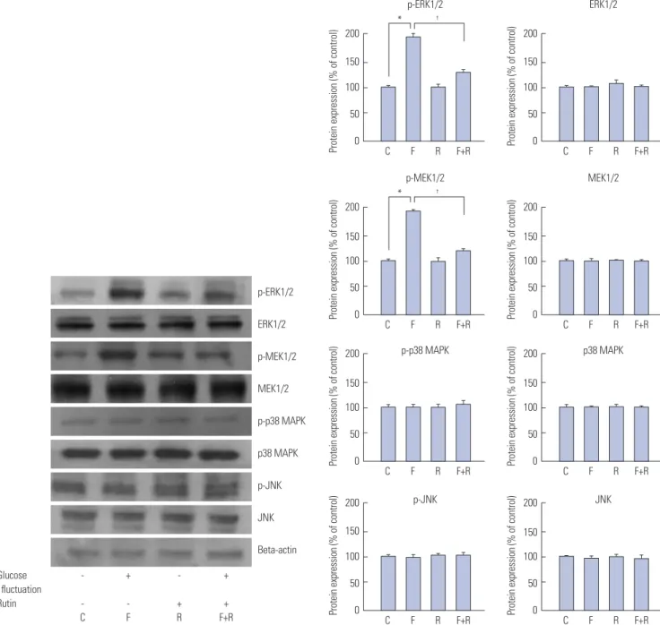
Rutin inhibits the proliferation of VSMCs by suppressing phosphorylation of MEK1/2 and ERK1/2 following exposure to glucose fluctuation
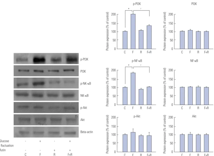
To investigate the influence of rutin on signal transduction pathways, cells were exposed to the fluctuating glucose condi- tion, and the protein levels of MEK1/2, ERK1/2, p38, JNK, PI3K, Akt, and NF-κB were measured. Protein levels of phospho- MEK1/2 (100±0.57% vs. 149±1.25%; p<0.05) and phospho- ERK1/2 (100±4.2% vs. 166±0.8%; p<0.05), were elevated in cells exposed to fluctuating glucose, compared with control, while those of phospho-p38 and phospho-JNK were unchanged (Fig. 2). Meanwhile, phospho-PI3K (100±3.22% vs. 193±2.62%;
p<0.05) and phospho-NF-κB (100±1.55% vs. 170±4%; p<0.05) protein levels were elevated in cells exposed to glucose fluctu- ation, compared with control, whereas those of phospho-Akt were unchanged (Fig. 3).
Fig. 1. Inhibitory effects of rutin on proliferation of vascular smooth muscle cells (VSMCs) from Otsuka Long-Evans Tokushima Fatty (OLETF) rats at fluctu- ating glucose concentrations. (A) Representative photomicrographs of morphology of OLETF rat VSMCs (magnification ×100). (B) Cell proliferation was evaluated by the methylthiazoletetrazolium assay and expressed as cell viability (%). VSMCs were incubated for 72 h with or without glucose fluctuation (alternating 5.5 and 25 mM every 12 h) and rutin (1, 10, 30, and 100 μM). Data are expressed as mean±SD from 5 separate experiments. *p<0.05 vs. control,
†p<0.05 vs. glucose fluctuations. Cont, control; Fluc, glucose fluctuations.
×100
72 h
200
150
100
50
0 Cont Fluc 30 1 10 30 100 (µM) Rutin
Fluctuation
* †
Cell viability (% of control)
A
B
The phospho-protein levels of MEK1/2, ERK1/2, PI3K, and NF-κB were reduced, while those of p38, JNK, and Akt were unaltered in cells pretreated with 30-µM rutin 72 h prior to glu- cose fluctuation. Together, our results indicate that MEK1/2, ERK1/2, and NF-κB are involved in the rutin-induced antipro- liferative effect on VSMCs. This effect was confirmed using ERK inhibitor PD98059 and PI3K inhibitor wortmannin (Fig. 4).
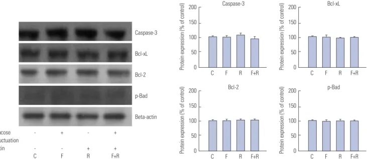
Rutin-induced antiproliferative effect is not related to apoptosis in VSMCs exposed to glucose fluctuation To identify the impact of rutin on expression of apoptosis-re-
lated proteins, cells were pretreated with rutin at a concentra- tion of 30 µM for 72 h prior to glucose fluctuation. Interestingly, no changes in the protein levels of caspase-3, Bcl-xL, Bcl-2, and phospho-Bad were observed, suggesting that apoptosis does not play a role in anti-proliferative effects in VSMCs exposed to glucose fluctuation, regardless of rutin pretreatment (Fig. 5).
Rutin inhibits migration of VSMCs with concomitant increase of BMK1 under conditions of glucose fluctuation
Since we demonstrated that glucose fluctuation caused VSMC
Glucose - + - + fluctuation
Rutin - - + +
C F R F+R
p-ERK1/2
ERK1/2
p-MEK1/2
MEK1/2
p-p38 MAPK
p38 MAPK
p-JNK
JNK
Beta-actin
Fig. 2. Inhibitory effects of rutin on proliferation of vascular smooth muscle cells (VSMCs) at fluctuating glucose concentrations through the mitogen-acti- vated protein kinase (MAPK) signaling pathway determined by western blot analysis. VSMCs were incubated for 72 h with or without glucose fluctuation (alternating 5.5 and 25 mM every 12 h) and rutin (30 μM). Data are expressed as mean±SD from 5 separate experiments. *p<0.05 vs. control, †p<0.05 vs. glu- cose fluctuations. C, control; ERK1/2, extracellular signal regulated kinase 1/2; F, glucose fluctuation; JNK, c-Jun N-terminal protein kinase; MEK1/2, mitogen- activated protein kinase kinase 1/2; R, rutin; p, phosphorylated; p38 MAPK, p38 mitogen-activated protein kinase.
200 150 100 50 0
200 150 100 50 0
200 150 100 50 0
200 150 100 50 0
200 150 100 50 0
200 150 100 50 0
200 150 100 50 0
200 150 100 50 0 C F R F+R
C F R F+R
C F R F+R
C F R F+R
C F R F+R
C F R F+R
C F R F+R
C F R F+R p-ERK1/2
p-MEK1/2
p-p38 MAPK
p-JNK
ERK1/2
MEK1/2
p38 MAPK
JNK
*
*
†
†
Protein expression (% of control)Protein expression (% of control)Protein expression (% of control)Protein expression (% of control) Protein expression (% of control)Protein expression (% of control)Protein expression (% of control)Protein expression (% of control)
proliferation, which was prevented by rutin, we chose to exam- ine whether glucose fluctuation in the presence or absence of rutin affected the migration of VSMCs. Cell migration (83.4±
12.4% vs. 45.1±19.2%; p<0.05) and phospho-BMK1 protein lev- els were higher in cells exposed to glucose fluctuation (Fig. 6).
The effect of fluctuation in glucose levels on cell migration and phospho-BMK1 protein levels was attenuated in cells pretreat- ed with 30-µM rutin for 72 h prior to glucose fluctuation (83.4±
12.4% vs. 35.7±7.5%; p<0.001). Our results reveal that rutin sup- presses the migration, as well as proliferation, of VSMCs ex- posed to glucose fluctuation in conjunction with an increase in BMK1.
Rutin inhibits ROS production in VSMCs under conditions of fluctuating glucose levels
To identify the inhibitory effect of rutin on production of reac- tive oxygen species (ROS), we analyzed ROS levels using fluo- rescence spectrophotometric analysis. ROS production was enhanced in cells exposed to glucose fluctuations, compared with control cells (61.5±21.0% vs. 145.3±13.7% in control cells
and cells exposed to fluctuations in glucose levels; p<0.05).
Elevated ROS levels induced by glucose fluctuation were de- creased by rutin treatment (145.3±13.7% vs. 74.2±11.9%; p<0.05) (Fig. 7).
DISCUSSION
In this study, we successfully demonstrated that intermittent hyperglycemia results in proliferation of VSMCs and that the antioxidant rutin inhibits the proliferation and migration of VS- MCs by suppressing the phosphorylation of MEK1/2, MAPK, PI3K, NF-κB, and BMK1, and production of ROS. To the best of our knowledge, this is the first study to report the inhibitory effects of rutin on VSMC proliferation and migration due to glycemic variability.
Rutin is a member of the flavonoid family, and to date, more than 2000 flavonoids have been described.12 Their positive ef- fects may be partially related to inhibitory actions in athero- sclerosis, brain ischemia, tumors, inflammation, and oxidative Glucose - + - +
fluctuation
Rutin - - + +
C F R F+R
p-PI3K
PI3K
p-NK-κB
NK-κB
p-Akt
Akt
Beta-actin
Fig. 3. Inhibitory effects of rutin on proliferation of vascular smooth muscle cells (VSMCs) at fluctuating glucose concentrations through nuclear factor kappa B (NF-κB) and phosphoinositide 3-kinase (PI3K)/Akt pathway determined by western blot analysis. VSMCs were incubated for 72 h with or without glucose fluctuation (alternating 5.5 and 25 mM every 12 h) and rutin (30 μM). Data are expressed as mean±SD from 5 separate experiments. *p<0.05 vs.
control, †p<0.05 vs. glucose fluctuations. C, control; F, glucose fluctuation; p, phosphorylated; R, rutin.
200 150 100 50 0
200 150 100 50 0
200 150 100 50 0
200 150 100 50 0
200 150 100 50 0
200 150 100 50 0 C F R F+R
C F R F+R
C F R F+R
C F R F+R
C F R F+R
C F R F+R p-NF-κB
p-PI3K
p-Akt
NF-κB PI3K
Akt
*
*
†
†
Protein expression (% of control)Protein expression (% of control)Protein expression (% of control) Protein expression (% of control)Protein expression (% of control)Protein expression (% of control)
stress.13-15 Rutin has been shown to have antioxidative, anti-in- flammatory, antiallergic, antiviral, and anti-cancer effects.16 Fur- thermore, a dose-dependent protective effect of flavonoids against the development of atherosclerosis may exist.13,17-20 However, the anti-atherogenic effects of rutin in diabetes with fluctuating glucose conditions are unknown, and there is in- sufficient data concerning the effects of rutin on the prolifera- tion of VSMCs.
Hyperglycemia enhances the proliferation of VSMCs, which is a critical step in the pathogenesis of atherosclerosis.21-23 Al- though the exact pathophysiologic mechanism linking hyper- glycemia to atherosclerosis remains unclear, diabetes increas- es the risk of myocardial infarction, stroke, amputation, and death.23,24 Compared with normal glucose-tolerant individu- als, those with impaired glucose tolerance and type 2 diabetes predominantly display greater intraday glucose fluctuations.
Fluctuation - + - + + +
Rutin - - + + - -
PD98059 10 µM 20 µM
Wortmannin 10 µM 20 µM
p-ERK1/2 p-MEK1/2 p-PI3K p-NF-κB Beta-actin
Fig. 4. Effect of ERK inhibitor (PD98059) and PI3K inhibitor (wortmannin) on proliferative protein expression of glucose fluctuation-induced vascular smooth muscle cell (VSMC) with or without rutin determined by western blot analysis. VSMCs were incubated for 72 h with or without glucose fluctuation (alternating 5.5 and 25 mM every 12 h) and rutin (30 μM). And PD98059 10 and 20 μM or wortmannin 10 and 20 μM were added in control (5.5 mM) and al- ternating (5.5 and 25 mM) glucose medium. Data are expressed as mean±SD from 5 separate experiments. *p<0.05. C, control; ERK1/2, extracellular sig- nal regulated kinase 1/2; F, glucose fluctuation; MEK1/2, mitogen-activated protein kinase kinase 1/2; NF-κB, nuclear factor kappa B; R, rutin; p, phosphor- ylated; PD, PD98059; PI3K, phosphoinositide 3-kinase; W, wortmannin.
200 150 100 50 0
200 150 100 50 0
200 150 100 50 0
200 150 100 50 0 C F R F+R PD10 PD20
C F R F+R W10 W20
C F R F+R PD10 PD20
C F R F+R W10 W20 p-ERK1/2
p-PI3K
p-MEK1/2
p-NF-κB
*
*
*
*
Protein expression (% of control)Protein expression (% of control) Protein expression (% of control)Protein expression (% of control)
Glucose - + - + fluctuation
Rutin - - + + C F R F+R
Caspase-3
Bcl-xL
Bcl-2
p-Bad
Beta-actin
Fig. 5. Inhibitory effect of rutin on proliferation of vascular smooth muscle cells (VSMCs) through apoptotic pathways determined by western blot analy- sis. VSMCs were incubated for 72 h with or without glucose fluctuation (alternating 5.5 and 25 mM every 12 h) and rutin (30 μM). Data are expressed as mean±SD from 5 separate experiments. Bcl-2, B-cell lymphoma 2; Bcl-xL, B-cell lymphoma-extra-large; C, control; F, glucose fluctuation; p, phosphory- lated; R, rutin.
200 150 100 50 0
200 150 100 50 0
200 150 100 50 0
200 150 100 50 0 C F R F+R
C F R F+R
C F R F+R
C F R F+R Caspase-3
Bcl-2
Bcl-xL
p-Bad Protein expression (% of control)Protein expression (% of control) Protein expression (% of control)Protein expression (% of control)
Patients with type 2 diabetes show increased postprandial glu- cose excursion, higher overnight glucose levels, and greater interday fluctuations.25 Kim, et al.26 showed that intermittent high glucose caused more apoptosis in insulinoma cell lines than continuous hyperglycemia, likely through an effect on forkhead box O-Sirtuin 1 (FOXO-SIRT) pathway. Monnier, et al.5 reported that glucose fluctuations during postprandial pe- riods exhibited a more specific triggering effect on oxidative stress than chronic sustained hyperglycemia in type 2 diabe- tes patients, which contributes to vascular damage.
Numerous studies have suggested that rutin has antiathero- genic effects. Rutin is an inhibitor of the protein disulfide isom- erase and significantly blocks thrombus formation in rats.27
Rodrigues, et al.14 have suggested that rutin is able to promote significant recovery of sensorimotor loss after cortical focal ischemia in rats. Meanwhile, research by Lee, et al.28 revealed that rutin inhibited lipopolysaccharide-induced barrier dis- ruption, expression of cell adhesion molecules, and the migra- tion of monocytes to human endothelial cells. Rutin also sup- pressed the production of tumor necrosis factor-α and sup- pressed the activation of NF-κB by lipopolysaccharide.29 In H9c2 cells treated with an apoptotic agent (H2O2), rutin de- creased expression of cleaved caspase-3, reduced the Bax/
Bcl-2 ratio, inhibited H2O2-induced apoptosis, and promoted cell survival via increased phosphorylation of ERK and Akt.29
Several signaling pathways are involved in the proliferation Fig. 6. Inhibitory effects of rutin on glucose fluctuation-induced vascular smooth muscle cell migration in Otsuka Long-Evans Tokushima Fatty rats. (A) The percentage inhibitory area of migratory cells was calculated using a microscope. Control (normal glucose, 5.5 mM), glucose fluctuations (alternating 5.5 mM and 25 mM every 12 h), glucose fluctuations with 30-μM rutin (rutin treatment with alternating 5.5 mM and 25 mM every 12 h). (B) Inhibitory effect of rutin on big mitogen-activated protein kinase 1 (BMK1; migration pathway) by western blot analysis. Five separate experiments were performed. *p<0.05 vs. control, †p<0.001 vs. glucose fluctuations. C, control; F, glucose fluctuation; p, phosphorylated; R, rutin.
p-BMK1 24 h 48 h 72 h
C
F
R
F+R
Glucose - + - + fluctuation
Rutin - - + +
C F R F+R
200
150
100
50
0
200
150
100
50
0
48 h 24 h
C F R F+R C
F R F+R
*
*
*
†
†
†
72 h
p-BMK1 Migrated cell percentage (%) Protein expression (% of control)
A
B
of VSMCs. Experiments performed by Choi, et al.6 in mouse cells lines demonstrated the inhibition of adhesion molecule expression in VSMCs, elicited through a down-regulation of the MAPK, Akt, and NF-κB signaling pathways. In OLETF rats, Park, et al.7 reported that rosiglitazone inhibited insulin-stim- ulated proliferation of VSMCs by inhibition of Akt-mTOR- P70S6K cascade, and this may be mediated by the MAPK and PI3K pathways. Yerneni, et al.8 demonstrated that NF-κB acti- vation in VSMCs in the hyperglycemic state was the key mech- anism for the production of VSMC factors mediating the vas- cular complications observed in diabetes. While Ruiz, et al.30 showed that kaempferol diminished 7β-hydroxycholesterol- induced apoptosis in rat VSMCs, rutin did not affect the apop- totic signaling pathway.
In the present study, we evaluated the effects of rutin on the proliferation and migration of VSMCs provoked by glucose fluctuations. Our findings revealed that increases in the prolif- eration and migration of VSMCs upon fluctuating glucose con- centrations are suppressed by rutin treatment, which also sup- pressed the phosphorylation of MEK1/2, MAPK, BMK1, PI3K, and NF-κB. However, JNK and Akt were not influenced by ru- tin treatment, and apoptotic signals (Bcl-xL, Bcl-2, caspase-3, and phospho-Bad) were unchanged by glucose fluctuation and rutin treatment. Similar to other reports,9,31 our experiments indicated that the ERK, anti-PI3K, and BMK1 pathway is pri- marily responsive to growth factors, and plays key roles in cell proliferation and migration. BMK1 is a newly identified mem- ber of the MAPK family and is known to be sensitive to oxida- tive stress and hyperglycemia, especially in mesangial cells and glomeruli of OLETF rats. Yoshizumi, et al.9 suggested that BMK1 is involved in the pathogenesis of atherosclerosis, par- ticularly in diabetic nephropathy, and that BMK1 activation is implicated in VSMC migration. Our findings also point to-
wards BMK1-dependent VSMC migration (Fig. 6).
The present study possesses several limitations. Firstly, our findings do not eliminate the possible involvement of alterna- tive transcription factors and signaling pathways. The present data suggest that the inhibitory effect of rutin on VSMCs is at least partially mediated through suppression of MAPK signal- ing. Secondly, we have not confirmed the effects of rutin in vivo in OLETF rats; however, we plan to investigate this in the future.
This study has demonstrated that the MAPK pathways play a role in the proliferation and migration of the VSMCs caused by hyperglycemic variability, which is representative of the physiologic diabetes condition. Rutin significantly reduces the proliferation and migration of VSMCs in obese type 2 diabetes rats caused by intermittent hyperglycemia via reduced phos- phorylation of MEK1/2, MAPK, BMK1, PI3K, and NF-κB. These findings emphasize a beneficial effect of rutin, and future stud- ies are necessary to determine the potential anti-atherosclerot- ic and anti-oxidative effects of rutin in patients and experimen- tal animals with diabetes.
ACKNOWLEDGEMENTS
This work was supported by the Korean Diabetes Association (SHY, 2010) and a grant from Hallym University Medical Cen- ter Research Fund (01-2011-14).
REFERENCES
1. Singh A, Donnino R, Weintraub H, Schwartzbard A. Effect of strict glycemic control in patients with diabetes mellitus on frequency of macrovascular events. Am J Cardiol 2013;112:1033-8.
2. Gaede P, Lund-Andersen H, Parving HH, Pedersen O. Effect of a multifactorial intervention on mortality in type 2 diabetes. N Engl J Med 2008;358:580-91.
3. Schwartz SM, Campbell GR, Campbell JH. Replication of smooth muscle cells in vascular disease. Circ Res 1986;58:427-44.
4. Bonora E, Muggeo M. Postprandial blood glucose as a risk factor for cardiovascular disease in Type II diabetes: the epidemiological evidence. Diabetologia 2001;44:2107-14.
5. Monnier L, Mas E, Ginet C, Michel F, Villon L, Cristol JP, et al. Acti- vation of oxidative stress by acute glucose fluctuations compared with sustained chronic hyperglycemia in patients with type 2 dia- betes. JAMA 2006;295:1681-7.
6. Choi KW, Park HJ, Jung DH, Kim TW, Park YM, Kim BO, et al. Inhi- bition of TNF-α-induced adhesion molecule expression by dios- genin in mouse vascular smooth muscle cells via downregulation of the MAPK, Akt and NF-κB signaling pathways. Vascul Pharma- col 2010;53:273-80.
7. Park S, Lim S, Chang W, Song H, Lee S, Song BW, et al. The inhibi- tion of insulin-stimulated proliferation of vascular smooth muscle cells by rosiglitazone is mediated by the Akt-mTOR-P70S6K path- way. Yonsei Med J 2008;49:592-600.
8. Yerneni KK, Bai W, Khan BV, Medford RM, Natarajan R. Hyper- glycemia-induced activation of nuclear transcription factor kap- paB in vascular smooth muscle cells. Diabetes 1999;48:855-64.
9. Yoshizumi M, Kyotani Y, Zhao J, Nagayama K, Ito S, Tsuji Y, et al.
Role of big mitogen-activated protein kinase 1 (BMK1) / extracellu- lar signal-regulated kinase 5 (ERK5) in the pathogenesis and pro-
* †
Fig. 7. Inhibitory effect of rutin on production of reactive oxygen species determined by fluorescence spectrophotometric analysis. VSMCs were incubated for 72 h with or without glucose fluctuation (alternating 5.5 and 25 mM every 12 h) and rutin (30 μM). Data are expressed as mean±SD from 5 separate experiments. *p<0.05 vs. control, †p<0.05 vs. glucose fluc- tuations. C, control; F, glucose fluctuation; p, phosphorylated; R, rutin;
VSMCs, vascular smooth muscle cells.
250
200
150
100
50
0 C F R F+R
Fluorescence intensity (A.U.)
gression of atherosclerosis. J Pharmacol Sci 2012;120:259-63.
10. Yoo HJ, Kozaki K, Akishita M, Watanabe M, Eto M, Nagano K, et al.
Augmented Ca2+ influx is involved in the mechanism of enhanced proliferation of cultured vascular smooth muscle cells from spon- taneously diabetic Goto-Kakizaki rats. Atherosclerosis 1997;131:
167-75.
11. Bochaton-Piallat ML, Ropraz P, Gabbiani F, Gabbiani G. Phenotyp- ic heterogeneity of rat arterial smooth muscle cell clones. Implica- tions for the development of experimental intimal thickening. Ar- terioscler Thromb Vasc Biol 1996;16:815-20.
12. Ramassamy C. Emerging role of polyphenolic compounds in the treatment of neurodegenerative diseases: a review of their intra- cellular targets. Eur J Pharmacol 2006;545:51-64.
13. Belcaro G, Cesarone MR, Ledda A, Cacchio M, Ruffini I, Ricci A, et al. 5-Year control and treatment of edema and increased capillary filtration in venous hypertension and diabetic microangiopathy using O-(beta-hydroxyethyl)-rutosides: a prospective compara- tive clinical registry. Angiology 2008;59 Suppl 1:14S-20S.
14. Rodrigues AM, Marcilio Fdos S, Frazão Muzitano M, Giraldi-Gui- marães A. Therapeutic potential of treatment with the flavonoid rutin after cortical focal ischemia in rats. Brain Res 2013;1503:53-61.
15. Procházková D, Boušová I, Wilhelmová N. Antioxidant and prooxi- dant properties of flavonoids. Fitoterapia 2011;82:513-23.
16. Araújo JR, Gonçalves P, Martel F. Chemopreventive effect of dietary polyphenols in colorectal cancer cell lines. Nutr Res 2011;31:77-87.
17. Kulisic´ T, Krisko A, Dragovic´-Uzelac V, Milos M, Pifat G. The effects of essential oils and aqueous tea infusions of oregano (Origanum vulgare L. spp. hirtum), thyme (Thymus vulgaris L.) and wild thyme (Thymus serpyllum L.) on the copper-induced oxidation of human low-density lipoproteins. Int J Food Sci Nutr 2007;58:
87-93.
18. Hsu IP, Jou HJ, Huang CW, Wang TA, Wu WH. The effects of soygerm extracts on blood lipoproteins, antioxidative capacity and urinary estrogen metabolites in postmenopausal women on hor- mone therapy. Int J Gynaecol Obstet 2007;98:29-33.
19. Botelho FV, Alvarez-Leite JI, Lemos VS, Pimenta AM, Calado HD, Matencio T, et al. Physicochemical study of floranol, its copper(II) and iron(III) complexes, and their inhibitory effect on LDL oxida- tion. J Inorg Biochem 2007;101:935-43.
20. Milde J, Elstner EF, Grassmann J. Synergistic effects of phenolics and carotenoids on human low-density lipoprotein oxidation. Mol Nutr Food Res 2007;51:956-61.
21. Alipui C, Ramos K, Tenner TE Jr. Alterations of rabbit aortic smooth muscle cell proliferation in diabetes mellitus. Cardiovasc Res 1993;
27:1229-32.
22. Natarajan R, Gonzales N, Xu L, Nadler JL. Vascular smooth muscle cells exhibit increased growth in response to elevated glucose. Bio- chem Biophys Res Commun 1992;187:552-60.
23. Beckman JA, Creager MA, Libby P. Diabetes and atherosclerosis:
epidemiology, pathophysiology, and management. JAMA 2002;
287:2570-81.
24. Natali A, Vichi S, Landi P, Severi S, L’Abbate A, Ferrannini E. Coro- nary atherosclerosis in Type II diabetes: angiographic findings and clinical outcome. Diabetologia 2000;43:632-41.
25. Wang C, Lv L, Yang Y, Chen D, Liu G, Chen L, et al. Glucose fluctu- ations in subjects with normal glucose tolerance, impaired glucose regulation and newly diagnosed type 2 diabetes mellitus. Clin En- docrinol (Oxf) 2012;76:810-5.
26. Kim M, Chung H, Yoon C, Lee E, Kim T, Kim T, et al. Increase of INS-1 cell apoptosis under glucose fluctuation and the involvement of FOXO-SIRT pathway. Diabetes Res Clin Pract 2012;98:132-9.
27. Jasuja R, Passam FH, Kennedy DR, Kim SH, van Hessem L, Lin L, et al. Protein disulfide isomerase inhibitors constitute a new class of antithrombotic agents. J Clin Invest 2012;122:2104-13.
28. Lee WH, Ku SK, Bae JS. Barrier protective effects of rutin in LPS- induced inflammation in vitro and in vivo. Food Chem Toxicol 2012;
50:3048-55.
29. Jeong JJ, Ha YM, Jin YC, Lee EJ, Kim JS, Kim HJ, et al. Rutin from Lonicera japonica inhibits myocardial ischemia/reperfusion-in- duced apoptosis in vivo and protects H9c2 cells against hydrogen peroxide-mediated injury via ERK1/2 and PI3K/Akt signals in vi- tro. Food Chem Toxicol 2009;47:1569-76.
30. Ruiz E, Padilla E, Redondo S, Gordillo-Moscoso A, Tejerina T.
Kaempferol inhibits apoptosis in vascular smooth muscle induced by a component of oxidized LDL. Eur J Pharmacol 2006;529:79-83.
31. Cobb MH. MAP kinase pathways. Prog Biophys Mol Biol 1999;71:
479-500.