A mucocele is a benign expansile paranasal sinus lesion which is most commonly found in the frontal sinus, and found occasionally in the ethmoid and sphenoid sinuses.1Maxillary sinus mucoceles are relatively rare, accounting for no more than 10% of all mucocele cases reported in the English literature.2However, maxillary sinus mucoceles are more commonly reported in Korea and Japan as a postoperative cheek cyst (POCC), typically occurring as a long-term compli- cation of the Caldwell-Luc procedure.3,4The explanation for this discrepancy is not immediately apparent.5Etiologies of mucoceles include chronic infection,
Comparison of Clinical Characteristics between Primary and Secondary Paranasal Mucoceles
Kyung Chul Lee and Nam Hoon Lee
Department of Otorhinolaryngology-Head and Neck Surgery, Kangbuk Samsung Hospital, Sungkyunkwan University School of Medicine, Seoul, Korea.
Purpose:Paranasal sinus mucocele is a benign, expansile mass which can occur as a result of trauma or spontaneous obstruction of a sinus tract. The purpose of this study was to describe and compare the clinical characteristics of primary mucoceles occurring in patients with no previous sinus surgery history or known cause of mucoceles and secondary mucoceles resulting as a complication following endoscopic sinus surgery or the Caldwell-Luc operation. Materials and Methods:We performed a retrospective chart review of 33 cases of primary mucoceles and 60 cases of secondary mucoceles which were diagnosed and surgically corrected between 1996 and 2008. Results: The most common presenting symptoms in primary mucoceles were nasal obstruction (19.4%) and rhinorrhea (17.7%). In secondary mucoceles, the most common symptoms were cheek pain (31.7%) and nasal obstruction (18.3%). The most common origins of primary mucoceles were the ethmoid sinus (45.5%) and the maxillary sinus (18.2%). In secondary mucoceles, the maxillary sinus was the most common site (86%), followed by the ethmoid sinus (7.1%). All patients with secondary muco- celes had a history of sinus surgery. Conclusion:The maxillary sinus was the most common site of secondary mucoceles while the ethmoid sinus was the most common origin of primary mucoceles. Cases of secondary mucoceles that occurred following sinus endoscopic surgery developed more frequently in the ethmoid sinus than in those following the Caldwell-Luc procedure, therefore, we suggest that the incidence of maxillary sinus mucoceles in the Asian population would decrease as the rate of endoscopic sinus surgery increases.
Key Words: Mucocele, sinusitis, endoscopy, paranasal sinus disease, iatrogenic disease
Received: October 6, 2009 Revised: November 22, 2009 Accepted: November 26, 2009
Corresponding author: Dr. Kyung Chul Lee, Department of Otorhinolaryngology-Head and Neck Surgery, Kangbuk Samsung Hospital, Sungkyunkwan University School of Medicine, 78 Saemunan-gil, Jongno-gu,
Seoul 110-746, Korea.
Tel: 82-2-2001-2268, Fax: 82-2-2001-2273 E-mail: [email protected]
∙The authors have no financial conflicts of interest.
© Copyright:
Yonsei University College of Medicine 2010 This is an Open Access article distributed under the terms of the Creative Commons Attribution Non- Commercial License (http://creativecommons.org/
licenses/by-nc/3.0) which permits unrestricted non- commercial use, distribution, and reproduction in any medium, provided the original work is properly cited.
INTRODUCTION
allergic sinonasal disease, trauma and prior sinus surgery, however, in many cases the cause remains unknown.6,7
The purpose of this study was to describe and compare the clinical characteristics of primary mucoceles occurring in patients with no known cause or history of previous sinus surgery and secondary mucoceles occurring as a compli- cation following endoscopic sinus surgery or the Caldwell- Luc procedure.
We carried out a retrospective chart review of 33 cases of primary mucoceles and 60 cases of secondary mucoceles which were diagnosed and surgically corrected between 1996 and 2008. The international review board at our institution approved this study and required neither patient approval nor informed consent for review of images and records. The group including primary mucoceles had no history of previous sinus surgery, while all cases of secon- dary mucoceles had undergone endoscopic sinus surgery or the Caldwell-Luc procedure. Almost all patients were treated with endoscopic approach, however, Caldwell-Luc operation was performed in other patients, because they were less amenable to endoscopic treatment due to a lateral-type mucocele, severe thickening of the medial bony wall, compartmentalization of the mucocele. Diagnosis was based on previous history of sinus surgery and physi- cal examination including nasal endoscopy, computed tomography (CT) and histopathologic findings. Mucoceles are defined as an expansion of an existing sinus with associated erosion of septations within the sinus and the walls of the sinus. Medical records were reviewed for pati- ent demographics, presenting symptoms, preoperative CT findings, surgical history, resolution of symptoms and need for revision surgery.
All diagnoses and operations were performed by one otorhinolaryngologist in the clinic. Preoperative and pos- toperative complications were also assessed.
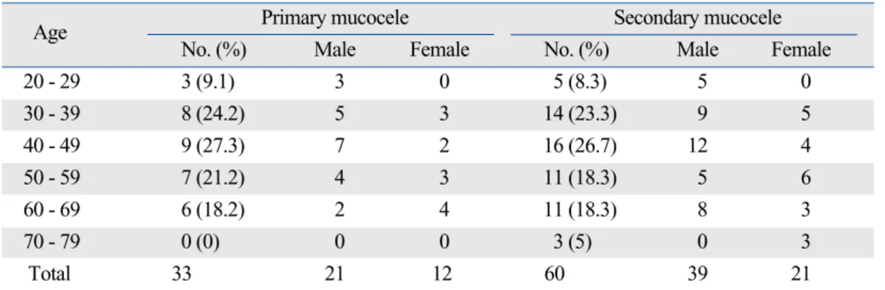
A total of 33 patients (21 men and 12 women) with a mean age of 44.7 years (range 20-69 years) were included in the primary mucocele group. The secondary mucocele group comprised 60 patients (39 men and 21 women) with a mean age of 48.6 years (range 20-78 years) (Table 1). All patients with secondary mucoceles had a history of sinus surgery (12 endoscopic sinus surgeries, 48 Caldwell-Luc opera- tions). The time elapsed between the initial procedure and the development of symptoms ranged from 2 to 42 years (average, 18.8 years). In the majority of cases (45%), 11-20 years had passed since the primary procedure (Fig. 1).
The most common presenting symptoms in primary mucoceles were nasal obstruction (19.4%), rhinorrhea (17.7%) and orbital pain (16.1%). Cheek pain (31.7%), nasal obstruction (18.3%) and posterior nasal drip (13.3%) were most frequently reported in patients with secondary mucoceles (Table 2). Preoperative CT scans were perform- ed in all patients. Mucocele which was located in maxill- ary sinus was 58.0% (n = 54) and 24.7% (n = 23) in ethmoid sinus. Location of mucocele in operative field was also checked, similar to previous CT finding. The origins of
MATERIALS AND METHODS
Table 1. Age and Sex Distribution of Primary and Secondary Mucocele
Age Primary mucocele Secondary mucocele
No. (%) Male Female No. (%) Male Female
20 - 29 3 (9.1) 3 0 5 (8.3) 5 0
30 - 39 8 (24.2) 5 3 14 (23.3) 9 5
40 - 49 9 (27.3) 7 2 16 (26.7) 12 4
50 - 59 7 (21.2) 4 3 11 (18.3) 5 6
60 - 69 6 (18.2) 2 4 11 (18.3) 8 3
70 - 79 0 (0) 0 0 3 (5) 0 3
Total 33 21 12 60 39 21
Fig. 1. Duration of periods after previous operation of secondary mucocele.
RESULTS
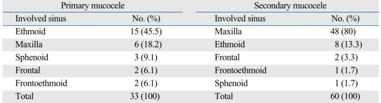
primary mucoceles were the ethmoid (45.5%), maxillary (18.2%), sphenoid (9.1%), frontal (6.1%), and frontoeth- moid sinus (6.1%). In secondary mucoceles, the maxillary sinus was the most common site (80%), followed by the ethmoid (13.3%), frontal (3.3%), sphenoid (1.7%), and frontoethmoid sinus (1.7%) (Table 3). However, the most common mucocele site in patients who had previously undergone endoscopic sinus surgery, as opposed to the Caldwell-Luc procedure, was the ethmoid sinus (58.3%) (Table 4).
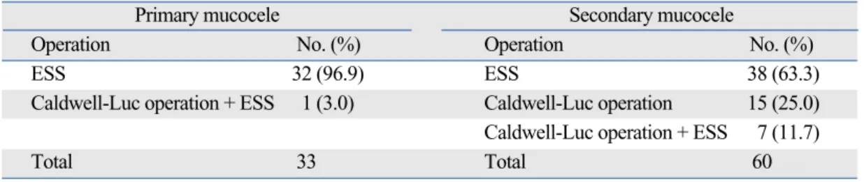
All patients (n = 93) underwent corrective sinus surgery between 1996 and 2008. The majority (96.9%) of patients
with primary mucoceles underwent endoscopic sinus surgery. Only one patient (3.0%) underwent both endosco- pic sinus surgery and the Caldwell-Luc procedure. In secondary mucoceles, 63.3% of patients underwent endos- copic sinus surgery, 25.0% were managed with the Cald- well-Luc operation, and 11.7% received both treatments (Table 5). The mean follow-up period from the initial operation in the secondary mucocele group was 4.7 years (range 1.4-7.3 years) and 5.5 years (range 1.2-9.8 years) in the primary mucocele group. There were no reported intra- or postoperative complications during admission.
A revision procedure was performed in 3 patients (9.1%) Table 2. Presenting Symptoms of Primary and Secondary Mucocele
Primary mucocele Secondary mucocele
Symptoms* No. (%) Symptoms* No. (%)
Nasal obstruction 12 (19.4) Cheek pain 38 (31.7)
Rhinorrhea 11 (17.7) Nasal obstruction 22 (18.3)
Orbital pain 10 (16.1) Posterior nasal drip 16 (13.3)
Orbital protrusion 9 (14.5) Cheek swelling 15 (12.5)
Headache 8 (12.9) Rhinorrhea 9 (7.5)
Posterior nasal drip 6 (9.6) Orbital pain 6 (5.0)
Visual disturbance 4 (6.5) Hyposmia 4 (3.3)
Hyposmia 1 (1.6) Headache 3 (2.5)
Toothache 1 (1.6) Orbital protrusion 3 (2.5)
Toothache 2 (1.7)
Fever 1 (0.8)
Visual disturbance 1 (0.8)
Total 62 (100) Total 120 (100)
*Multiple responses were allowed for each patient.
Table 3. Involved Sinuses of Primary and Secondary Mucocele
Primary mucocele Secondary mucocele
Involved sinus No. (%) Involved sinus No. (%)
Ethmoid 15 (45.5) Maxilla 48 (80)
Maxilla 6 (18.2) Ethmoid 8 (13.3)
Sphenoid 3 (9.1) Frontal 2 (3.3)
Frontal 2 (6.1) Frontoethmoid 1 (1.7)
Frontoethmoid 2 (6.1) Sphenoid 1 (1.7)
Total 33 (100) Total 60 (100)
Table 4. Involved Sinuses of Secondary Mucocele According to Surgical Method
Previous Caldwell-Luc operation Previous endoscopic sinus surgery
Involved sinus No. (%) Involved sinus No. (%)
Maxilla 47 (97.9) Ethmoid 7 (58.3)
Ethmoid 1 (2.1) Frontal 2 (16.7)
Maxilla 2 (16.7)
Sphenoid 1 (8.3)
Total 48 (100) Total 12 (100)
with primary mucoceles due to persistent sinusitis, as evi- denced by residual symptoms and lesions on CT. Twelve patients (20.0%) with secondary mucoceles experienced recurrence necessitating a revision procedure. The origin of recurrence of secondary mucoceles was predominantly the maxillary sinus (11 maxillary, 1 ethmoid), while all pri- mary mucocele recurrences originated in the ethmoid sinus.
All three cases of primary mucocele recurrence achieved symptom relief by revision endoscopic sinus surgery after several years of follow-up. In the secondary mucocele group, 4 patients (33.3%) underwent revision Caldwell- Luc operation, including patients that had formerly under- gone endoscopic sinus surgery. The remaining 8 patients (66.6%) underwent an endoscopic revision procedure.
Mucoceles of the paranasal sinus are benign, cyst-like, expansile lesions lined with pseudostratified columnar epithelium.1A mucocele is a mucoid-filled mass that devel- ops following obstruction of the sinus ostium and drainage pattern, which explains the high incidence of mucoceles in the frontal sinus caused by variations in the nasofrontal duct.8Most mucoceles occur in the frontal sinus (60%) followed by the ethmoid sinus (30%), maxillary sinus (10%) and rarely in the sphenoid sinus.8Maxillary sinus mucoceles are more commonly reported in Korea and Japan as POCC,3,4accounting for more than 50% of all paranasal sinus mucoceles in Korean literature9and 131 of 132 cases of sinus mucoceles in Japan.3The explanation for the difference in incidence between Asian and Cauca- sian populations remains unclear.5Mucoceles that develop following the Caldwell-Luc operation are presumed to result from entrapped sinus mucosa.4Secondary mu- coceles following sinus surgery generally develop as a delayed complication, typically 10 to 30 years postopera- tively.10 Similar to previous reports, the mean duration between the initial surgery and the development of secon- dary mucoceles was 18.8 years in the present study. In the majority of cases (45%), 11 to 20 years had passed since the previous sinus surgery, with the longest interval being 42 years. Diagnosis of mucoceles was made on the basis of
symptoms, imaging, surgical exploration and histological confirmation. The most informative radiologic evaluation is CT, which shows a mucocele as a homogenous lesion, isodense with brain tissue and without contrast enhance- ment, unless infected.1
Traditional surgical management of maxillary sinusitis requires complete drainage through an open approach.
Depending on the extent of the sinusitis, this might include either a simple sublabial approach or a lateral rhinotomy incision.11 With the increasing popularity of minimally invasive surgery, functional endoscopic sinus surgery has become the standard procedure for most surgical cases of chronic sinusitis. Therefore, the use of the Caldwell-Luc operation is on the decline and is now only recommended in selected cases.12This trend has increased the relative incidence of frontal and ethmoid sinus mucoceles. The causes of this change are presumed to be narrow sinus orifice, poor surgical view and outflow tract stenosis after endoscopic widening.4,13Likewise, in this study, although the maxillary sinus was the most common site of muco- celes, the most common site of primary mucoceles was the ethmoid sinus. Cases of secondary mucoceles following previous sinus endoscopic surgery, as opposed to the Caldwell-Luc operation, also developed more frequently in the ethmoid sinus.
Despite advances in endoscopic equipment and techni- ques, several indications for the Caldwell-Luc operation remain. Although the most common cause of failure in endoscopic sinus surgery is an obstructed ostium, some patients have pathologic mucosal disease.14,15In this select- ed patient population, the Caldwell-Luc operation may be necessary to treat the underlying mucosal disease. Compa- rison of the results of different surgical techniques is hea- vily affected by many confounding factors including, but not limited to, different study populations, inaccurate diag- nostics and disparate follow-up care. Similar to the delayed occurrence of POCC following the Caldwell-Luc operation, long-term observation is necessary for patients undergoing endoscopic sinus surgery to monitor for recurrence.
Based on the results of this study, we suggest that the incidence of maxillary sinus mucoceles in Asian popula- tions will decrease as the practice of endoscopic sinus sur- gery increases.
Table 5. Surgical Method for Primary and Secondary Mucocele
Primary mucocele Secondary mucocele
Operation No. (%) Operation No. (%)
ESS 32 (96.9) ESS 38 (63.3)
Caldwell-Luc operation + ESS 1 (3.0) Caldwell-Luc operation 15 (25.0) Caldwell-Luc operation + ESS 7 (11.7)
Total 33 Total 60
ESS, endoscopic sinus surgery.
DISCUSSION
This study does not have any financial relationship with certain organizations. But, we want to acknowledge alumni of Department of Otolaryngorhinology, Kangbuk Sam- sung Hospital, for various supports.
1. Busaba NY, Salman SD. Maxillary sinus mucoceles: clinical presentation and long-term results of endoscopic surgical treat- ment. Laryngoscope 1999;109:1446-9.
2. Caylakli F, Yavuz H, Cagici AC, Ozluoglu LN. Endoscopic sinus surgery for maxillary sinus mucoceles. Head Face Med 2006;
2:29.
3. Busaba NY, Kieff D. Endoscopic sinus surgery for inflammatory maxillary sinus disease. Laryngoscope 2002;112:1378-83.
4. Kim SS, Kang SS, Kim KS, Yoon JH, Lee JG, Park IY. Clinical characteristics of primary paranasal sinus mucoceles and their surgical treatment outcomes. Korean J Otolaryngol-Head Neck Surg 1998;41:1436-9.
5. Hasegawa M, Kuroishikawa Y. Protrusion of postoperative max- illary sinus mucocele into the orbit: case reports. Ear Nose Throat J 1993;72:752-4.
6. Lund VJ, Milroy CM. Fronto-ethmoidal mucoceles: a histopa- thological analysis. J Laryngol Otol 1991;105:921-3.
7. Marks SC, Latoni JD, Mathog RH. Mucoceles of the maxillary sinus. Otolaryngol Head Neck Surg 1997;117:18-21.
8. Arrué P, Kany MT, Serrano E, Lacroix F, Percodani J, Yardeni E, et al. Mucoceles of the paranasal sinuses: uncommon location. J Larlyngol Otol 1998;112:840-4.
9. Song HM, Park HW, Chung YS, Jang YJ, Lee BJ. Primary mucoceles of the maxillary sinus. Korean J Otolaryngol-Head Neck Surg 2006;49:47-51.
10. Kaneshiro S, Nakajima T, Yoshikawa Y, Iwasaki H, Tokiwa N.
The postoperative maxillary cyst: report of 71 cases. J Oral Surg 1981;39:191-8.
11. Athevino CC, Atherino TC. Maxillary sinus mucocele. Arch Otolaryngol 1984;110:200-2.
12. Barzilai G, Greenberg E, Uri N. Indications for the Caldwell-Luc approach in the endoscopic era. Otolaryngol Head Neck Surg 2005;132:219-20.
13. Har-El G, Balwally AN, Lucente FE. Sinus mucoceles: is marsupialization enough? Otolaryngol Head Neck Surg 1997;
117:633-40.
14. Cutler JL, Duncavage JA, Matheny K, Cross JL, Miman MC, Oh CK. Results of Caldwell-Luc after failed endoscopic middle meatus antrostomy in patients with chronic sinusitis. Laryngos- cope 2003;113:2148-50.
15. Richtsmeier WJ. Top 10 reasons for endoscopic maxillary sinus surgery failure. Laryngoscope 2001;111:1952-6.