Silent brain infarction (SBI) is defined as an asymptomatic infarction, and is often incidentally detected on computed tomography (CT) or magnetic resonance imaging (MRI) in subjects with no history of stroke. It is relatively common in the elderly, and the incidence of SBI significantly increases with older age.1,2The presence of SBI has been identified as an independent risk factor for the development of symptomatic infarction.3-7Therefore, population-based studies have been performed to identify risk factors for SBI, as well as factors that promote the progression of SBI to other symptomatic cerebrovascular disorders.8
It has been suggested that hyperhomocysteinemia is a risk factor for a number of occlusive vascular diseases, including SBI.2,9-12The methylenetetrahydrofolate reductase (MTHFR) gene has genetic variants (677C>T and 1298A>C) that reduce the enzyme activity, thereby elevating plasma total homocysteine (tHcy) levels.13-17 Most previous studies have concentrated on the MTHFR 677C>T polymorphisms
Association of Methylenetetrahydrofolate Reductase (MTHFR 677C>T and 1298A>C) Polymorphisms
and Haplotypes with Silent Brain Infarction and Homocysteine Levels in a Korean Population
In Bo Han,
1,2*Ok Joon Kim,
3*Jung Yong Ahn,
4Doyeun Oh,
2Sun Pyo Hong,
5Ryoong Huh,
1Sang Sup Chung,
1and Nam Keun Kim
21Department of Neurosurgery, 2Institute for Clinical Research, and 3Department of Neurology, School of Medicine, CHA University, Seongnam;
4Department of Neurosurgery, Yonsei University College of Medicine, Seoul; 5Genematrix Co., Ltd. Yongin, Korea.
Purpose:Methylenetetrahydrofolate reductase (MTHFR) is the main regulatory enzyme for homocysteine metabolism. In the present study, we evaluated whether the MTHFR 677C>T and 1298A>C gene polymorphisms are associated with SBI and plasma homocysteine concentration in a Korean population. Materials and Methods:
We enrolled 264 patients with SBI and 234 healthy controls in South Korea. Fasting plasma total homocysteine (tHcy) concentrations were measured, and genotype analysis of the MTHFR gene was carried out. Results:The plasma tHcy levels were significantly higher in patients with SBI than in healthy controls. Despite a significant association between the MTHFR 677TT genotype and hyperhomocysteinemia, the MTHFR 677C>T genotypes did not appear to influence susceptibility to SBI. However, odds ratios of the 1298AC and 1298AC + CC genotypes for the 1298AA genotype were significantly different between SBI patients and normal controls. The frequencies of 677C-1298A and 677C-1298C haplotypes were significantly higher in the SBI group than in the control group.
Conclusion:This study demonstrates that the MTHFR 1298A>C polymorphism is a risk factor for SBI in a Korean population. The genotypes of 677C>T and 1298A>C polymorphisms interact additively, and increase the risk of SBI in Korean subjects.
Key Words: Methylenetetrahydrofolate reductase, polymorphism, haplotype, silent brain infarction
Received: March 17, 2009 Revised: June 8, 2009 Accepted: July 1, 2009
Corresponding author: Dr. Nam Keun Kim, Institute for Clinical Research,
School of Medicine, CHA University, 351 Yatap-dong, Bundang-gu, Seongnam 463-712, Korea.
Tel: 82-31-780-5762, Fax: 82-31-780-5766 E-mail: [email protected]
*These authors contributed equally to this work.
∙The authors have no financial conflicts of interest.
© Copyright:
Yonsei University College of Medicine 2010
INTRODUCTION
associated with stroke, cancer, birth defects, recurrent abor- tion, and cardiovascular disorders.15,18-24However, only a few studies have been conducted to assess the association of a second common polymorphism, 1298A>C, with ischemic stroke, cancer, and heart disease.25-31No association studies of the MTHFR 1298A>C polymorphism with SBI were conducted.
Therefore, we conducted this study to investigate the association between the MTHFR 677C>T and 1298A>C polymorphisms and the risk of SBI in a Korean population.
Patients
The study population was composed of 264 patients (121 males, 143 females; mean age ± SD, 62.26 ± 11.95 years) with SBI and 227 control subjects (109 males, 118 females;
mean age ± SD, 59.62 ± 11.67 years). Patients with SBI were enrolled and recruited between July 2000 and February 2005 in the Bundang CHA Hospital.
Patients with known history of stroke or cardiovascular diseases were excluded. MRI was performed on a 1.5-T superconducting magnet (Siemens Magnetom Symphony, Erlangen, Germany). Transverse T1-weighted, T2-weight- ed, and FLAIR images were obtained with a slice thickness of 7 mm. The diagnosis of SBI was made as follows: 1) spotty areas ≥ 3 mm in diameter in the area supplied by deep perforating arteries, showing high intensity in the T2 and FLAIR images and low intensity in the T1 image; 2) ab- sence of neurological signs and symptoms corresponding to the MRI lesions; 3) no history of clinical stroke, including transient ischemic attack. Small punctate hyperintensity lesions (1 to 2 mm in diameter) were more likely to repre- sent a dilated perivascular space, and were not considered in the present study. The diagnosis of SBI was made when two independent researchers agreed on the diagnosis.
Controls (n = 227), which showed no evidence of SBI on MRI, were recruited from subjects who visited the hospital for a health examination. They had no past history of stroke or cardiovascular disease. Our subjects were clas- sified into two age groups: subjects aged < 65 years and subjects aged ≥ 65 years. Baseline demographic data and a history of conventional vascular risk factors were obtained from each control subject. Detailed information on medical history was obtained from all study subjects. The institu- tional review committee of Bundang CHA Hospital appro- ved this study in June 2000. Informed consent was obtained from all participants.
Biochemical measurements
Overnight fasting (12 hours) blood samples were collected
in EDTA-containing tubes and immediately placed on ice.
After centrifugation at 2000 rpm for 15 minutes, plasma samples were kept at -20˚C until analysis. Plasma total homocysteine (tHcy) levels were determined by fluores- cence polarization immunoassay (IMx, Abbott Laboratories, North Chicago, IL, USA).
Genetic analysis
Genomic DNA was extracted from leukocytes by using a DNA extraction kit (QIAmp blood kit, Qiagen) according to the protocol of the manufacturer. MTHFR 677C>T and 1,298A>C genotypes were identified as described previou-
sly.15,17Regions containing the two polymorphisms were
amplified separately. For the nucleotide 677 polymorphi- sm, the primers 5’-GCA CTT GAA GAG AAG GTG TC- 3’ (forward) and 5’-AGG ACG GTG CGG TGA GAG TG- 3’ (reverse) were used, and for the nucleotide 1298 poly- morphism, 5’-CTT TGG GGA GCT GAA GGA CTACTA C-3’ (forward) and 5’-CAC TTT GTG ACC ATT CCG GTT TG-3’ (reverse) were used. Human genomic DNA (200 ng) was amplified with 100 pmol of each forward and reverse primer, 1.5 mM MgCl2, 0.2 M each deoxynucleo- tide triphosphate, and 1 unit Taq polymerase (Takara, Madison, Wisconsin, USA) in a total volume of 100 uL.
PCR conditions were as follows: denaturation at 94˚C for 5 min, followed by 35 cycles at 94˚C for 30s, 51˚C for 30s, and 72˚C for 30s, and a final terminal elongation at 72˚C for 5 min. PCR products were digested with HinfI (for nucleo- tide 677) or Fnu4HI (for nucleotide 1,298) for 2 hours at 37˚C.
Amplification success was monitored by 3.0% agarose electrophoresis. For the nucleotide 677, an undigested PCR product (203 bp) indicated a homozygous wild-type, three bands of 203, 173, and 30 bp indicated the heterozy- gous genotype, and two bands of 170 and 30 bp indicated the homozygous genotype. For nucleotide 1,298, a single band of 138 bp indicated a wild-type, and two fragments of 119 and 19 bp indicated the homozygous genotype.
Statistical analysis
To estimate the relative risk for SBI for the various geno- types, an odds ratio (OR) and 95% confidence interval (CI) were calculated. Differences between the patient and control groups were assessed by the χ2test for categorical variables (sex, hypertension, and diabetes mellitus) and the two-sample t-test for continuous variables (age and tHcy level). For the multivariate analysis, logistic regression an- alysis was used to adjust for possible confounders, including age, sex, hypertension, and diabetes mellitus. The plasma tHcy levels differences between patients and controls were examined by analysis of covariance (ANCOVA), adjusted for age and sex. Haplotypes analysis was performed using
MATERIALS AND METHODS
case-control haplotype analysis-permutation test.
The analysis was performed using GraphPad Prism 4.0 (GraphPad Software Inc., San Diego, CA, USA), Stats- Direct Statistical Software Version 2.4.4 (StatsDirect Ltd., Altrincham, UK), Medcalc (version 7.4 for Windows;
Frank. Schoonjans, Belgium) and SAS 9.1 for Windows (SAS Institute Inc., Cary, NC, USA).
Table 1 shows the distributions of clinical characteristics of patients with SBI and control subjects. Patients had a significantly higher prevalence of hyperlipidemia, but not of diabetes mellitus or hypertension, compared to the con- trols. To evaluate the pure effects of the MTHFR genotypes
on SBI, we adjusted the OR for age, sex, hypertension, and diabetes mellitus. Significant elevation of pHcy concen- trations was found in patients compared to controls (11.34
± 6.385 µmol/L vs. 9.676 ± 3.959 µmol/L, p = 0.0007).
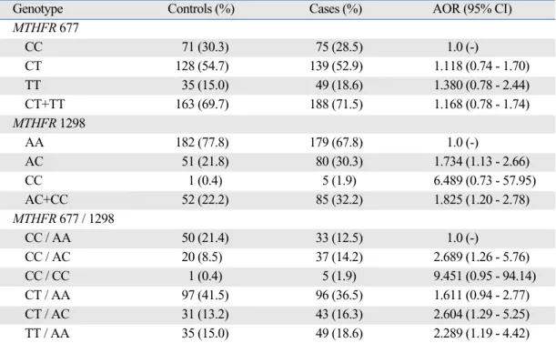
In this study, all of the study populations were in Hardy- Weinberg equilibrium. The 1298AC genotype was found to show a significant 1.734-fold increased risk of develop- ing SBI (AOR = 1.734, 95% CI = 1.13-2.66), and 1298AC + CC genotypes were significantly associated with a 1.825- fold increased risk for SBI (AOR = 1.825, 95% CI = 1.20- 2.78), whereas for the MTHFR 677C>T polymorphisms in SBI patients there were no statistically significant differences in the effects of the MTHFR genotypes on SBI between the patients and the controls (Table 2).
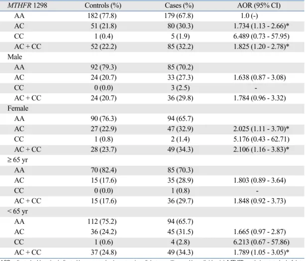
For the MTHFR 1298A>C polymorphisms, significant difference was found in terms of the risk for SBI in females
RESULTS
Table 1. Baseline Characteristics in Silent Brain Infarction (SBI) Patients and Control Subjects
Control (%) SBI (%) p value
Male (%) 116 (49.6) 121 (45.8) 0.420
Age (yrs)* 59.55 ± 11.75 62.38 ± 11.90 0.008
tHcy (µmol/L)* 9.68 ± 3.96 11.34 ± 6.39 0.001
Folate (nmol/L)* 9.85 ± 6.61 9.70 ± 5.99 0.845
Hypertension 118 (50.6) 141 (53.4) 0.598
Diabetes mellitus 42 (18.1) 38 (14.4) 0.273
Hyperlipidemia 43 (18.4) 86 (32.8) 0.0003
p-values are Chi-square test for categorical data, and two sample t-test for the continuous data.
*Values are mean ±SD (standard deviation).
Table 2. Comparison of Genotype Frequencies in the MTHFR 677C>T and 1298A>C Polymorphisms between Patients with Silent Brain Infarction (SBI) and Control Subjects
Genotype Controls (%) Cases (%) AOR (95% CI)
MTHFR 677
CC 71 (30.3) 75 (28.5) 1.0 (-)
CT 128 (54.7) 139 (52.9) 1.118 (0.74 - 1.70)
TT 35 (15.0) 49 (18.6) 1.380 (0.78 - 2.44)
CT+TT 163 (69.7) 188 (71.5) 1.168 (0.78 - 1.74)
MTHFR 1298
AA 182 (77.8) 179 (67.8) 1.0 (-)
AC 51 (21.8) 80 (30.3) 1.734 (1.13 - 2.66)
CC 1 (0.4) 5 (1.9) 6.489 (0.73 - 57.95)
AC+CC 52 (22.2) 85 (32.2) 1.825 (1.20 - 2.78)
MTHFR 677 / 1298
CC / AA 50 (21.4) 33 (12.5) 1.0 (-)
CC / AC 20 (8.5) 37 (14.2) 2.689 (1.26 - 5.76)
CC / CC 1 (0.4) 5 (1.9) 9.451 (0.95 - 94.14)
CT / AA 97 (41.5) 96 (36.5) 1.611 (0.94 - 2.77)
CT / AC 31 (13.2) 43 (16.3) 2.604 (1.29 - 5.25)
TT / AA 35 (15.0) 49 (18.6) 2.289 (1.19 - 4.42)
AOR, adjusted odds ratios (adjusted by age, gender, hypertension, diabetes mellitus, and hyperlipidemia); MTHFR, methylenetetrahydrofolate reductase.
(AC, AOR = 2.205, 95% CI = 1.11-3.70; AC + CC, AOR = 2.106, 95% CI = 1.16-3.83). When this population was separated into two age groups, 1298AC + CC genotypes showed a 1.789-fold increased risk for SBI in patients aged
< 65 years, and this relationship showed statistical signi- ficance (AOR = 1.789; 95% CI = 1.05-3.05) (Table 3). In addition, when stratified by age of 55 years, genotype frequencies were significantly different for SBI in females and older than 55 years (in females, AC, AOR = 2.025, 95% CI = 1.11-3.70, AC + CC, AOR = 2.106, 95% CI = 1.16-3.83; in ≥ 55 years, AC, AOR = 1.987, 95% CI = 1.18-3.34, AC + CC, AOR = 2.088, 95% CI = 1.25-3.50).
MTHFR 677C>T polymorphism in subgroup analysis of the SBI patients was not statistically significant such as genders or age groups (p > 0.05; data not shown).
As seen in Table 2, nine combinations are possible.
However, no individual with 677CT/1298CC, 677TT/
1298AC, and 677TT/1298CC was detected.32-34The compound genotype of 677CC/1298AC showed a 2.689- fold increased risk for SBI (AOR=2.689, 95% CI = 1.26-
5.76), and the compound genotype of 677CT/1298AC revealed a 2.604-fold increased risk for SBI (AOR = 2.604, 95% CI = 1.29-5.25). The 677TT/1298AA compound genotype was approximately 2.3 times more prone to developing SBI than the controls (AOR = 2.289; 95% CI = 1.19-4.42) (Table 2).
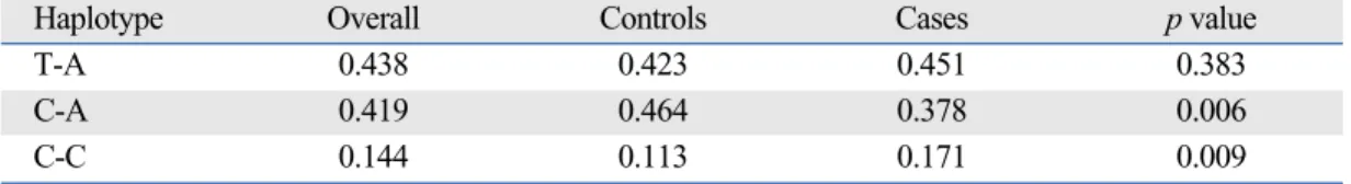
Four haplotypes are possible, but the 677T-1298C haplotype was not present in our patient population. When the cases of SBI were compared with the controls, there were significant differences in the distribution frequency between cases and controls for the 677C-1298A (p = 0.006) and 677C-1298C (p = 0.009) haplotypes (Table 4).
The 677C-1298A haplotype showed a lower frequency in patients with SBI than in the controls. However, the 677C- 1298C haplotype was more frequently observed in patients than in controls.
The tHcy levels were higher in patients with heterozy- gous 677CT and 677CT + TT genotypes compared to the corresponding control population. A statistically significant elevation of tHcy levels was observed for genotypes
Table 3. Comparison of Genotype Frequencies in the MTHFR 1298A>C Polymorphism between Patients with Silent Brain Infarction (SBI) and Control Subjects, Stratified by Sex and Age
MTHFR 1298 Controls (%) Cases (%) AOR (95% CI)
AA 182 (77.8) 179 (67.8) 1.0 (-)
AC 51 (21.8) 80 (30.3) 1.734 (1.13 - 2.66)*
CC 1 (0.4) 5 (1.9) 6.489 (0.73 - 57.95)
AC + CC 52 (22.2) 85 (32.2) 1.825 (1.20 - 2.78)*
Male
AA 92 (79.3) 85 (70.2)
AC 24 (20.7) 33 (27.3) 1.638 (0.87 - 3.08)
CC 0 (0.0) 3 (2.5) -
AC + CC 24 (20.7) 36 (29.8) 1.784 (0.96 - 3.32)
Female
AA 90 (76.3) 94 (65.7)
AC 27 (22.9) 47 (32.9) 2.025 (1.11 - 3.70)*
CC 1 (0.8) 2 (1.4) 5.176 (0.43 - 62.71)
AC + CC 28 (23.7) 49 (34.3) 2.106 (1.16 - 3.83)*
≥ 65 yr
AA 70 (82.4) 85 (70.3)
AC 15 (17.6) 35 (28.9) 1.803 (0.89 - 3.64)
CC 0 (0.0) 1 (0.8) -
AC + CC 15 (17.6) 36 (29.7) 1.848 (0.92 - 3.73)
< 65 yr
AA 112 (75.2) 94 (65.7)
AC 36 (24.2) 45 (31.5) 1.665 (0.97 - 2.87)
CC 1 (0.6) 4 (2.8) 6.213 (0.67 - 57.86)
AC + CC 37 (24.8) 49 (34.3) 1.789 (1.05 - 3.05)*
AOR, adjusted odds ratios (adjusted by age, gender, hypertension, diabetes mellitus and hyperlipidemia); MTHFR, methylenetetrahydrofolate reductase.
*p < 0.05.
1298AA, 1298AC, and 1298AC + CC. The tHcy levels were higher in patients with 1298CC than in the controls, but did not reach statistical significance due to the limited number of cases with this genotype. In combined geno- types, the tHcy levels were significantly higher in patients with 677CT/1298AC than in the controls, and 677TT/
1298AA genotypes showed an increase in tHcy levels compared to controls, with marginal significance (Table 5).
Hyperhomocysteinemia is a well-known risk factor for vascular diseases, including stroke, and it has been sug- gested that this condition may be a risk factor for the development of SBI.8,9,12The tHcy levels can potentially be elevated by either environmental (including diet) and/or
genetic factors.
The common polymorphism in the MTHFR 677C>T can reduce enzyme activity, resulting in hyperhomocystei- nemia, and can also increase the risks of cardiovascular diseases, certain types of cancer, and birth defects.31 The MTHFR 1298A>C polymorphism has also been found to reduce MTHFR enzyme activity, to a lesser extent than those with the 677C>T mutation, but conflicting results have been reported with respect to the association between 1298A>C polymorphism and tHcy levels. It has been reported that the MTHFR 1298A>C polymorphism may be associated with ischemic stroke, and may also play a protective role against colorectal cancer and acute lympho- cytic leukemia.35
In this study, the tHcy levels were significantly higher in patients with MTHFR 677TT than in patients with other MTHFR 677C>T genotypes, while no correlation between Table 5. Levels of Plasma Total Homocysteine According to MTHFR 677C>T and 1298A>C Genotypes in SBI Patients and Control Subjects
Controls (µmol/L)* Cases (µmol/L)* p value
Total 9.75 ± 0.347 11.28 ± 0.325 0.001
MTHFR 677C>T
CC 9.32 ± 0.000 10.09 ± 0.365 0.147
CT 9.67 ± 0.367 10.76 ± 0.351 0.034
TT 10.69 ± 1.581 14.69 ± 1.316 0.056
CT + TT 9.95 ± 0.466 11.75 ± 0.432 0.005
MTHFR 1298A>C
AA 9.99 ± 0.435 11.81 ± 0.439 0.004
AC 8.84 ± 0.450 10.17 ± 0.349 0.022
CC 12.79 ± 3.289 9.36 ± 1.216 0.456
AC + CC 8.87 ± 0.440 10.16 ± 0.334 0.022
MTHFR 677 / 1298
CC / AA 9.34 ± 0.416 9.74 ± 0.514 0.554
CC / AC 9.34 ± 0.824 10.37 ± 0.593 0.320
CC / CC 12.79 ± 3.288 9.37 ± 1.216 0.456
CT / AA 10.04 ± 6.335 11.10 ± 6.336 0.107
CT / AC 8.49 ± 2.825 10.02 ± 2.809 0.027
TT / AA 10.69 ± 9.339 14.69 ± 9.298 0.056
MTHFR, methylenetetrahydrofolate reductase.
*Mean ±standard error.
p values are calculated by an analysis of covariance (ANCOVA). Homocysteine levels are adjusted by age and gender.
DISCUSSION
Table 4. Comparison of Haplotype Frequencies in the MTHFR 677C>T and 1298A>C Polymorphisms between Patients with Silent Brain Infarction (SBI) and Control Subjects
Haplotype Overall Controls Cases p value
T-A 0.438 0.423 0.451 0.383
C-A 0.419 0.464 0.378 0.006
C-C 0.144 0.113 0.171 0.009
MTHFR, methylenetetrahydrofolate reductase.
p-values are calculated by the permutation test.
tHcy and MTHFR 1298A>C genotypes was found. Recent studies have demonstrated that 1298A>C, in combination with 677C>T, may be associated with decreased MTHFR activity resulting in hyperhomocysteinemia, which is con- sistent with our results (Table 5).14,36-38MTHFR 1298AC and MTHFR 1298AC + CC individuals were more prone to an increased risk for SBI than the corresponding controls, while the polymorphism of 677C>T at the MTHFR gene did not significantly influence the risk of SBI. When com- bined with the 1298A>C genotypes, however, we found that MTHFR 677C>T could have an additive effect on the variation at the MTHFR 1298A>C gene, because indi- viduals carrying the predisposing variants (677CC/1298AC, 677CT/1298AC, 677TT/1298AA) at the two loci showed a higher risk of the development of SBI than any AOR individuals registered for MTHFR 1298A>C genotypes (Table 2).
Furthermore, we investigated the associations between SBI and allele haplotypes. The C-C haplotype increased the risk of SBI, whereas the C-A haplotype, a normal allele- combined haplotype, decreased the risk of the develop- ment of SBI. These results suggested that 1298A>C may be a more important genetic risk factor for SBI than 677C>T, and the MTHFR 1298C allele-containing haplo- type may have the potential to be a predictive marker of the development of SBI in the Korean population.
However, the relationships between MTHFR poly- morphisms and multifactorial diseases, especially cardio- vascular disease and stroke, remain highly controversial, and genetic influences of MTHFR polymorphisms have not been observed in a number of populations. There was a racial difference in the frequency of the 1298C allele:
Canadian, 0.36, French, 0.33, English, 0.32, German, 0.30, American, 0.29, South African, 0.21, Japanese, 0.19, Chinese, 0.17, Korean, 0.125, Korean, 0.178, and Korean, 0.163.25,28,32,36,37,39-42From the literature, the MTHFR 1298C allele frequency was 0.125 to 0.178 in the Korean popula- tions. The 1298CC genotype is present at a much lower frequency, at 1.4-3.7%, in Asian populations, compared to 7.2-12.6% in the Caucasian population; this information is consistent with our results.25,27-29,32,36,39In the current study, the frequencies of the 1298AA, AC, and CC genotypes in the control group were 77.8%, 21.8%, and 0.4%, respecti- vely. The corresponding frequencies in the patient group were 67.8%, 30.3%, and 1.9%, respectively. The frequency of the MTHFR 1298C allele was significantly higher in the SBI patients than in the controls. These findings indicate that there are ethnic variations in terms of the 1298A>C polymorphism, as well as a difference in the occurrence of SBI between the Asian population and the Caucasian pop- ulation. It is conceivable that the contributions of MTHFR polymorphisms to SBI may vary in different ethnic groups.
In addition, no statistically significant difference in the risk for SBI was found between MTHFR genotypes in subjects over 65 years of age. We found that the 1298AC and 1298AC + CC genotypes showed an increased risk for SBI in patients under 65 years of age. SBI, a cerebrovascular disease, has an age-dependent nature. The causes of SBI are multifactorial, and additional environmental risk factors of SBI may develop with age. In this study, logistic regres- sion, adjusting for possible confounders such as age, hypertension, and diabetes mellitus, showed a significant relationship between the 1298A>C polymorphism and early-onset SBI under 65 years of age. Prior to this study, Kohara, et al.2reported that the MTHFR 677TT genotype is an independent risk factor for SBI and white matter lesions in the general Japanese population, especially in elderly subjects over 60 years of age. Since genetic poly- morphisms often vary among ethnic groups or geographical areas, further studies are needed to clarify the association between MTHFR polymorphisms and SBI in diverse ethnic populations.
This study has limitations because it was conducted in a hospital-based population. The other possible limitations are related to exposure to different environmental factors, such as daily folate intake, additional genetic effects such as methionine synthase (MTR) and methionine synthase reductase (MTRR), and ethnic differences. Large, com- munity-based random sampling is needed in order to resolve these limitations.
Despite these limitations, this study is unique in that it focused on the relationship between the MTHFR 1298A>C polymorphism and SBI in a Korean population. This study presents evidence that the 1298A>C polymorphism, but not the 677C>T polymorphism, acts as an independent risk factor for SBI, especially in patients under 65 years of age.
In addition, it indicates that the polymorphisms of MTHFR 677C>T and 1298A>C interact additively, resulting in an increased risk of SBI in a Korean population.
This work was partly supported by the Korea Research Foundation Grant, funded by the Korean Government (MOEHRD) (KRF-2008-521-E00121) and partly support- ed by a grant of the Healthcare Technology R & D Project, Ministry for Health, Welfare & Family Affairs, Republic of Korea (A084923).
1. Kobayashi S, Okada K, Yamashita K. Incidence of silent lacunar
REFERENCES
ACKNOWLEDGEMENTS
lesion in normal adults and its relation to cerebral blood flow and risk factors. Stroke 1991;22:1379-83.
2. Kohara K, Fujisawa M, Ando F, Tabara Y, Niino N, Shimokata H; NILS-LSA Study. MTHFR gene polymorphism as a risk factor for silent brain infarcts and white matter lesions in the Japanese general population: The NILS-LSA Study. Stroke 2003;34:1130-5.
3. Bernick C, Kuller L, Dulberg C, Longstreth WT Jr, Manolio T, Beauchamp N, et al. Silent MRI infarcts and the risk of future stroke: the cardiovascualr health study. Neurology 2001;57:1222-9.
4. Kobayashi S, Okada K, Koide H, Bokura H, Yamaguchi S. Sub- cortical silent brain infarction as a risk factor for clinical stroke.
Stroke 1997;28:1932-9.
5. Uehara T, Tabuchi M, Mori E. Risk factors for silent cerebral infarcts in subcortical white matter and basal ganglia. Stroke 1999;30:378-82.
6. Szolnoki Z. Chemical events behind leukoaraiosis: medicinal chemistry offers new insight into a specific microcirculation dis- turbance in the brain (a chemical approach to a frequent cerebral phenotype). Curr Med Chem 2007;14:1027-36.
7. Szolnoki Z. Pathomechanism of leukoaraiosis: a molecular bridge between the genetic, biochemical, and clinical processes (a mito- chondrial hypothesis). Neuromolecular Med 2007;9:21-33.
8. Notsu Y, Nabika T, Park HY, Masuda J, Kobayashi S. Evaluation of genetic risk factors for silent brain infarction. Stroke 1999;30:
1881-6.
9. Boushey CJ, Beresford SA, Omenn GS, Motulsky AG. A quanti- tative assessment of plasma homocysteine as a risk factor for vascular disease. Probable benefits of increasing folic acid intakes.
JAMA 1995;274:1049-57.
10. Kim NK, Choi BO, Jung WS, Choi YJ, Choi KG. Hyperhomo- cysteinemia as an independent risk factor for silent brain infarction.
Neurology 2003;61:1595-9.
11. Matsui T, Arai H, Yuzuriha T, Yao H, Miura M, Hashimoto S, et al. Elevated plasma homocysteine levels and risk of silent brain infarction in elderly people. Stroke 2001;32:1116-9.
12. Vermeer SE, van Dijk EJ, Koudstaal PJ, Oudkerk M, Hofman A, Clarke R, et al. Homocysteine, silent brain infarcts, and white matter lesions: The Rotterdam Scan Study. Ann Neurol 2002;51:
285-9.
13. Clarke R, Daly L, Robinson K, Naughten E, Cahalane S, Fowler B, et al. Hyperhomocysteinemia: an independent risk factor for vascular disease. N Engl J Med 1991;324:1149-55.
14. Friedman G, Goldschmidt N, Friedlander Y, Ben-Yehuda A, Selhub J, Babaey S, et al. A common mutation A1298C in human methylenetetrahydrofolate reductase gene: association with plasma total homocysteine and folate concentrations. J Nutr 1999;129:
1656-61.
15. Frosst P, Blom HJ, Milos R, Goyette P, Sheppard CA, Matthews RG, et al. A candidate genetic risk factor for vascular disease: a common mutation in methylenetetrahydrofolate reductase. Nat Genet 1995;10:111-3.
16. Lievers KJ, Boers GH, Verhoef P, den Heijer M, Kluijtmans LA, van der Put NM, et al. A second common variant in the methy- lenetetrahydrofolate reductase (MTHFR) gene and its relation- ship to MTHFR enzyme activity, homocysteine, and cardiovas- cular disease risk. J Mol Med 2001;79:522-8.
17. van der Put NM, Gabreëls F, Stevens EM, Smeitink JA, Trijbels FJ, Eskes TK, et al. A second common mutation in the methy- lenetetrahydrofolate reductase gene: an additional risk factor for
neural-tube defects? Am J Hum Genet 1998;62:1044-51.
18. Choi BO, Kim NK, Kim SH, Kang MS, Lee S, Ahn JY, et al.
Homozygous C677T mutation in the MTHFR gene as an inde- pendent risk factor for multiple small-artery occlusions. Thromb Res 2003;111:39-44.
19. Girelli D, Martinelli N, Pizzolo F, Friso S, Olivieri O, Stranieri C, et al. The interaction between MTHFR 677C-->T genotype and folate status is a determinant of coronary atherosclerosis risk. J Nutr 2003;133:1281-5.
20. Heijmans BT, Boer JM, Suchiman HE, Cornelisse CJ, Westen- dorp RG, Kromhout D, et al. A common variant of the methy- lenetetrahydrofolate reductase gene (1p36) is associated with an increased risk of cancer. Cancer Res 2003;63:1249-53.
21. Kim NK, Choi YK, Kang MS, Choi DH, Cha SH, An MO, et al.
Influence of combined methylenetetrahydrofolate reductase (MTHFR) and thymidylate synthase enhancer region (TSER) polymorphisms to plasma homocysteine levels in Korean patients with recurrent spontaneous abortion. Thromb Res 2006;117:653-8.
22. Ko KH, Kim NK, Yim DJ, Hong SP, Park PW, Rim KS, et al.
Polymorphisms of 5,10-methylenetetrahydrofolate reductase (MTHFR C677T) and thymidylate synthase enhancer region (TSER) as a risk factor of cholangiocarcinoma in a Korean population. Anticancer Res 2006;26:4229-33.
23. Nelen WL, Blom HJ, Steegers EA, den Heijer M, Eskes TK.
Hyperhomocysteinemia and recurrent early pregnancy loss: a meta-analysis. Fertil Steril 2000;74:1196-9.
24. Ou CY, Stevenson RE, Brown VK, Schwartz CE, Allen WP, Khoury MJ, et al. 5,10 Methylenetetrahydrofolate reductase genetic polymorphism as a risk factor for neural tube defects. Am J Med Genet 1996;63:610-4.
25. Matsuo K, Suzuki R, Hamajima N, Ogura M, Kagami Y, Taji H, et al. Association between polymorphisms of folate-and methio- nine-metabolizing enzymes-and susceptibility to malignant lymp- homa. Blood 2001;97:3205-9.
26. Sazci A, Ergul E, Tuncer N, Akpinar G, Kara I. Methylenetet- rahydrofolate reductase gene polymorphisms are associated with ischemic and hemorrhagic stroke: Dual effect of MTHFR poly- morphisms C677T and A1298C. Brain Res Bull 2006;71:45-50.
27. Shen H, Xu Y, Zheng Y, Qian Y, Yu R, Qin Y, et al. Polymor- phisms of 5,10-methylenetetrahydrofolate reductase and risk of gastric cancer in a Chinese population: a case-control study. Int J Cancer 2001;95:332-6.
28. Song C, Xing D, Tan W, Wei Q, Lin D. Methylenetetrahydrofolate reductase polymorphisms increase risk of esophageal squamous cell carcinoma in a Chinese population. Cancer Res 2001;61:
3272-5.
29. Stegmann K, Ziegler A, Ngo ET, Kohlschmidt N, Schröter B, Ermert A, et al. Linkage disequilibrium of MTHFR genotypes 677C/T-1298A/C in the German population and association studies in probands with neural tube defects (NTD). Am J Med Genet 1999;87:23-9.
30. van der Put NM, Gabreëls F, Stevens EM, Smeitink JA, Trijbels FJ, Eskes TK, et al. A second common mutation in the methylen- etetrahydrofolate reductase gene: an additional risk factor for neural-tube defects? Am J Hum Genet 1998;62:1044-51.
31. Weisberg I, Tran P, Christensen B, Sibani S, Rozen R. A second genetic polymorphism in methylenetetrahydrofolate reductase (MTHFR) associated with decreased enzyme activity. Mol Genet Metab 1998;64:169-72.
32. Isotalo PA, Wells GA, Donnelly JG. Neonatal and fetal methyle-
netetrahydrofolate reductase genetic polymorphisms: an exami- nation of C677T and A1298C mutations. Am J Hum Genet 2000;67:986-90.
33. Zetterberg H, Regland B, Palmér M, Ricksten A, Palmqvist L, Rymo L, et al. Increased frequency of combined methylenetetrahy- drofolate reductase C677T and A1298C mutated alleles in spon- taneously aborted embryos. Eur J Hum Genet 2002;10:113-8.
34. Bae J, Shin SJ, Cha SH, Choi DH, Lee S, Kim NK. Prevalent genotypes of methylenetetrahydrofolate reductase (MTHFR C677T and A1298C) in spontaneously aborted embryos. Fertil Steril 2007;87:351-5.
35. Krajinovic M, Lamothe S, Labuda D, Lemieux-Blanchard E, Theoret Y, Moghrabi A, et al. Role of MTHFR genetic polymor- phisms in the susceptibility to childhood acute lymphoblastic leukemia. Blood 2004;103:252-7.
36. Chango A, Potier De Courcy G, Boisson F, Guilland JC, Barbé F, Perrin MO, et al. 5,10-methylenetetrahydrofolate reductase com- mon mutations, folate status and plasma homocysteine in healthy French adults of the Supplementation en Vitamines et Mineraux Antioxydants (SU.VI.MAX) cohort. Br J Nutr 2000;84:891-6.
37. Kim NK, Kang GD, Kim HJ, Kim SH, Nam YS, Lee S, et al.
Genetic polymorphisms of 5,10-methylenetetrahydrofolate reduc- tase (MTHFR C677T and A1298C) in healthy Korean. Korean J
Genetics 2002;24:227-34.
38. Kluijtmans LA, Young IS, Boreham CA, Murray L, McMaster D, McNulty H, et al. Genetic and nutritional factors contributing to hyperhomocysteinemia in young adults. Blood 2003;101:
2483-8.
39. Kim HN, Lee IK, Kim YK, Tran HT, Yang DH, Lee JJ, et al.
Association between folate-metabolizing pathway polymorphism and non-Hodgkin lymphoma. Br J Haematol 2008;140:287-94.
40. Wiemels JL, Smith RN, Tayler GM, Eden OB, Alexander FE, Greaves MF; United Kingdom Childhood Cancer Study investi- gators. Methylenetetrahydrofolate reductase (MTHFR) poly- morphisms and risk of molecularly defined subtypes of childhood acute leukemia. Proc Natl Acad Sci U S A 2001;98:4004-9.
41. Gebhardt GS, Scholtz CL, Hillermann R, Odendaal HJ. Combined heterozygosity for methylenetetrahydrofolate reductase (MTHFR) mutations C677T and A1298C is associated with abruptio placentae but not with intrauterine growth restriction. Eur J Obstet Gynecol Reprod Biol 2001;97:174-7.
42. Kim JK, Kim S, Han JH, Kim HJ, Chong SY, Hong SP, et al.
Polymorphisms of 5,10-methylenetetrahydrofolate reductase and risk of stomach cancer in a Korean population. Anticancer Res 2005;25:2249-52.