Further Increases in Carbapenem-, Amikacin-, and Fluoroquinolone-Resistant Isolates of Acinetobacter spp. and
P. aeruginosa in Korea: KONSAR Study 2009
Kyungwon Lee,
1Mi-Na Kim,
2Jae-Seok Kim,
3Hye Lim Hong,
4Jung Oak Kang,
5Jong Hee Shin,
6Yeon-Joon Park,
7Dongeun Yong,
1Seok Hoon Jeong,
1Yunsop Chong,
1and The KONSAR Group
1Department of Laboratory Medicine, Yonsei University College of Medicine, Seoul; 2Department of Laboratory Medicine,
University of Ulsan College of Medicine, Asan Medical Center, Seoul; 3Department of Laboratory Medicine, Hallym University College of Medicine, Seoul;
4Department of Laboratory Medicine, Seoul Clinical Laboratories, Seoul; 5Department of Laboratory Medicine, Hanyang University College of Medicine, Seoul; 6Department of Laboratory Medicine, Chonnam National University Medical School, Gwangju;
7Department of Laboratory Medicine, School of Medicine, The Catholic University of Korea, Seoul, Korea.
Received: April 20, 2011 Revised: June 2, 2011 Accepted: June 8, 2011
Corresponding author: Dr. Yunsop Chong, Department of Laboratory Medicine, Yonsei University College of Medicine, 50 Yonsei-ro, Seodaemun-gu, Seoul 120-752, Korea.
Tel: 82-2-2228-2446, Fax: 82-2-313-0908 E-mail: [email protected]
∙ The authors have no financial conflicts of interest.
© Copyright:
Yonsei University College of Medicine 2011 This is an Open Access article distributed under the terms of the Creative Commons Attribution Non- Commercial License (http://creativecommons.org/
licenses/by-nc/3.0) which permits unrestricted non- commercial use, distribution, and reproduction in any medium, provided the original work is properly cited.
Purpose: The increasing prevalence of antimicrobial resistant bacteria has become a serious worldwide problem. The aim of this study was to analyze antimicrobial resistance data generated in 2009 by hospitals and commercial laboratories partici- pating in the Korean Nationwide Surveillance of Antimicrobial Resistance pro- gram. Materials and Methods: Susceptibility data were collected from 24 hospi- tals and two commercial laboratories. In the analysis, resistance did not include intermediate susceptibility. Duplicate isolates were excluded from the analysis of hospital isolates, but not from the commercial laboratory isolates. Results: Among the hospital isolates, methicillin-resistant Staphylococcus aureus, penicillin G-non- susceptible Streptococcus pneumoniae based on meningitis breakpoint, and ampi- cillin-resistant Enterococcus faecium remained highly prevalent. The proportion of vancomycin-resistant E. faecium gradually increased to 29%. Ceftazidime-resistant Escherichia coli and Klebsiella pneumoniae increased to 17% and 33%, respective- ly, and fluoroquinolone-resistant K. pneumoniae, Acinetobacter spp. and Pseudo- monas aeruginosa increased to 33%, 67% and 39%, respectively. Amikacin-resis- tant Acinetobacter spp. increased to 48%. Imipenem-resistant Acinetobacter spp.
and P. aeruginosa increased to 51% and 26%, respectively. Higher resistance rates were observed in intensive care unit (ICU) isolates than in non-ICU isolates among the isolates from hospitals. Resistance rates were higher in hospital isolates than in clinic isolates among the isolates from commercial laboratories. Conclusion:
Among the hospital isolates, ceftazidime-resistant K. pneumoniae and fluoroquino- lone-resistant K. pneumoniae, Acinetobacter spp., and P. aeruginosa further in- creased. The increase in imipenem resistance was slight in P. aeruginosa, but dras- tic in Acinetobacter spp. The problematic antimicrobial-organism combinations were much more prevalent among ICU isolates.
Key Words: Antimicrobial resistance surveillance, fluoroquinolone resistance, imipenem resistance, KONSAR, Staphylococcus, Acinetobacter spp., P. aeruginosa
resistant Enterococcus faecium and imipenem-resistant Aci- netobacter spp., was also found. In addition, third-generation cephalosporin-resistant E. coli and K. pneumoniae, and imi- penem-resistant P. aeruginosa were highly prevalent among the commercial laboratory (Com Lab) tested isolates.
In the present study, the trends of resistance of the afore- mentioned antimicrobial-organism combinations were de- termined. Stenotrophomonas maltophilia was also added to the surveillance study, because this organism is naturally re- sistant to quite a few classes of antimicrobial agents, and because it has been increasingly detected. Because inten- sive care units (ICUs) are a major source of resistant bacte- ria, resistance rates of isolates from ICUs and non-ICUs were compared using the data generated at six large hospi- tals with >1,000 beds. Resistance rates of isolates from sec- ondary-care hospitals and from primary care clinics were also compared using data from a Com Lab.
MATERIALS AND METHODS
Antimicrobial susceptibility data were generated in 2009 from participating KONSAR hospitals and two Com Labs.
Among the data collected, those from 24 hospitals were an- alyzed, excluding hospitals with poor quality performance.
Two Com Labs processed specimens requested by institu- tions with no in-house microbiology laboratory, i.e. second- ary care hospitals and primary-care clinics.
Responses to a questionnaire revealed that methods of sus- ceptibility testing used by the participants were: for E. coli (representing Gram-negative bacilli), the CLSI disk diffu- sion12 in two laboratories, the commercial broth microdilu- tion methods in 19, and both methods in three; for S. aureus (representing Gram-positive cocci), the CLSI disk diffusion in three laboratories, the broth microdilution method in 17, and both methods in four. Fluoroquinolone susceptibility was tested using either ciprofloxacin or levofloxacin. Cefotaxime and ceftazidime susceptibility was interpreted according to the previous CLSI breakpoints.12 For all Streptococcus pneu- moniae isolates, the oxacillin disk test was used to screen penicillin nonsusceptible isolates for meningitis interpreta- tion, because the majority of the serotypes in the respiratory tract can cause meningitis. The MIC of penicillin G was used to determine meningitis breakpoint-based susceptibility.13
The majority of the laboratories used WHONET soft- ware14 to analyze routine test data. Duplicate isolates were excluded from the analysis of hospital isolates, but not from
INTRODUCTION
The increasing prevalence of antimicrobial resistant bacte- ria has become a serious worldwide problem. The preva- lence of antibiotic-resistant bacteria varies greatly from country to country, because it is greatly influenced by anti- microbial usage and failure to control the spread of resistant bacteria. Antimicrobial-resistant bacteria have been rela- tively more prevalent in Asian countries1 and in some Euro- pean countries such as Spain, Italy and France (www.ecdc.
europa.eu). Although bacterial resistance has generally been rare in Scandinavian countries, rare antimicrobial-or- ganism combinations are being reported with increasing frequency, mostly due to importation from other countries.2
In general, resistant bacteria have been less prevalent in the U.S. and in Europe; however, vancomycin-resistant entero- cocci were first reported in the U.K. and France in 1988.3,4 Following the first discovery of highly vancomycin-resis- tant VanA-producing Staphylococcus aureus in the U.S., nine more isolates were subsequently detected, while only one of each was isolated in India and Iran.5 The KPC type carbapenemase-producing Klebsiella pneumoniae emerged in the U.S.6 and spread to other countries.
Resistance of Gram-positive cocci was once considered to be a more serious problem, but the emergence of carbapen- em-resistant Gram-negative bacilli, either due to class A β- lactamase, KPC,6 or class B β-lactamases, IMP, VIM, recent- ly identified NDM-1, etc.7,8 raised great concern once again about the resistance of Gram-negative bacilli, because these enzymes can inactivate the most potent class of β-lactam an- tibiotics, i.e. carbapenems.
An antimicrobial resistance surveillance study is a funda- mental means of appreciating trends in resistance, develop- ing accurate treatment guidelines, and evaluating the efficacy of intervention (www.who.int/drugresistance/en/). Monitor- ing the temporal trend of resistance is an essential element of the detection of subtle variations in antimicrobial resis- tance.9 The Korean Nationwide Surveillance of Antimicro- bial Resistance (KONSAR) program has been conducted for the purpose of surveillance since 1997.10 The study from 200711 showed continued prevalence of methicillin-resis- tant S. aureus (MRSA), third-generation cephalosporin-re- sistant K. pneumoniae, and fluoroquinolone-resistant Esch- erichia coli, Pseudomonas aeruginosa and Acinetobacter spp. A high amikacin resistance rate in Acinetobacter spp., and, more importantly, a gradual increase in vancomycin-
Antimicrobial resistance
The antimicrobial resistance rates of Gram-negative bacilli isolated in 2009 are shown in Table 2. The resistance rates of hospital-tested E. coli isolates were 67% to ampicillin, 19% to cefotaxime, 4% to piperacillin-tazobactam, 2% to amikacin, 35% to fluoroquinolone, and 36% to cotrimoxa- zole. The resistance rates of the Com Lab-tested E. coli iso- lates were slightly higher than those of the hospital isolates.
The cefoxitin resistance rates of the hospital and Com Lab isolates were 8% and 15%, respectively. The resistance rates of hospital-isolated K. pneumoniae were 33% to cef- tazaidime, 15% to piperacillin-tazobactam, 25% to cefoxi- tin, 15% to amikacin, and 33% to fluoroquinolone. The ES- BL-positive rates of hospital and Com Lab-isolated E. coli were 20% and 34%, respectively, and those of K. pneumoni- ae were 33% and 44%, respectively (Table 2, footnote).
The resistance rates of hospital-isolated Enterobacter clo- acae and S. marcescens were 32% and 14% to ceftazidime, 11% and 13% to cefepime, 5% and 10% to amikacin, and 11% and 16% to fluoroquinolone. The resistance rates of hospital-isolated S. maltophilia were 46% to ceftazidime, 9% to fluoroquinolone, and 12% to cotrimoxazole. The re- sistance rates of Acinetobacter spp. isolated by hospitals and Com Labs were 57% and 47% to ampicillin-sulbactam, 64% and 66% to cefepime, 51% and 48% to imipenem, 48%
and 25% to amikacin, 67% and 71% to fluoroquinolone, re- spectively. The resistance rates of P. aeruginosa isolated by hospitals and Com Labs were 23% and 34% to ceftazidime, among the Com Lab isolates. As in the 2007 study,11 the re-
sistance rates did not include intermediate susceptibility, and the mean resistance rates were calculated by averaging the resistance rate at each hospital to avoid the influence of large numbers of isolates at large hospitals. If a hospital tested less than ten isolates of an organism, they were exclud- ed from the analysis to avoid biasing the rates.15,16 Statistical significance of the differences in resistance rates was not de- termined in this surveillance as it has been a common prac- tice in large scale and continuous surveillance programs.17,18
RESULTS
Rank order of bacteria
The total numbers of bacterial isolates tested in 2009 were:
142,107 by 24 hospitals, and 68,391 by two Com Labs (Ta- ble 1). The five most prevalent bacteria tested by the hospi- tals were E. coli (21.1%), S. aureus (16.6%), coagulase-nega- tive staphylococci (CNS, 13.8%), P. aeruginosa (9.7%), and K. pneumoniae (9.4%). The 6th to 8th most prevalent organ- isms were E. faecalis (7.4%), Acinetobacter spp. (6.6%), and E. faecium (6.1%). The five most prevalent organisms test- ed by the Com Lab were E. coli (26.8%), P. aeruginosa (18.1%), K. pneumoniae (11.7%), S. aureus (11.1%), and E.
faecalis (7.4%). The 6th to 8th most prevalent organisms were CNS (7.3%), Acinetobacter spp. (5.7%), and Serratia marcescens (3.2%).
Table 1. Number, Proportion, and Rank Order of Clinically Important Bacteria Isolated in 2007 and 2009 Bacteria
Hospitals Commercial laboratories
No. (%) of isolates Rank order No. (%) of isolates Rank order
2009 2009 2007 2009 2009 2007
Escherichia coli 30,005 (21.1) 1 1 18,308 (26.8) 1 1
Klebsiella pneumoniae 13,343 (9.4) 5 5 7,997 (11.7) 3 4
Enterobacter cloacae 4,353 (3.1) 9 9 1,730 (2.5) 10 9
Serratia marcescens 2,142 (1.5) 12 10 2,208 (3.2) 8 7
Stenotrophomonas maltophilia 3,326 (2.3) 10 NT 1,567 (2.3) 11 NT
Acinetobacter spp. 9,426 (6.6) 7 8 3,899 (5.7) 7 6
Pseudomonas aeruginosa 13,755 (9.7) 4 4 12,404 (18.1) 2 2
Haemophilus influenzae 651 (0.5) 13 13 30 (0.04) 14 13
Non-typhoidal Salmonella 506 (0.4) 14 12 226 (0.3) 13 12
Staphylococcus aureus 23,578 (16.6) 2 2 7,567 (11.1) 4 3
Coag-Neg staphylococci 19,578 (13.8) 3 3 5,018 (7.3) 6 8
Enterococcus faecalis 10,516 (7.4) 6 6 5,038 (7.4) 5 5
E. faecium 8,641 (6.1) 8 7 1,895 (2.8) 9 10
Streptococcus pneumoniae 2,287 (1.6) 11 11 504 (0.7) 12 11
Total 142,107 (100) 68,391 (100)
Coag-Neg, coagulase negative; NT, not tested.
resistant to penicillin G at the CLSI meningitis breakpoint.
Trends of resistance
MRSA, suspected penicillin G-nonsusceptible S. pneumoni- ae by the oxacillin disk screening test for meningitis interpre- tation, and ampicillin-resistant E. faecium have remained highly prevalent over the past 13 years (Fig. 1). The propor- tion of vancomycin-resistant E. faecium gradually increased from 4% in 1997 to 29% in 2009.
Among the Gram-negative bacilli tested, the resistance rates of K. pneumoniae to ceftazidime and cefoxitin gradu- ally increased, reaching 33% and 25% in 2009, respective- ly; the resistance rate to fluoroquinolone, on the other hand, increased rapidly from 8% in 1997 to 33% in 2009 (Fig. 2).
The amikacin resistance rate of K. pneumoniae reached 24% in 2005, then decreased to 15% in 2009.
The high resistance rates of Acinetobacter spp. in 1999 to fluoroquinolone, amikacin, and ceftazidime decreased until 2007, but the rates increased again in 2009 to 67%, 66%, and 26% and 31% to imipenem, 19% and 37% to amikacin,
and 39% and 53% to fluoroquinolone, respectively.
The resistance rates of nontyphoidal Salmonella isolates from hospitals and Com Labs were 20% and 43% to ampi- cillin, 14% and 6% to cefotaxime, and 1% and 0% to fluoro- quinolone, respectively. Among 651 H. influenzae isolates from hospitals, 44% were resistant to ampicillin (Table 2, footnote).
The resistance rates of Gram-positive cocci are shown in Table 3. The resistance rates to oxacillin (or to cefoxitin for MRSA detection), and clindamycin were: 69% and 60%
among the hospital isolates, and 74% and 57% among the Com Lab isolates, respectively. The resistance rates of E. fae- calis hospital isolates to ampicillin and vancomycin were 6%
and 0.7%, respectively, but those of E. faecium were 92%
and 29%, respectively. The resistance rates of E. faecium Com Lab isolates to ampicillin and vancomycin were 86%
and 24%, respectively. Among the S. pneumoniae strains, 39% of hospital isolates and 44% of Com Lab isolates were
Table 2. Antimicrobial Resistance of Clinically Important Gram-Negative Bacilli Isolated at Hospitals and at Commercial Labo- ratories in 2009*
Antimicrobial agents
Resistance (%) of isolates (hospital/commercial laboratory) E. coli K. pneu-
moniae E. cloacae S. marce-
scens Non-T
Salmonella S. maltophilia Acineto-
bacter spp. P. aeru- ginosa 30,005/18,308† 13,343/7,997 4,353/1,730 2,142/2,208 506/226 3,326/1,567 9,426/3,899 13,755/12,404
Ampicillin 67/74 -‡ - - 20/43 - - -
Ampicillin-
sulbactam 30/11 51/42 - - - - 57/47 -
Cephalothin 29/38 40/53 - - - - - -
Cefotaxime 19/31 30/43 32/20 25/20 14/6 - 77/- 70/-
Ceftazidime 17/31 33/46 32/23 14/5 11/6 46/27 66/73 23/34
Cefepime 17/30 27/40 11/6 13/2 - - 64/66 24/38
Aztreonam 18/30 35/46 31/21 17/14 12/6 - 77/83 28/35
Cefoxitin 8/15 25/32 - - - - - -
Piperacillin 52/65 78/100 35/34 37/41 - - 65/74 30/45
Piperacillin-
tazobactam 4/9 15/- 16/16 15/6 - - 55/68 24/43
Imipenem 0.1/0 0.5/0.5 0.6/0.5 1.3/0 - - 51/48 26/31
Meropenem 0.1/0 0.8/0.5 0.7/0.5 1.3/0 - - 56/60 23/36
Amikacin 2/4 15/26 5/5 10/19 - - 48/25 19/37
Gentamicin 26/34 19/28 13/14 25/38 - - 64/70 29/47
Tobramycin 18/20 27/38 18/21 33/44 - - 54/47 28/46
Fluoroquinolone 35/48 33/39 11/11 16/19 1/0 9/5 67/71 39/53
Cotrimoxazole 36/- 18/- 23/- 19/- 9/- 12/- 67/- -
Tetracycline 48/51 20/20 21/18 45/32 5/- 8/2 59/50 -
Non-T, non-typhoidal.
*Data not shown in the table: ESBL-positive rates of E. coli 20% and 34%, and K. pneumoniae 33% and 44% for hospitals and commercial laboratories isolates; ampicillin-resistance rates of H. influenzae 44% among 651 isolates at hospitals, and 63% among 30 isolates at commercial laboratories.
†Number of isolates from hospitals/commercial laboratories.
‡not tested.
imipenem-resistant P. aeruginosa, oxacillin-resistant S. au- reus, and vancomycin-resistant E. faecium were more preva- lent among ICU isolates than among non-ICU isolates (Fig.
5). It was observed that the imipenem resistance rate of P.
aeruginosa ICU isolates was almost two-fold higher (39%) than that of the non-ICU isolates (20%).
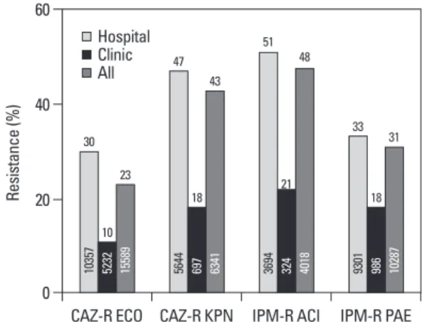
The proportions of antimicrobial-organism combinations detected by a Com Lab from hospital patients and from clinic patients are shown in Fig. 6. The isolates from hospital patients showed a 3-fold higher rate of ceftazidime-resistant E. coli, more than 2-fold higher rates of ceftazidime-resistant K. pneu- moniae, and imipenem-resistant Acinetobacter spp., and a nearly 2-fold higher rate of imipenem-resistant P. aeruginosa.
48%, respectively (Fig. 3). The proportion of imipenem-re- sistant isolates steadily increased from 1% in 1997 to 22%
in 2007, and then drastically increased, reaching 51% in 2009. The high fluoroquinolone resistance rates of P. aeru- ginosa fluctuated between 33% and 42% during the period of 1997-2009, while the amikacin resistance rates steadily declined from 33% in 1997 to 19% in 2009 (Fig. 4). The cef- tazidime and imipenem resistance rates slowly increased from 16% to 23% and from 17% to 26%, during the study period, respectively.
Analysis of the data from six hospitals with >1,000 beds showed that cefotaxime-resistant E. coli, ceftazidime-resis- tant K. pneumoniae, imipenem-resistant Acinetobacter spp.,
Table 3. Antimicrobial Resistance of Clinically Important Gram-Positive Cocci Isolated at Hospitals and in Commercial Laboratories in 2009
Antimicrobial agents
Resistance (%) of isolates (hospital/commercial laboratory) S. aureus* Coag-Neg
Staphylococcus* E. faecalis E. faecium S. pneumoniae 23,578/7,567 19,578/5,018 10,516/5,038 8,641/1,895 2,287/504
Penicillin/ampicillin† 94/97 89/94 6/- 92/86 39/44
Gentamicin 56/58 47/33 - - -
Ciprofloxacin 57/54 44/37 30/37 91/88 6/6
Clindamycin 60/57 49/34 - - 60/-
Erythromycin 67/64 60/52 72/74 96/86 70/73
Fusidic acid 49/36 42/57 - - -
Oxacillin/cefoxitin 69/74 73/74 - - 70/-
Tetracycline 55/58 31/31 85/87 22/22 69/73
Teicoplanin 0.1/0 1.3/0 0.9/1 22/14 -
Vancomycin 0/0 0.02/0 0.7/2 29/24 -
Coag-Neg, coagulase-negative; -, not tested.
*4% and 32% of hospital-isolated S. aureus and Coag-Neg Staphylococcus, respectively, were resistant to cotrimoxazole.
†Penicillin for Staphylococcus spp., and S. pneumoniae, and ampicillin for Enterococcus spp.
Fig. 1. Resistance trends of gram-positive cocci isolated in 1997- 2009 from the participating hospitals. Oxacillin-resistant S. aureus (OXA-R SAU), penicillin G-nonsusceptible S. pneumoniae (PEN- NS SPN), and ampicillin-resistant E. faecium (AMP-R EFM) re- mained highly prevalent. However, the proportion of vancomycin- resistant E. faecium (VAN-R EFM) gradually increased from 4% in 1997 to 29% in 2009.
Fig. 2. The resistance trends of K. pneumoniae isolated in 1997-2009 from the participating hospitals. The resistance rates to ceftazidime (CAZ-R), cefoxitin (FOX-R) gradually increased and reached 33%
and 25%, respectively, in 2009, whereas the resistance rate to fluo- roquinolone (FQN-R) increased rapidly from 8% in 1997 to 33% in 2009. The amikacin resistance rate (AMK-R) increased to 24% in 2005 and then decreased to 15% in 2009.
0 10 20 30 40
Resistance (%)
1997 1999 2001 2003 2005 2007 2009 CAZ-R FOX-R FQN-R AMK-R 0
20 40 80 60 100
Resistance (%)
1997 1999 2001 2003 2005 2007 2009 OXA-R SAU
PEN-NS SPN AMP-R EFM VAN-R EFM
75
84 88 88 85 86 92
72
78 72 70
64 69 70
70 69 70 68
59 64
69
4 16 20
15 20 21
29
26 24
27 25
32 29
33
16 22 20 23
30
27 25
8 10 12
19
24 21
15
6
10 13
18
laboratory or by analyzing the susceptibility data generated by each hospital laboratory. It was considered that the pres- ent need is for a supranational resistance surveillance system including non-invasive isolates, preferably based on all rou- tine susceptibility data obtained from local laboratories.19
The present KONSAR study showed that the majority of hospital laboratories used commercial broth microdilution methods for susceptibility testing of E. coli and S. aureus, which can represent Gram-negative and Gram-positive bac- teria, respectively. Automation and rapid results are some of the advantage of these methods, but they may not perform optimally for the detection of some antimicrobial-organism
DISCUSSION
The prevalence of resistant organisms varies significantly by time, country, and patient population. Therefore, antimicro- bial resistance surveillance studies are very important to monitor levels of endemic or emerging resistance. In fact, with rapid increases in resistant bacteria, we need to devote more surveillance efforts to help delay the increasing trend of resistance, although it is impossible to completely over- come the problem. An antimicrobial surveillance study is done either by testing collected isolates at a coordinating
Fig. 3. The resistance trends of Acinetobacter spp. isolated in 1997- 2009 from the participating hospitals. The high resistance rates in 1999 to fluoroquinolone (FQN-R), amikacin (AMK-R), and ceftazi- dime (CAZ-R) decreased by 2007, but the rates increased again in 2009 to 67%, 66%, and 48%, respectively. The proportion of imipen- em-resistant (IPM-R) isolates steadily increased from 1% in 1997 to 22% in 2007, and after that the increase was drastic, reaching 51%
in 2009.
Fig. 4. The resistance trends of P. aeruginosa isolated in 1997-2009 from the participating hospitals. The high fluoroquinolone (FQN-R) resistance rates fluctuated between 33% and 42% during the study period. The amikacin resistance rate (AMK-R) declined steadily from 33% in 1997 to 19% in 2009. The resistance rates to ceftazi- dime (CAZ-R) and imipenem (IPM-R) increased slowly from 16% to 23% and from 17% to 26%, respectively, during the study period.
Fig. 5. Comparison of resistance rates of ICU vs. non-ICU isolates from six hospitals with >1,000 beds. Cefotaxime-resistant E. coli (CTX-R ECO), ceftazidime-resistant K. pneumoniae (CAZ-R KPN), imi- penem-resistant Acinetobacter spp. (IPM-R ACI), imipenem-resistant P. aeruginosa (IPM-R PAE), oxacillin-resistant S. aureus (OXA-R SAU), and vancomycin-resistant E. faecium (VAN-R EFM) were much more prevalent among ICU isolates than among non-ICU iso- lates. The imipenem-resistance rate of P. aeruginosa ICU isolates was almost two-fold higher (39%) than that of non-ICU isolates (20%).
Fig. 6. The high prevalence of four antimicrobial agent-organism combinations among commercial laboratory tested isolates from hospital patients compared to those from clinic patients. The isolates from hospital patients showed a 3-fold higher rate of ceftazidime-re- sistant (CAZ-R) E. coli, more than 2-fold higher rates of CAZ-R K.
pneumoniae and imipenem-resistant (IPM-R) Acinetobacter spp., and nearly 2-fold higher rate of IPM-R P. aeruginosa. All numbers of isolates are shown in the bars, and the proportions (%) of antimicro- bial-organism combinations are shown on the bars.
0 0
0
20 20
60 80
60
40 40
40
20
80 100
60
Resistance (%) Resistance (%)Resistance (%)
1997 CTX-R
ECO
CAZ-R ECO CAZ-R KPN IPM-R ACI IPM-R PAE CAZ-R
KPN IPM-R ACI IPM-R
PAE OXA-R SAU VAN-R 1999 2001 2003 2005 2007 2009 EFM
64 68
65
58 56
51 50
67
55
45 66 50
34
30
47
51
33 51
74
39 77
44
23
10
18 21
18 23
43
48
31 31
53
20 57
28
38110357 5644 3694 9301
787 1264 978 1430 517
120885232 697 324 986
15589 6341 4018 10287
4501 2148 4747 7188 3268
65 61
54
43 37
48
1 5 16
6 13
22 51
FQN-R AMK-R CAZ-R IPM-R
0 10 20 40 30 50
Resistance (%)
1997 1999 2001 2003 2005 2007 2009
42 39
28
40 40
34
20 33
39
21 23
33
20 26
21 20
25 22
19 16
17
17 19
19 19 19
26
FQN-R AMK-R CAZ-R IPM-R
ICUNon-ICU
Hospital Clinic All
was 3.3% and rank order was No. 8.22 In our study, the rank order of S. maltophilia was similar, i.e., No. 10.
Our present Com Lab data showed that isolates from sec- ondary-care hospitals were much more frequently resistant than those from primary-care clinics, suggesting a higher prevalence of nosocomially acquired pathogens among the hospital isolates (Fig. 6). In Germany, hospitals with their own microbiology laboratory reported higher frequencies of health care-associated infections than did hospitals with- out in-house laboratory services.23
In a recent review, Peleg and Hooper24 expressed concern about hospital-acquired Gram-negative bacterial infections.
In the U.S., it was estimated that a total of 1.7 million hospi- tal-acquired infections occurred (4.5 per 100 admissions) in 2002, and hospital-acquired infections were the sixth leading cause of death. It is of great concern that only approximately one third of hospital-acquired infections are preventable.
ICUs are reservoirs of resistant bacteria. The U.S. Na- tional Healthcare Safety Network (NHSN) indicated that Gram-negative bacteria caused more than 30% of hospital- acquired infections, and these bacteria predominated in cas- es of ventilator-associated pneumonia (47%) and urinary tract infections (45%). In ICUs, Gram-negative bacteria ac- counted for about 70% of these types of infections.24 An in- ternational study showed that resistance rates of ICU iso- lates from Latin America, Asia, Africa, and Europe were higher than those in the U.S.: MRSA 84.1% vs. 56.8%, K.
pneumoniae to ceftazidime or ceftriaxone 76.1% vs. 27.1%, A. baumannii to imipenem 46.3% vs. 29.2%, and P. aerugi- nosa to piperacillin 78.0% vs. 20.2%, respectively.25 Howev- er, it is noteworthy that in the U.S., the proportion of MRSA was high, and those of other organisms were not low.
Resistance is reported to have increased throughout Eu- rope, among Gram-negative bacilli in particular.19 Gram- negative bacterial infections have features that are of partic- ular concern. These organisms are highly efficient at up- regulating or acquiring resistance genes.24 In our present study, the ceftazidime resistance rate of hospital isolates of K. pneumoniae was 33%. The rate was much higher than the 0% among 27 isolates in Iceland, but much lower than the 68.7% found in Greece (www.ecdc.europa.eu). Among the Gram-negative bacilli associated with intra-abdominal infections in 2008 from 11 Asia-Pacific countries, the high- est rates of ESBL-producing E. coli and K. pneumoniae were observed in India (61.2% and 46.8%, respectively);
the rates were 30.4% and 25.0% in Korea, respectively.1 Currently the most feared resistance is those to carbapen- combinations. With the increasing prevalence of multidrug
resistant (MDR) Acinetobacter spp. and P. aeruginosa, poly- myxins and tigecycline susceptibility testing have become a requirement, but these were not included in the present study mainly because of their unavailability in the majority of commercial systems.
In 2010, the CLSI13 lowered the resistance breakpoints of cefotaxime, ceftazidime, and aztreonam to ≥4 μg/mL, ≥16 μg/mL, and ≥16 μg/mL, respectively, and eliminated the necessity of ESBL testing of E. coli, K. pneumoniae spp., and P. mirabilis. In our present study, the previous CLSI breakpoints12 were used for these antimicrobial agents, and some laboratories differentiated ESBL-producing isolates.
In the analysis of laboratory generated susceptibility data, the CLSI20 recommends including only the first isolate if there were multiple isolates from one patient, for the pur- pose of guiding clinicians in the empirical selection of ap- propriate antimicrobial agents. The WHONET software14 can be used to exclude duplicate isolates. In the present study, the Com Labs were not able to exclude duplicate iso- lates due to difficulty identifying relevant patients. This ap- parently raised the resistance rates of some organisms to certain antimicrobial agents. However, if only the first iso- late is included, and particularly if the analysis is made only once a year, it may underestimate resistance acquired dur- ing hospitalization, as well as the emergence of resistance due to mutations during therapy.19 A study showed that not all U.S. hospitals excluded duplicate isolates.21 In fact, to detect developing resistance during therapy, the CLSI13 rec- ommends testing the susceptibility of subsequent isolates three to four days after the initial isolation of Enterobacter, Citrobacter, and Serratia spp. to third-generation cephalo- sporins, of P. aeruginosa to all antimicrobial agents and of staphylococci to quinolones.
In 2009, as in 2007, E. coli, S. aureus, CNS, P. aerugino- sa, and K. pneumoniae were the five most prevalent organ- isms among those isolated by hospitals (Table 1). Among the Com Lab isolates, E. coli, P. aeruginosa, and E. faeci- um were ranked numbers 1, 2, and 5, respectively, in both 2007 and 2009, but the rank orders of S. aureus and K. pneu- moniae were reversed (3 and 4 in 2007; 4 and 3 in 2009, re- spectively). The rank orders in the U.S. were largely simi- lar. The SENTRY Program in 2004-2008 showed that the five most frequently isolated bacteria from hospitalized pa- tients with pneumonia were S. aureus (36.3%), P. aerugino- sa (19.7%), Klebsiella spp. (8.5%), E. coli (4.6%) and Aci- netobacter spp. (4.8%). The proportion of S. maltophilia
and ventilator-associated bacterial pneumonia in North America, 54% were nonsusceptible to oxacillin.22 Vancomy- cin-resistant enterococci are also important Gram-positive nosocomial pathogens. In the U.S., hospitalizations with van- comycin-resistant enterococcal infections increased from 3.16 to 6.51 per 10,000 hospitalizations during 2003-2006.32 The U.S. NHSN data33 showed that 80% of E. faecium iso- lates in 2006 and 2007 from central line-associated blood- stream infections, catheter-associated urinary tract infections, ventilator-associated pneumonia, and surgical site infections were vancomycin resistant.
The EARS-Net 2009 report (www.ecdc.europa.eu) showed that the prevalence of invasive MRSA and vancomycin-resis- tant E. faecium varied greatly by country. For example, the proportions of MRSA were 0% of 59 isolates in Iceland, and 49.1% of 1,824 isolates in Portugal, and those of vancomycin- resistant E. faecium were 0% of 243 isolates in Finland, and 37.8% of 386 isolates in Ireland. The report also showed that the resistance rates varied greatly depending on hospitals even within a country: the mean prevalence of MRSA from Ger- many was 18.7%, but ranged from a minimum of 0% to a maximum of 60%. In our present study, 39% of hospital iso- lates of S. pneumoniae were resistant to penicillin G at the CLSI meningitis breakpoint.13 This rate did not differ greatly from those of a U.S. study, 28.4% in 1998 and 41.0% in 2009.34 Until 2008, penicillin G susceptibility of all S. pneu- moniae isolates was interpreted according to CLSI breakpoint for meningitis, because a non-meningitis breakpoint did not exist. In our present report, we continued to include oxacillin screening test results for respiratory isolates, because the ma- jority of S. pneumoniae serotypes recovered from the respira- tory tract are those causing meningeal infections, too, and be- cause the result could show a resistance trend.
With increasing international air travel and import of ani- mal produce, it has become very difficult to control the spread of antimicrobial-resistant bacteria. Carbapenemases IMP-1 and VIM-2, initially detected in Japan35 and in France,7 respectively, are now widespread globally, including in Korea. Scandinavia is a region with a low prevalence of antimicrobial resistance, but import and local clonal expan- sion of VIM-2-producing P. aeruginosa has been reported.2 Also, blaSPM-1, which was initially found in Brazil, was de- tected recently in a P. aeruginosa isolate in Switzerland.36 An isolate of K. pneumoniae, producing a new MBL, NDM-1, was first found in Sweden in a patient transferred from In- dia.8 K. pneumoniae isolates producing a class A carbapene- mase, KPC-2, were first found in New York.6 K. pneumoniae ems. In our present study, 26% of P. aeruginosa were resistant
to imipenem (Table 2, Fig. 4). This rate is much higher than the 0% of 16 isolates found in Iceland, but much lower than the 44% found in Greece (www.ecdc.europa.eu). Of over 800 β-lactamases identified from Gram-negative bacilli, at least 120 have been detected in P. aeruginosa. IMP- and VIM-type MBLs are predominantly found in P. aeruginosa and in Aci- netobacter spp.26 The proportions of imipenem-resistant iso- lates in the 1997 vs. 2009 KONSAR studies were: P. aerugi- nosa 17% vs. 26%, and Acinetobacter spp. 1% vs. 51%, respectively (Figs. 3 and 4). The drastic increase in imipenem- resistant Acinetobacter spp. was probably due to OXA-type carbapenemase production. It is interesting that almost all car- bapenem-resistant A. baumannii carried blaOXA-23-like or ISA- ba1-activated blaOXA-51-like genes, while the majority of non- baumannii Acinetobacter carried MBL genes.27
The terms MDR, extreme drug resistance (XDR), and pandrug resistance (PDR), are very useful to attract atten- tion, but there has been no consensus as to their definition.
The term PDR in A. baumannii and P. aeruginosa in partic- ular is thought to have been used inappropriately.28 In one study, MDR was defined as nonsusceptibility to one or more antimicrobials in three or more antimicrobial classes (antipseudomonal penicillins, third-generation cephalospo- rins, carbapenems, fluoroquinolones, and aminoglyco- sides). The definition did not include colistin and tigecy- cline susceptibility.29 The study showed that the proportions of MDR isolates of A. baumannii and P. aeruginosa report- ed to the U.S. NHSN systems in 2008 were 74% and 17%, respectively, but XDR isolates (nonsusceptible to all anti- microbials including polymyxins and tigecycline) remained rare. Of the 344 blaKPC-positive ICU isolates of Enterobac- teriaceae, 91% and 99% were susceptible to colistin and ti- gecycline, respectively. Only two isolates were nonsuscepti- ble to both antimicrobial agents. In Korea, colistin-resistant A. baumannii isolates were detected.30 Therefore, it is nec- essary to include colistin and tigecycline susceptibility in future surveillance studies in Korea.
In our present study, 44% of hospital isolates of H. influ- enzae were resistant to ampicillin. Bae, et al.31 reported that among 544 nationwide collections of respiratory isolates of H. influenzae, 58.5% were ampicillin resistant, and 52.4%
were β-lactamase producers.
In Korea, hospital-associated MRSA has been a serious problem (Fig. 1). MRSA has become a problem organism in North America as well. The SENTRY Program showed that among the S. aureus isolates in 2008 from hospital-acquired
Yang, Kosin University Gospel Hospital, Busan; Gyoung- Yim Ha, Dongguk University Gyeongju Hospital, Gyeongju;
Chulhun L. Chang, Pusan National University Hospital, Busan; Ki-hyung Park, Busan Medical Center, Busan; Jeey- oung Ahn, Soonchunhyang University Gumi Hospital, Gumi;
Wee Gyo Lee, Ajou University Hospital, Suwon; Chae-Hoon Lee, Yeungnam University Medical Center, Daegu; Joseph Jeong, Ulsan University Hospital, Ulsan; Ji-Hyun Cho, Wonk- wang University Hospital, Iksan; Seok-Il Hong, Korea Can- cer Center Hospital, Seoul; Young Uh, Yonsei University Wonju Christian Hospital, Wonju; Jin Ju Kim, Inha Universi- ty Hospital, Incheon; Hye Soo Lee, Chonbuk National Uni- versity Hospital, Jeonju; Sook Jin Jang, Chosun University Hospital, Gwangju; Chang Hyun Rhim, Wallace Memorial Baptist Hospital, Busan; Myung Hee Lee, Seoul Veterans Hospital, Seoul; Wonkeun Song, Gangnam Sacred Heart Hospital, Seoul; Tae Yeal Choi, Hanyang University Hospi- tal, Seoul; Seong Geun Hong, CHA Bundang Medical Cen- ter, Seongnam; Young Ah Kim, National Health Insurance Corporation Ilsan Hospital, Goyang; Hyukmin Lee, Myongji Hospital, Goyang; Dong Hee Whang, Inje University Seoul Paik Hospital, Seoul; Miae Lee, Ewha Womans University Mokdong Hospital, Seoul; Hee Bong Shin, Soonchunhyang University Bucheon Hospital, Bucheon; Seonghee Lee, Han- maeum Hospital, Jeju; Seong-Gyu Lee, Bundang Jesaeng Hospital, Seongnam; Seok Lae Chae, Dongguk University Ilsan Hospital, Goyang; Yong-kyun Kim, Samkwang Medi- cal Laboratories, Seoul.
REFERENCES
1. Hsueh PR, Badal RE, Hawser SP, Hoban DJ, Bouchillon SK, Ni Y, et al. Epidemiology and antimicrobial susceptibility profiles of aerobic and facultative Gram-negative bacilli isolated from pa- tients with intra-abdominal infections in the Asia-Pacific region:
2008 results from SMART (Study for Monitoring Antimicrobial Resistance Trends). Int J Antimicrob Agents 2010;36:408-14.
2. Samuelsen O, Toleman MA, Sundsfjord A, Rydberg J, Leegaard TM, Walder M, et al. Molecular epidemiology of metallo-β- lactamase-producing Pseudomonas aeruginosa isolates from Nor- way and Sweden shows import of international clones and local clonal expansion. Antimicrob Agents Chemother 2010;54:346-52.
3. Leclercq R, Derlot E, Duval J, Courvalin P. Plasmid-mediated re- sistance to vancomycin and teicoplanin in Enterococcus faecium.
N Engl J Med 1988;319:157-61.
4. Uttley AH, Collins CH, Naidoo J, George RC. Vancomycin-resis- tant enterococci. Lancet 1988;1:57-8.
5. Périchon B, Courvalin P. VanA-type vancomycin-resistant Staphy- lococcus aureus. Antimicrob Agents Chemother 2009;53:4580-7.
6. Yigit H, Queenan AM, Anderson GJ, Domenech-Sanchez A, Bid-
isolates, carrying NDM-1 (unpublished information), and carrying KPC-237 were detected in Korean patients in 2010.
We now know that use of antimicrobial agents is the cause of the emergence and spread of resistant bacteria. However, we should acknowledge that antimicrobial resistant bacteria existed before the clinical use of antimicrobial agents. We also should know that stopping the prescription of antimicro- bial agents to outpatients with colds may not reduce the emergence of certain resistances such as to vancomycin and carbapenems. It has been hoped that resistant bacteria could be reduced by stopping overuse of antimicrobial agents.
However, it is difficult to judge the overuse of antimicrobial agents at a hospital or ward. A study in France in 2007 showed the level of antimicrobial consumption to be only 60 defined daily doses (DDD)/1,000 patient-days (PD) in psy- chiatric wards compared to 1,466 DDD/1,000 PD in ICUs.
Glycopeptides and carbapenems were mostly used in cancer patients and in teaching hospitals.38 Sykes39 stated that how- ever hard we try and however clever we are, bacteria that have adapted to survive under the most extreme conditions for the past three billion years, and will overcome whatever we do to control them. The emergence of antibiotic resis- tance is inevitable, but we must seek to decrease its impact and to prolong the effectiveness of the agents available to us.40 For this purpose, continued surveillance study of antimi- crobial resistance is needed more than ever before to deter- mine the trends of already prevalent resistant organisms and to detect the recently imported KPC-2 and NDM-1-produc- ing Gram-negative bacilli.
In conclusion, the present surveillance study in 2009 showed that among hospital laboratory isolated bacteria, the prevalence of ceftazidime-resistant K. pneumoniae, fluoro- quinolone-resistant K. pneumoniae, Acinetobacter spp., and P. aeruginosa further increased, and imipenem-resistant Acinetobacter spp. increased drastically. It was also noted that methicillin-resistant S. aureus remained highly prevalent, and vancomycin-resistant E. faecium further increased.
Problematic antimicrobial-organism combinations were much more prevalent among ICU isolates, and were also prevalent among isolates tested at Com Labs.
ACKNOWLEDGEMENTS
This work was supported by a grant from bioMeriux Korea.
Other KONSAR program participants were: Sunjoo Kim, Gyeongsang National University Hospital, Jinju; Heeyoung
negative bacteria. N Engl J Med 2010;362:1804-13.
25. Rosenthal VD, Maki DG, Jamulitrat S, Medeiros EA, Todi SK, Gomez DY, et al. International Nosocomial Infection Control Consortium (INICC) report, data summary for 2003-2008, issued June 2009. Am J Infect Control 2010;38:95-104.e2.
26. Zhao WH, Hu ZQ. β-lactamases identified in clinical isolates of Pseudomonas aeruginosa. Crit Rev Microbiol 2010;36:245-58.
27. Lee K, Kim MN, Choi TY, Cho SE, Lee S, Whang DH, et al. Wide dissemination of OXA-type carbapenemases in clinical Acineto- bacter spp. isolates from South Korea. Int J Antimicrob Agents 2009;33:520-4.
28. Falagas ME, Koletsi PK, Bliziotis IA. The diversity of definitions of multidrug-resistant (MDR) and pandrug-resistant (PDR) Aci- netobacter baumannii and Pseudomonas aeruginosa. J Med Mi- crobiol 2006;55:1619-29.
29. Kallen AJ, Srinivasan A. Current epidemiology of multidrug resis- tant gram negative bacilli in the United States. Infect Control Hosp Epidemiol 2010;31 Suppl 1:S51-4.
30. Ko KS, Suh JY, Kwon KT, Jung SI, Park KH, Kang CI, et al.
High rates of resistance to colistin and polymyxin B in subgroups of Acinetobacter baumannii isolates from Korea. J Antimicrob Chemother 2007;60:1163-7.
31. Bae S, Lee J, Lee J, Kim E, Lee S, Yu J, et al. Antimicrobial resis- tance in Haemophilus influenzae respiratory tract isolates in Ko- rea: results of a nationwide acute respiratory infections surveil- lance. Antimicrob Agents Chemother 2010;54:65-71.
32. Ramsey AM, Zilberberg MD. Secular trends of hospitalization with vancomycin-resistant Enterococcus infection in the United States, 2000-2006. Infect Control Hosp Epidemiol 2009;30:184-6.
33. Hidron AI, Edwards JR, Patel J, Horan TC, Sievert DM, Pollock DA, et al. NHSN annual update: antimicrobial-resistant pathogens associated with healthcare-associated infections: annual summary of data reported to the National Healthcare Safety Network at the Centers for Disease Control and Prevention, 2006-2007. Infect Control Hosp Epidemiol 2008;29:996-1011.
34. Jones RN, Sader HS, Moet GJ, Farrell DJ. Declining antimicrobial susceptibility of Streptococcus pneumoniae in the United States:
report from the SENTRY Antimicrobial Surveillance Program (1998-2009). Diagn Microbiol Infect Dis 2010;68:334-6.
35. Watanabe M, Iyobe S, Inoue M, Mitsuhashi S. Transferable imi- penem resistance in Pseudomonas aeruginosa. Antimicrob Agents Chemother 1991;35:147-51.
36. Salabi AE, Toleman MA, Weeks J, Bruderer T, Frei R, Walsh TR.
First report of the metallo-β-lactamase SPM-1 in Europe. Antimi- crob Agents Chemother 2010;54:582.
37. Rhee JY, Park YK, Shin JY, Choi JY, Lee MY, Peck KR, et al.
KPC-producing extreme drug-resistant Klebsiella pneumoniae isolate from a patient with diabetes mellitus and chronic renal fail- ure on hemodialysis in South Korea. Antimicrob Agents Che- mother 2010;54:2278-9.
38. Dumartin C, L’Hériteau F, Péfau M, Bertrand X, Jarno P, Boussat S, et al. Antibiotic use in 530 French hospitals: results from a sur- veillance network at hospital and ward levels in 2007. J Antimi- crob Chemother 2010;65:2028-36.
39. Sykes R. The 2009 Garrod lecture: the evolution of antimicrobial resistance: a Darwinian perspective. J Antimicrob Chemother 2010;65:1842-52.
40. Woodford N, Livermore DM. Infections caused by Gram-positive bacteria: a review of the global challenge. J Infect 2009;59 Suppl 1:S4-16.
dle JW, Steward CD, et al. Novel carbapenem-hydrolyzing β-lactamase, KPC-1, from a carbapenem-resistant strain of Klebsi- ella pneumoniae. Antimicrob Agents Chemother 2001;45:1151-61.
7. Walsh TR, Toleman MA, Poirel L, Nordmann P. Metallo-β- lactamases: the quiet before the storm? Clin Microbiol Rev 2005;
18:306-25.
8. Yong D, Toleman MA, Giske CG, Cho HS, Sundman K, Lee K, et al. Characterization of a new metallo-β-lactamase gene, blaNDM-1, and a novel erythromycin esterase gene carried on a unique genet- ic structure in Klebsiella pneumoniae sequence type 14 from In- dia. Antimicrob Agents Chemother 2009;53:5046-54.
9. Morris AK, Masterton RG. Antibiotic resistance surveillance: action for international studies. J Antimicrob Chemother 2002;49:7-10.
10. Chong Y, Lee K, Park YJ, Jeon DS, Lee MH, Kim MY, et al. Ko- rean Nationwide Surveillance of Antimicrobial Resistance of bac- teria in 1997. Yonsei Med J 1998;39:569-77.
11. Lee K, Lee MA, Lee CH, Lee J, Roh KH, Kim S, et al. Increase of ceftazidime- and fluoroquinolone-resistant Klebsiella pneu- moniae and imipenem-resistant Acinetobacter spp. in Korea: anal- ysis of KONSAR study data from 2005 and 2007. Yonsei Med J 2010;51:901-11.
12. Clinical and Laboratory Standards Institute. Performance stan- dards for antimicrobial susceptibility testing; nineteenth informa- tional supplement, M100-S19. Wayne, PA: CLSI; 2009.
13. Clinical and Laboratory Standards Institute. Performance stan- dards for antimicrobial susceptibility testing; twenty-first informa- tional supplement, M100-S21. Wayne, PA: CLSI; 2011.
14. O’Brien TF, Stelling JM. WHONET: removing obstacles to the full use of information about antimicrobial resistance. Diagn Mi- crobiol Infect Dis 1996;25:162-8.
15. Fridkin SK, Hill HA, Volkova NV, Edwards JR, Lawton RM, Gaynes RP, et al. Temporal changes in prevalence of antimicrobial resistance in 23 US hospitals. Emerg Infect Dis 2002;8:697-701.
16. Van Beneden CA, Lexau C, Baughman W, Barnes B, Bennett N, Cassidy PM, et al. Aggregated antibiograms and monitoring of drug-resistant Streptococcus pneumoniae. Emerg Infect Dis 2003;
9:1089-95.
17. Sahm DF, Marsilio MK, Piazza G. Antimicrobial resistance in key bloodstream bacterial isolates: electronic surveillance with the Sur- veillance Network Database-USA. Clin Infect Dis 1999;29:259-63.
18. Felmingham D, Grüneberg RN. The Alexander Project 1996- 1997: latest susceptibility data from this international study of bacterial pathogens from community-acquired lower respiratory tract infections. J Antimicrob Chemother 2000;45:191-203.
19. Giske CG, Cornaglia G, ESCMID Study Group on Antimicrobial Resistance Surveillance (ESGARS). Supranational surveillance of antimicrobial resistance: the legacy of the last decade and propos- als for the future. Drug Resist Updat 2010;13:93-8.
20. Clinical and Laboratory Standards Institute. Analysis and presenta- tion of cumulative antimicrobial susceptibility test data; Approved guideline-Third Edition. M39-A3. Wayne, PA: CLSI; 2008.
21. Halstead DC, Gomez N, McCarter YS. Reality of developing a community-wide antibiogram. J Clin Microbiol 2004;42:1-6.
22. Jones RN. Microbial etiologies of hospital-acquired bacterial pneumonia and ventilator-associated bacterial pneumonia. Clin Infect Dis 2010;51 Suppl 1:S81-7.
23. Llata E, Gaynes RP, Fridkin S. Measuring the scope and magni- tude of hospital-associated infection in the United States: the value of prevalence surveys. Clin Infect Dis 2009;48:1434-40.
24. Peleg AY, Hooper DC. Hospital-acquired infections due to gram-