INTRODUCTION
According to the World Health Organization, the world’s first and second highest causes of mortality in 2016 were coronary artery disease (CAD) and stroke. Because both diseases result from preexisting atherosclerosis that remains asymptomatic
over a long period of time,1 it is difficult to prevent the first car- diovascular event. Carotid artery stenosis (CAS) is a common cause of stroke and can easily be assessed by readily available tests. A number of previous studies have demonstrated a cor- relation between CAD and CAS, as they share similar risk fac- tors.2,3 Thus, it is important to identify the risk for CAS or stroke in patients with CAD.
There are several ways to detect CAS, including ultrasound of carotid intima media thickness, computed tomography (CT), or catheter angiography. Compared to ultrasound and CT, catheter angiography results in an excellent image and pro- vides additional anatomical details, including the percentage of stenosis, the location of the bifurcation in relation to the an- gle of the jaw, the extent of plaque, and the status of contralat- eral carotid and collateral flow.4
Nevertheless, most studies have evaluated carotid artery im- ages obtained by ultrasound or CT. We examined the preva-
Prevalence and Clinical Outcomes of Asymptomatic Carotid Artery Stenosis in Patients Undergoing
Concurrent Coronary and Carotid Angiography
Jongkwon Seo*, Gwang Sil Kim*, Hye Young Lee, Young Sup Byun, In Hyun Jung, Kun Joo Rhee, and Byung Ok Kim
Division of Cardiology, Department of Internal Medicine, Sanggye Paik Hospital, Inje University College of Medicine, Seoul, Korea.
Purpose: The prevalence and clinical outcomes of asymptomatic carotid artery stenosis (CAS) in patients with coronary artery disease (CAD) have not been thoroughly studied. We examined the prevalence and predictors of asymptomatic CAS detected by carotid angiography and determined the impact of concomitant CAS on prognosis in patients undergoing coronary angiography (CAG) due to CAD.
Materials and Methods: Between January 2013 and July 2015, 395 patients who underwent carotid digital subtraction angiogra- phy to screen for CAS during CAG were analyzed. The presence of CAS was defined as angiographically significant stenosis (≥50%). Major adverse cardiac and cerebrovascular event (MACCE) rates were compared between patients with and without CAS. MACCEs included a composite of cardiac death, cerebrovascular death, acute myocardial infarction, and stroke.
Results: Of the 395 patients, 101 (25.5%) patients had significant CAS. The independent predictors of CAS were age, male sex, hy- pertension, diabetes, and multi-vessel disease. In patients with CAD, the presence of CAS was as an independent predictor for MACCEs after adjusting for confounding factors (hazard ratio 2.47, 95% confidence interval 1.16–5.24, p=0.018).
Conclusion: Asymptomatic CAS was documented in up to 25% of patients with CAD. The presence of CAS in patients with CAD was associated with a higher rate of MACCEs. Therefore, detection of CAS by carotid angiography during CAG may be important for risk stratification for CAD patients, particularly those with multi-vessel disease
Key Words: Carotid stenosis, coronary artery disease, coronary angiography, digital subtraction angiography
pISSN: 0513-5796 · eISSN: 1976-2437
Received: November 19, 2018 Revised: March 28, 2019 Accepted: April 15, 2019
Corresponding author: Byung Ok Kim, MD, PhD, FESC, Division of Cardiology, Department of Internal Medicine, Sanggye Paik Hospital, Inje University College of Medicine, 1342 Dongil-ro, Nowon-gu, Seoul 01757, Korea.
Tel: 82-2-950-1266, Fax: 82-2-950-1248, E-mail: [email protected]
*Jongkwon Seo and Gwang Sil Kim contributed equally to this work.
•The authors have no potential conflicts of interest to disclose.
© Copyright: Yonsei University College of Medicine 2019
This is an Open Access article distributed under the terms of the Creative Com- mons Attribution Non-Commercial License (https://creativecommons.org/licenses/
by-nc/4.0) which permits unrestricted non-commercial use, distribution, and repro- duction in any medium, provided the original work is properly cited.
Yonsei Med J 2019 Jun;60(6):542-546 https://doi.org/10.3349/ymj.2019.60.6.542
lence and predictors of asymptomatic CAS detected by carot- id angiography and evaluated the impact of concomitant CAS on prognosis for patients undergoing coronary angiography (CAG) because of angina.
MATERIALS AND METHODS
Patents
Between January 2013 and July 2015, a total of 395 patients who underwent carotid digital subtraction angiography (DSA) to screen for CAS during CAG at Sanggye Paik Hospital in Seoul, South Korea were analyzed. The cohort comprised consecu- tive patients who underwent CAG by two of five intervention- ists during the period, and this accounted for 30% of all patients in the center during the period. All patients underwent CAG because of stable angina, unstable angina, or myocardial in- farction.
Procedure to obtain DSA images
In case of a trans-right radial artery approach, after diagnostic angiography of the left coronary artery with a 4 Fr Judkins left catheter, we located the tip of the catheter at the ostium of the left common carotid artery. DSA projection was performed at a left anterior oblique angle of 60°. A right carotid angiogram was performed using a 4 Fr Judkins right catheter at a right anteri- or oblique angle of 60°. A total of 12 cc contrast was used on a shot of 6 cc. We used a Judkin right catheter only when we ap- plied a left radial approach and both femoral approaches.
Assessment of stenosis
We evaluated the degree of stenosis of the carotid artery using the North American Symptomatic Carotid Endarterectomy Trial (NASCET) method, which compares stenosis to the dis- tal normal post-stenotic internal carotid artery diameter.5
The presence of significant CAS was defined as ≥50% steno- sis on the DSA image. Maximum stenosis was defined as the greatest stenosis observed in the common carotid artery and bifurcation of the internal carotid artery, and the severity of CAS was classified as 1) moderate stenosis (<70%) or 2) severe stenosis (≥70%). If carotid artery angiography showed ≥50%
stenosis, we performed another angiography at other angle for accurate evaluation. CAD was defined as angiographically significant stenosis (≥50%) of the coronary arteries, and the severity of CAD was classified as minimal and one-, two-, or three-vessel disease.
Follow up
The major adverse cardiac and cerebrovascular event (MAC- CE) rate was compared between patients with and without CAS. MACCE included a composite of cardiac death, cerebro- vascular death, acute myocardial infarction, and stroke during the follow-up period. If the patient had multiple events, we
included the first event in MACCE. The secondary endpoint was major adverse cardiac events (MACE), which included cardiac death, myocardial infarction, and target vessel revas- cularization (TVR).
Statistical analyses
The Statistical Package of Social Science for Window, release 25.0 (IBM Corp., Armonk, NY, USA) was used for all statistical analyses. The t test was used to compare continuous variables, and the chi-square test was used to compare frequency vari- ables. Logistic regression analyses were used to analyze the independent factors of CAS, and Cox proportional analyses were used to evaluate the prognostic factors of MACCEs. In all analyses, a p value <0.05 was considered significant.
RESULTS
From January 2013 to July 2015, a total of 395 patients were enrolled in this study, and the mean follow-up duration was 23 months. Baseline characteristics according to the presence of CAS are shown in Table 1. Significant CAS was observed in 101 patients (25.5%). CAS was more common in males and older adults. There were no significant differences according to disease presentation, although multi-vessel disease was significantly more frequent in patients with CAS. Patients with CAS were more likely to have an underlying disease, such as hypertension and diabetes, and to undergo coronary artery bypass surgery. Laboratory findings showed higher high-sen- sitivity C-reactive protein (hs-CRP) and lower high-density li- poprotein cholesterol (HDL-C) levels in the CAS group than in the non-CAS group.
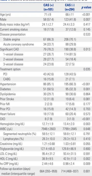
More than half of patients with CAS have multi-vessel dis- ease, and CAS was confirmed in more than half of all patients with three-vessel disease in this study (Fig. 1). MACCE oc- curred in 14% of patients in the CAS group (Table 2). Although MACE was also more prevalent in the CAS group, it was not statistically significant. In univariate analysis, factors affecting the presence of CAS were age, male sex, the presence of hy- pertension, diabetes, and multi-vessel disease. Significance was maintained in multivariate analysis (Table 3).
As shown in Table 4, CAS and multi-vessel disease were in- dependent factors associated with MACCEs [hazard ratio (HR) 2.47, 95% confidence interval (CI) 1.16–5.24, p value 0.018;
HR 3.34, 95% CI 1.41–7.56, p value 0.007, respectively]. Kaplan- Meier curves for the 3-year follow-up period also showed sta- tistically significant differences in outcomes depending on the presence or absence of CAS (Fig. 2).
DISCUSSION
The prevalence of CAS in patients with CAD was 25.5% in our
study. This prevalence is not significantly different from the 19.6% reported by carotid ultrasound in CAD patients in Ja- pan.6 Asymptomatic CAS was found in up to 50% of patients with multi-vessel disease and was more prevalent in patients with an underlying disease, including hypertension, diabetes, and chronic kidney disease (CKD). In addition, the presence of CAS was associated with a 2.47-fold higher risk for MACCEs after adjusting for several well-known risk factors, including age, hypertension, and CKD.
CAS causes approximately 10–20% of strokes,4 and early de-
tection of CAS is important for stroke prevention. Moreover, there have been reports that the incidence of CAS increases with CAD. Jeevarethinam, et al.7 reported that carotid plaque and carotid intima-media thickness assessed by ultrasound are associated with CAD. Cappelletti, et al.8 showed a relation- ship between significant carotid disease and CAD severity.
Cohen, et al.2 indicated that carotid disease is associated with CAD, as assessed via CT angiograms. Considering these stud- ies, it is important to confirm the presence of CAS in patients with CAD. However, most previous studies have used carotid ultrasound to detect CAS.
The strength and unique features of our study are that we in- vestigated the relationship between these two diseases based on angiography of the carotid artery during the recording of coronary angiograms. This method has several advantages over carotid ultrasound. First, DSA has higher accuracy than ultrasound in detecting CAS.9,10 Second, ultrasound can only detect atherosclerosis located in a bifurcation lesion, whereas DSA can be used to obtain not only the bifurcation lesion but also information that can affect treatment options.
Considering the relationship between CAD and CAS, it is reasonable to consider the presence of CAS in patients with CAD. In addition, this strategy could be time-saving for pa- Table 1. Baseline Characteristics according to the Presence of CAS
CAS (+) (n=101)
CAS (-)
(n=294) p value
Age (yrs) 71±9 66±11 <0.001
Male 58 (57.4) 123 (41.8) 0.007
Body mass index (kg/m²) 24.1±2.7 24.4±3.3 0.417 Current smoking status 18 (17.8) 37 (12.6) 0.145
Disease presentation 0.533
Stable angina 67 (66.3) 206 (70.1)
Acute coronary syndrome 34 (33.7) 88 (29.9)
Significant CAD 79 (78.2) 190 (38.8) 0.013
1-vessel disease 27 (26.7) 114 (81.5)
2-vessel disease 28 (27.7) 54 (18.4)
3-vessel disease 24 (23.8) 22 (7.5)
Treatment option 0.035
PCI 43 (42.6) 128 (43.5)
CABG 16 (15.8) 21 (7.1)
Hypertension 86 (85.1) 195 (66.3) <0.001
Diabetes 51 (50.5) 95 (32.3) 0.001
Dyslipidemia 30 (29.7) 90 (30.6) 0.864
Prior Stroke 12 (11.8) 19 (6.4) 0.081
Prior MI 2 (2.0) 17 (5.8) 0.177
Prior PCI 16 (15.8) 42 (14.3) 0.703
Heart failure 18 (17.8) 60 (20.4) 0.573
CKD 8 (7.9) 3 (1.0) <0.001
Hemoglobin (mg/dL) 12.7±1.9 13.4±2.0 0.003
WBC (/μL) 7946±2603 7799±2845 0.648
Segmented neutrophils (%) 59.5±12.1 59.8±12.1 0.791
Lymphocytes (%) 28.3±10.0 29.3±10.3 0.410
Creatinine (mg/dL) 1.21±0.68 1.03±0.81 0.055
Triglyceride (mg/dL) 127.4±65.0 129.8±86.9 0.800
LDL-C (mg/dL) 95.4±31.2 93.4±31.5 0.591
HDL-C (mg/dL) 38.9±9.5 42.9±11.0 0.002
hs-CRP (mg/dL) 2.44±4.6 0.98±2.4 0.009
Follow-up duration (days)
median (interquartile range) 664 (355–959) 714 (468–937) 0.601 CAS, carotid artery stenosis; CABG, coronary artery bypass grafting; CAD, coronary artery disease; CKD, chronic kidney disease; hs-CRP, high sensitivity C-reactive protein; HDL-C, high-density lipoprotein cholesterol; LDL-C, low- density lipoprotein cholesterol; MI, myocardial infarction; PCI, percutaneous coronary intervention; WBC, white blood cell.
Values are presented as a mean±standard deviation or numbers (percentage) unless otherwise noticed.
Fig. 1. Distribution of significant CAS according to the severity of CAD.
CAS, carotid artery stenosis; CAD, coronary artery disease.
60
40
20
0 1-VD CAD 2-VD CAD
Presence of CAS Moderate stenosis Sever stenosis
3-VD CAD
Prevalence of CAS (%)
Table 2. Incidence of Baseline Coronary Artery and MACCE during Follow-Up
Clinical event CAS (+) (n=101)
CAS (-)
(n=294) p value Composite event
MACCE 14 (14) 23 (7.8) 0.022
MACE 16 (15.8) 28 (9.5) 0.098
Individual events
Stroke 6 (5.9) 7 (2.4) 0.105
Myocardial infarction 5 (5.0) 8 (2.7) 0.331
TVR 6 (5.9) 13 (4.4) 0.284
Cardio/cerebrovascular death 6 (5.9) 11 (3.7) 0.394 CAS, carotid artery stenosis; MACCE, major adverse cardiac and cerebrovas- cular event; MACE: major adverse cardiac event; TVR, target vessel revascu- larization.
Values are numbers (percentage).
tients and reduce inconvenience with performing additional tests to evaluate CAS. Our center detected approximately 25%
of asymptomatically significant CAS patients using this ap- proach. Of these, 50 patients (12.7%) had more than 70% se- vere lesions. Further intervention, such as carotid stenting or endarterectomy, was considered after cooperation with the neurologist. We also took into consideration the patient’s con- dition. Overall, carotid stenting was performed in 15 patients (30%). During the follow-up period, six patients with CAS de- veloped stroke, three of whom with severe CAS were recom- mended for carotid stent implantation but refused.
In our study, CAS was associated with older age, a higher prevalence of multi-vessel CAD, hypertension, diabetes, and CKD. In addition, HDL-cholesterol levels were lower and hs- CRP levels were higher in CAS patients than in the non-CAS group. Possible explanations for these observations are as fol- lows: 1) Patients with diabetes, hypertension, and low HDL- cholesterol may develop systemic atherosclerosis due to lim- ited glycemic control, altered shear stress, and/or an impaired cholesterol reuptake mechanism. 2) hs-CRP may enhance the instability of plaques to induce plaque rupture. 3) Patients Table 3. Multivariate Analysis of the Prevalence of Carotid Artery Stenosis
Non-adjusted Adjusted
OR (95% CI) p value OR (95% CI) p value
Age ≥65 2.38 (1.44–3.93) 0.001 1.78 (1.04–3.06) 0.034
Male 1.88 (1.19–2.96) 0.007 1.98 (1.21–3.25) 0.006
BMI ≥25 kg/m² 1.02 (0.63–1.64) 0.952
Hypertension 2.91 (1.60–5.30) <0.001 2.51 (1.29–4.86) 0.006
Diabetes 2.14 (1.35–3.39) 0.001 1.74 (1.06–2.88) 0.029
Multi-vessel disease 3.06 (1.91–4.89) <0.001 3.03 (1.85–4.96) <0.001
Heart failure 0.84 (0.47–1.52) 0.574
Dyslipidemia 0.95 (0.59–1.57) 0.864
Current smoker 1.70 (0.81–2.79) 0.192
OR, odds ratio; CI, confidence interval; BMI, body mass index.
Table 4. Multivariate Analysis of the Prevalence of Major Adverse Cardiac and Cerebrovascular Event in Patients with Significant Coronary Artery Disease
Non-adjusted Adjusted
HR (95% CI) p value HR (95% CI) p value
Age ≥65 2.42 (1.03–5.64) 0.041 1.68 (0.69–4.09) 0.250
Male 1.25 (0.60–2.58) 0.546
Acute coronary syndrome 0.93 (0.44–1.96) 0.841
Multi-vessel disease 4.07 (1.56–10.59) 0.004 3.34 (1.41–7.56) 0.007
BMI ≥25 kg/m2 0.54 (0.23–1.28) 0.166
Hypertension 2.43 (0.85–6.98) 0.097 1.67 (0.56–4.99) 0.356
Diabetes 1.56 (0.76–3.21) 0.220
Chronic kidney disease 2.98 (0.90–9.84) 0.073 1.69 (0.49–5.76) 0.401
Carotid artery stenosis 3.12 (1.52–6.39) 0.002 2.47 (1.16–5.24) 0.018
Heart failure 0.95 (0.38–2.33) 0.916
Dyslipidemia 0.90 (0.41–1.98) 0.805
Current smoker 1.44 (0.60–3.45) 0.413
HR, hazard ratio; CI, confidence interval; BMI, body mass index.
Fig. 2. Clinical outcomes according to the presence of CAS. CAS, carotid artery stenosis; MACCE, major adverse cardiac and cerebrovascular event.
30
20
10
0
0 1 2 3 Follow-up duration (yr)
p=0.009 by log-rank
CAS (+)
CAS (-)
Numer at risk
CAS+ 101 95 77 63
CAS- 294 202 183 145
MACCE rate (%)
with multi-vessel artery lesions may be more vulnerable to plaque rupture, which increases the risk for overt atheroscle- rotic disease. In multivariate analyses, the presence of CAS was associated with older age, sex, hypertension, and diabe- tes. In addition, multi-vessel disease showed a three-fold inci- dence of CAS, and the presence of CAS was identified as an independent risk factor for MACCEs. Thus, patients with mul- tiple underlying diseases or multi-vessel disease should be carefully evaluated for CAS.
There were some limitations to this study. First, because our study was a nonrandomized, observational study, selection bias and unmeasured confounding factors could not be elimi- nated. Second, as a single-center study, patients in our study may not reflect the general population of a large region, and the small sample size may have weakened the statistical pow- er. Patients enrolled by two interventionists were consecutive during the index period; however, there was a possibility of selection bias because we did not target all of the patients in our center. Third, the fact initial screening carotid artery DSA was only one projection, which could lead to underestimation of the arteries that have asymmetrical eccentric stenosis.
Fourth, concurrent coronary and carotid angiography leads increased consumption of contrast dye, fluoroscopy time, and increases the probability of contrast induced nephropathy.
In conclusion, asymptomatic CAS was found in up to 25%
of patients with CAD. The presence of CAS in patients with CAD was associated with a higher rate of MACCEs. Therefore, the detection of CAS by carotid angiography during CAG may be important for risk stratification of these patients, particu- larly those with multi-vessel disease.
AUTHOR CONTRIBUTIONS
J.K.S and G.S.K conceptualized the hypothesis, designed the study, and drafted the manuscript. H.Y.L, Y.S.B and I.H.J collected and inter- preted the data. B.O.K and K.J.R conceptualized the hypothesis and revised the manuscript.
ORCID iDs
Jongkwon Seo https://orcid.org/0000-0001-9179-1992 Gwang Sil Kim https://orcid.org/0000-0001-5206-8187 Hye Young Lee https://orcid.org/0000-0003-3187-3667
Young Sup Byun https://orcid.org/0000-0002-6360-6400 In Hyun Jung https://orcid.org/0000-0002-1793-215X Kun Joo Rhee https://orcid.org/0000-0003-1064-7337 Byung Ok Kim https://orcid.org/0000-0002-7920-2750
REFERENCES
1. Yamagishi M, Terashima M, Awano K, Kijima M, Nakatani S, Dai- koku S, et al. Morphology of vulnerable coronary plaque: insights from follow-up of patients examined by intravascular ultrasound before an acute coronary syndrome. J Am Coll Cardiol 2000;35:
106-11.
2. Cohen GI, Aboufakher R, Bess R, Frank J, Othman M, Doan D, et al. Relationship between carotid disease on ultrasound and coro- nary disease on CT angiography. JACC Cardiovasc Imaging 2013;
6:1160-7.
3. Akosah KO, McHugh VL, Barnhart SI, Schaper AM, Mathiason MA, Perlock PA, et al. Carotid ultrasound for risk clarification in young to middle-aged adults undergoing elective coronary angi- ography. Am J Hypertens 2006;19:1256-61.
4. Grotta JC. Clinical practice. Carotid stenosis. N Engl J Med 2013;
369:1143-50.
5. North American Symptomatic Carotid Endarterectomy Trial Col- laborators, Barnett HJM, Taylor DW, Haynes RB, Sackett DL, Peerless SJ, Ferguson GG, et al. Beneficial effect of carotid endar- terectomy in symptomatic patients with high-grade carotid ste- nosis. N Engl J Med 1991;325:445-53.
6. Tanimoto S, Ikari Y, Tanabe K, Yachi S, Nakajima H, Nakayama T, et al. Prevalence of carotid artery stenosis in patients with coronary artery disease in Japanese population. Stroke 2005;36:2094-8.
7. Jeevarethinam A, Venuraju S, Dumo A, Ruano S, Rosenthal M, Nair D, et al. Usefulness of carotid plaques as predictors of obstructive coronary artery disease and cardiovascular events in asymptom- atic individuals with diabetes mellitus. Am J Cardiol 2018;121:
910-6.
8. Cappelletti A, Astore D, Godino C, Bellini B, Magni V, Mazzavil- lani M, et al. Relationship between Syntax Score and prognostic localization of coronary artery lesions with conventional risk fac- tors, plasma profile markers, and carotid atherosclerosis (CAPP Study 2). Int J Cardiol 2018;257:306-11.
9. Anzidei M, Napoli A, Zaccagna F, Di Paolo P, Saba L, Cavallo Marincola B, et al. Diagnostic accuracy of colour Doppler ultraso- nography, CT angiography and blood-pool-enhanced MR angi- ography in assessing carotid stenosis: a comparative study with DSA in 170 patients. Radiol Med 2012;117:54-71.
10. Nederkoorn PJ, van der Graaf Y, Hunink MG. Duplex ultrasound and magnetic resonance angiography compared with digital sub- traction angiography in carotid artery stenosis: a systematic re- view. Stroke 2003;34:1324-32.