고혈압심장질환의 최신지견
정 중 화
조선대학교 의학전문대학원 내과학교실 순환기내과
Changing Concept of Hypertensive Heart Disease
Joong-Wha Chung, MD
Division of Cardiology, Department of Internal Medicine, Chosun University School of Medicine, Gwangju, Korea 1)
❙ABSTRACT❙
Arterial hypertension leads to both structural and functional changes of the heart. Hypertensive heart disease (HHD) is characterized by complex changes in myocardial structure (e.g., enhanced cardiomyocyte growth, excessive cardiomyocyte apoptosis, accumulation of interstitial and perivascular collagen fibers, disruption of endomysial and perimysial collagen network) that cause the remodeling of the myocardium. In the 1970s, hypertrophic growth of cardiomyocytes is compensatory to reduce wall stress on the ventricular wall imposed by pressure overload. Recent data from animal studies suggest that inhibition of ventricular hypertrophy was not associated with ventricular dilatation or reduced wall motion despite elevated wall stress. The genetic complexity (gene-gene and/or gene-environment interactions) may modulate left ventricular mass and transcriptional regulators are participated in pathologic myocardial growth. Many hormones and cytokines lead to a profibrotic and inflammatory environment. Excess of ventricular collagen in hypertensive patients is the result of both increased collagen synthesis by fibroblasts and stimulated myofibroblasts, and unchanged or decreased collagen degradation by matrix metalloproteinase. Several biochemical markers of myocardial remodeling will prove to be useful. The development of noninvasive methods like echoreflectivity, cardiac magnetic resonance imaging, speckle tracking echocardiography, and cardiac molecular imaging would enable broader application. Meta-analysis showed that there was a significant difference among medication classes in decreasing left ventricular mass.
(J Korean Soc Hypertens 2012;18(4):137-145) Key Words: Heart diseases; Hypertension; Ventricular remodeling; Ventricular hypertrophy
서 론
수축기혈압의 상승은 그 자체로도 심장 합병증의 위험
논문접수일: 2012.9.6, 수정완료일: 2012.9.18, 게재승인일: 2012.9.21 교신저자: 정중화
주소: 광주시 동구 필문대로 365 조선대학교병원 심혈관센터 Tel: 062) 220-3013, Fax: 062) 222-3858
E-mail: [email protected]
을 증가시키지만 다른 심혈관계 위험인자, 유전적 요인이 나 만성신장질환 등과 만나게 되면 좌심실비대(left ven- tricular hypertrophy)의 발생이 커지게 된다.1) 좌심실비대 는 고혈압 환자의 15–20%에서 발생하며 향후 발생되는 심혈관계 사건의 강력한 예측인자이기도 하다.2) 전통적 으로 좌심실비대는 심실벽의 긴장을 정상화시켜 정상적 인 심박출량을 유지시키기 위한 보상기전으로 이해되었
Proremodeling molecules Antiremodeling molecules
Vasoactive substances (norepinephrine, angiotensin II) Vasoactive substances (nitric oxide, prostacyclin, angiotensin-[1–7]) Hormones (thyroid hormone, aldosterone) Hormones (glucocorticoids)
Growth factors (transforming growth factor-β) Growth factors (insulin-like growth factor 1)
Cytokines (cardiotrophin 1) Cytokines (tumor necrosis factor-α)
Other (reactive oxygen species, endogenous peroxisome
proliferator-activated receptor-γligands) Other (endogenous peroxisome proliferator-activated receptor-α ligands)
Table 1. Molecules involved in myocardial remodeling in hypertensive heart disease (From Diez J, et al. Hypertension. 2010;
55:1-8)10)
으나3) 최근에는 좌심실비대가 고혈압에 대한 부적응의 단계이며, 심부전과 부정맥 같은 심혈관계 사건을 증가시 키는 위험한 질환으로 이해되고 있다.1,4) 최근 여러 동물 연구들에 의하면 후부하(afterload)가 증가된 상황에서 좌 심실비대를 예방하여도 심실확장이나 심부전이 발생되지 않는다고 한다.5) 따라서 고혈압의 조기 단계에서 좌심실 비대를 예방하는 것이 중요할 것으로 생각된다. 고혈압심 장질환의 주된 치료방침은 약물을 사용하여 간질섬유증 (interstitial fibrosis)을 개선시켜 두꺼워진 심근을 퇴행시 키는 것이었으나 최근에는 여러 가지 조기 진단법의 개발 로 인해 고혈압심장질환의 발생 가능성이 높을 것으로 생 각되는 환자를 조기에 진단하여 심근이 두꺼워지기 전에 치료를 시작하는 것으로 개념 전환이 되고 있다. 고혈압 은 이환율과 사망률을 증가시키는 중요한 질환이며 오랜 기간 동안에 걸친 지속적인 혈압상승은 심근이나 관상동 맥과 같은 심장의 구조에 변화를 가져오게 되는데,6) 이러 한 변화들을 종합하여 고혈압심장질환이라고 한다.
고혈압심장질환의 병태생리
고혈압은 심근과 동맥의 리모델링을 유발하여 심장과 혈관의 정상기능에 악영향을 미치게 되어 심혈관계질환 의 발생을 증가시키게 된다. 후부하의 증가는 심장을 신 장시켜서 세포 내의 신호전달(signaling cascades)을 자극 하여 근섬유분절(sarcomere)과 관련된 유전자발현과 단 백질합성을 활성시켜 심근세포의 크기를 증가시킨다.7) 고혈압심장질환에서 안지오텐신 I형 수용체 유전자의 A1166C 다형태(polymorphism)는 I형 콜라겐의 합성을
가속시켜 심근섬유화를 조장하며8) 근섬유모세포는 심장 내의 섬유화를 가속시키기 위한 유전자 전개프로그램 (gene unfolding programs)을 발현하여 심근세포의 병적 인 성장에 관여하는 유전프로그램(fetal program)이 가동 되면서 Nkx2.5/Csx와 같은 전사인자(transcriptional fac- tor)들이 활성화되어 결국 심근 리모델링을 유발한다. 이 러한 다양한 기전에 의해 세포외기질(extracellular ma- trix)의 콜라겐 회전율(turnover)에 변화를 주어 결국 두껍 고 단단한 콜라겐 I형과 III형의 침착이 증가하게 된다.9) 고혈압심장질환의 초기 과정부터 섬유모세포(fibroblast) 에서 근섬유모세포(myofibroblast)로의 변이가 진행되는 데 여기에는 레닌-안지오텐신-알도스테론계통, 엔도텔린 -1 (endothelin-1), 종양괴사인자-β1 (transforming growth factor-β1)이 관여한다(Table 1).10) 좌심실비대가 동반된 고혈압 환자에서 정상 혈압 군과 좌심실비대가 동반되지 않은 군에 비해 혈장 내의 레닌활성도와 알도스테론 농도 가 증가되어 있다.11) 아교섬유(collagen fiber)는 섬유모세 포에서 합성되고, 합성된 아교섬유의 분해는 섬유모세포 나 내피세포에 의해 분비되는 금속단백분해효소(matrix metalloproteinase, MMP)-1/2/9에 의해 이뤄진다. MMP 의 활성도는 MMP의 억제인자인 tissue inhibitors of met- alloproteinases (TIMP)-1-4에 의해 조절된다. 정상적으로 는 아교섬유의 생성과 분해가 평형을 유지하는데, 고혈압 이 있는 경우에는 TIMP의 상대적인 과잉으로 심근과 혈 관벽의 리모델링을 초래하게 된다. 증가된 콜라겐은 탄력 소(elastin)와 교차결합을 하여 튼튼한 아교섬유를 형성하 게 되는데 이 과정은 산화를 통한 효소적 교차결합과, 당
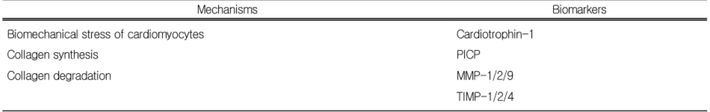
Mechanisms Biomarkers
Biomechanical stress of cardiomyocytes Cardiotrophin-1
Collagen synthesis PICP
Collagen degradation MMP-1/2/9
TIMP-1/2/4 PICP, procollagen type I C-terminal propeptide; MMP, matrix
metalloproteinase; TIMP, tissue inhibitors of metalloproteinases.
Table 2. Circulating biomarkers of hypertensive heart disease 화반응(glycation)을 통해 advanced glycation and product (AGE) 등을 형성하는 비효소적 교차결합 2가지로 나눠
진다.12,13) 고혈압심장질환의 구조적 변화는 심근세포의 비
대, 심근세포의 세포자멸사(apoptosis), 간질섬유증 (interstitial fibrosis), 그리고 혈관벽의 리모델링으로 정리 해 볼 수 있겠다.14,15) 결국 이러한 구조적 변화는 심장에 기능변화를 초래하여 수축과 확장기능의 장애를 초래하 며 좌심실 기능부전과 부정맥을 발생시키고 혈관벽에서 는 동맥경직의 발생으로 인해 말초혈관 저항이 증가하고 장기의 허혈을 유발한다. 여러 연구결과들에 의하면 고혈 압심장질환에서 콜라겐 부피 분율(collagen volume frac- tion)과 확장기능장애(diastolic dysfunction) 사이에 밀접 한 상관관계가 있다고 하며 심근 섬유화가 더욱 진행되면 심실 박출률(ejection fraction)이 감소하게 된다. 심부전이 동반된 고혈압 환자에서 확장기 심부전에 비해 수축기 심 부전에서 콜라겐 부피 분율이 더욱 증가한다. 최근 자가 포식현상(autophagy)이 심부전 발생의 기전이 될 수 있다 는 연구들이 보고되고 있다.16) 좌심실비대에서 안정 시 관상동맥 혈류는 대개 정상이지만 좌심실비대의 정도가 커지면 감소하게 된다. 혈관 주위 콜라겐 부피 분율과 관 상동맥예비력(coronary flow reserve) 사이에 역상관관계 가 존재한다.17) 심근 내 섬유화는 비정상적인 구조 즉 해 부학적인 기질(anatomical substrate)을 제공하여 회귀 (reentry)현상에 의한 부정맥의 발생을 증가시키는데 심 방세동이 대표적이며 좌심실질량과 심방세동의 발생에는 유의한 상관관계가 있다.18) 또한 좌심실질량이 증가하면 돌연사의 발생도 증가한다.19)
고혈압심장질환의 진단
근래에 고혈압심장질환을 초기에 진단하기 위한 생물 표지자(biomarker)와 초기 단계의 치료제 개발에 대한 연 구들이 활발하게 이루어지고 있다.20) 안지오텐신전환효소 (angiotensin converting enzyme) 유전자에 위치하고 있는 삽 입/결실(insertion/deletion) 다형성(polymorphism) 중 D 대립 유전자가 좌심실비대의 표지자로 사용 가능하다는 연구 가 있으며21) 말초혈액세포를 이용해 좌심실비대와 관련된 심근의 유전자 발현(myocardial gene expression)을 알아볼 수 있는 분자 진단(molecular diagnosis)이 있는데 GATA4 와 myocardin의 발현이 좌심실질량과 상관관계가 있다는 연구가 여기에 해당된다.22) 다음으로 고혈압심장질환의 조기 진단에 사용될 수 있는 생물표지자는 크게 세 가지 로 나눠볼 수 있다(Table 2).23,24) 혈청 cardiotrophin-1은 좌심실비대 발생 가능성이 높은 고혈압 환자를 조기 예측 하는데 유용하며25) 혈청 procollagen type I C-terminal propeptide (PICP)는 좌심실비대 환자에서 심부전으로의 진행을 예측하는데 도움을 줄 수 있다.10) 특히 좌심실비대 가 심부전으로 진행하는 것을 예측하는 데는 뇌나트륨이 뇨펩티드(brain natriuretic peptide)보다 MMP-2가 진 단적 가치가 크다.26) 최근의 메타분석자료를 살펴보면 1) 심부전을 동반하지 않은 고혈압 환자에서는 혈장 내 MMP-9과 TIMP-1이 증가되어 있으며, 2) 좌심실비대가 있는 환자에서는 혈장 내 TIMP-1이 증가되어 있고, 3) 확 장기 심부전 환자에서는 혈장 내 MMP-2가 증가되어 있 다고 한다.27) 이외에도 annexin A5와 같은 섬유소용해와 세포자멸사와 관련된 단백질을 이용한 연구들도 진행 중
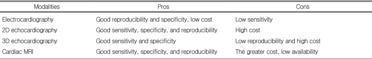
Modalities Pros Cons Electrocardiography Good reproducibility and specificity, low cost Low sensitivity
2D echocardiography Good sensitivity, specificity, and reproducibility High cost
3D echocardiography Good sensitivity and specificity Low reproducibility and high cost Cardiac MRI Good sensitivity, specificity, and reproducibility The greater cost, low availability 2D, 2-dimensional; 3D, 3-dimensional; MRI, magnetic resonance imaging.
Table 3. Imaging modalities to assess hypertensive heart disease
인데 고혈압에 의한 심부전 환자에서 수축기능의 악화와 상관관계가 있다고 한다.28) 고혈압심장질환을 진단하는 데는 심장 생검을 통한 조직섬유화 계측이 가장 정확한 방법이지만 침습적이기 때문에 현실적으로 심장섬유화나 좌심실비대를 평가하는 데는 심전도, 심장초음파, 심장 magnetic resonance imaging (MRI) 등이 이용되는데 자 세한 내용은 Table 3에 정리하였다.29,30) 심전도상의 좌심 실비대 소견과 심초음파에서의 좌심실질량이 심혈관계 이환율과 사망률의 독립적인 위험인자임은 여러 연구들 을 통해 증명되었다. 특히 심전도는 좌심실비대를 진단하 는데 있어 낮은 민감도의 문제점을 가지고 있지만31) 고혈 압 환자에서 P파 분산으로 심방세동의 발생을 예측할 수 있다는 연구를 비롯한 다양한 연구들이 진행 중이다.32) 심장초음파를 이용한 좌심실의 구조변화가 심해질수록 박출률이 감소하지만 정상 범위 이하로 감소하는 경우는 드물기 때문에 최근에는 좌심실분절의 국소 수축기능을 평가하는 Ecc (peak circumferential strain)를 이용하여 수 축기능저하의 진행을 판단하는 연구들이 있다.33) 심비대 를 동반한 모든 고혈압 환자는 심근섬유화(myocardial fib- rosis)가 증가되어 있으며 이로 인해 심근의 탄성이 감소 되어 결국 좌심실이완기능장애가 발생된다. 도플러를 이 용한 심초음파로 좌심실이완기능장애를 확인할 수 있으 며 심장섬유화 정도와 역상관관계를 가지고 있다고 알려 져 있다.34,35) 치료받고 있지 않은 고혈압 환자의 20% 정 도에서는 좌심실비대가 없어도 확장기능장애가 동반되어 있다고 하며 최근의 연구결과에 의하면 좌심실비대에서 는 심초음파로 측정한 이면성 반점추적(speckle tracking) 과 등용적 이완기의 꼬임풀기(untwisting)가 지연된다고 한 다.36) 수축기 후 종축 심근단축(longitudinal myocardial
shortening)을 대변하는 수축기 후 변형률(postsystolic strain index, PSI)의 증가가 좌심실의 이완기능장애와 독 립적인 상관관계를 가지며 증가된 PSI는 혈장 PICP와 상 관관계가 있다는 연구가 있다.37)
고혈압심장질환의 치료
1970년대 항고혈압제의 사용이 증가하면서 치료받고 있는 고혈압 환자에서의 좌심실비대 발생 빈도가 감소하 는 현상이 발견되었는데38) 이러한 관찰연구를 바탕으로 최근까지 고혈압심장질환 치료의 주된 방향은 좌심실비 대를 찾아내어 치료하는 것이다. 일정 수준 이상의 좌심 실비대는 관상동맥의 죽상경화판 파열(plaque disruption) 을 증가시키며39) 좌심실비대가 있는 환자에서 관상동맥 질환이 동반되어 있으면 심부전으로의 진행이 원활하 다.40) 반대로 좌심실비대를 퇴행시키면 예후가 개선된다 는 치료에 관한 연구결과들이 발표되는데41,42) 한 연구결 과를 보면 약물을 사용하여 혈압을 조절하더라도 혈압의 조절 정도와는 무관하게 좌심실비대의 퇴행이 있는 군에 서 없는 군에 비해 사망률이 개선되었다.43,44) 이렇듯 효 과적인 혈압조절은 좌심실질량을 개선시키고 생존율을 향상시키지만 일련의 자료들을 살펴보면 고혈압의 초기 단계부터 좌심실비대의 위험도를 예측하여 치료에 반영 하는 것이 중요해 보인다. 과거의 메타분석을 보더라도 병원에서 측정한 혈압(office blood pressure, OBP)과 좌 심실질량과의 상관관계가 크지 않기 때문에(상관계수 <
0.50) 활동혈압측정(ambulatory blood pressure monitor- ing, ABPM)을 통해 야간혈압이 적절하게 감소하지 않거 나(nondipping) 아침 혈압이 올라가는 경우(early morning
blood pressure surge)와 같이 좌심실비대의 발생이 증가 할 수 있는 경우를 구별해야 하겠다.45,46) 항고혈압제에 의한 좌심실비대의 퇴행(regression)을 예측하는데 있어 서도 OBP에 비해 ABPM이 우수하다.47) Lisinopril을 이 용한 연구를 보면 좌심실비대가 개선되지 않더라도 좌심 실 확장기능과 콜라겐 부피 분율의 개선이 수반되었다.
2001년에 발표된 Heart outcomes prevention evaluation study (HOPE) 연구에 의하면 라미프릴(ramipril)이 다른 항고혈압제에 비해 좌심실비대의 퇴행효과가 크다는 사 실을 알 수 있다.48) 이후 발표된 Losartan intervention for endpoint reduction (LIFE) 연구에서는 베타차단제인 아 테놀롤(atenolol)에 비해 로사탄(losartan)이 좌심실비대 개선효과가 크다고 보고하고 있는데49) 특히 심전도에서 R파와 S파의 합으로 계측되는 Sokolow-Lyon지수가 10.5 mm씩 개선될 때마다 심혈관계 사건이 17%씩 감소하였 다.50) 이처럼 연속된 심전도의 측정을 통해 고혈압심장질 환의 예후를 알아낼 수 있다.41) 이외에도 좌심실질량지수 (left ventricular mass index)가 10%씩 개선될 때마다 좌 심방의 크기가 감소하고 early (E) and late atrial (A) mi- tral filling velocity (E/A) 비와 감속시간(deceleration time)이 개선된다는 보고가 있다.51-53) 심장초음파를 이용 한 한 연구를 보면 칸데사탄(candesartan)과 에날라프릴 (enalapril)의 좌심실질량지수 개선효과는 비슷하였으며54) 심장 MRI를 이용한 연구를 보면 텔미사탄(telmisartan)이 카르베딜롤(carvedilol)보다 좌심실질량지수 개선효과가 좋 다.55) 광물부신겉질호르몬(mineralocorticoid) 길항제인 에 플레레논(eplerenone)과 에날라프릴의 좌심실질량 개선효 과는 유사하지만 단독요법에 비해 에날라프릴과 에플레 레논의 병합요법에서 좌심실비대 개선효과가 보다 좋 다.56) 좌심실비대를 개선시키면 심방세동과 심실조기박 동의 발생이 감소하며 심장 돌연사(sudden cardiac death) 와 심부전의 발생도 감소한다.57-60) 그간 항고혈압제 간의 항섬유화(antifibrotic) 효과가 다르기 때문에 좌심실비대 개선에 대한 효과가 차이가 있는데 여러 연구들을 종합해 보면 다른 약제에 비해 안지오텐신전환효소억제제, 안지 오텐신수용체차단제 그리고 칼슘통로차단제의 항섬유화 효과가 뛰어남을 보고하였다.10,61,62) 최근에는 OPB-9195
와 같은 AGE와 관련된 비효소적 교차결합과정에 작용하 는 억제제 등이 연구 중에 있어 향후 고혈압심장질환의 초기 단계부터 적용할 수 있는 약제들이 개발될 것으로 기대된다.63,64)
결 론
고혈압심장질환은 고혈압이 있으면서 유전자 다형태와 같은 선행요인이 있는 상태에서 세포 내외의 기질(matrix) 에서 전사 프로파일링(transcriptional profiling)과 단백질 의 양적 변화 및 변형(proteomic profiling)이 발생하여 심 근의 구조적 변화를 가져오게 되고 결국 이로 인한 합병 증을 유발하게 된다. Pressioni arteriose monitorate e loro associazioni (PAMELA) 연구65)를 보면 약물을 이용하여 고혈압을 치료하게 되면 치료받지 않는 고혈압 환자에 비 해 좌심실질량이 낮지만 정상인에 비해서는 여전히 높게 유지되기 때문에 항고혈압제 외의 다른 치료법의 개발이 절실해 보인다. 좌심실비대는 심근세포의 크기가 증가하 면서 섬유화를 동반하는 세포 외 기질의 변화가 수반되는 특징을 가진다. 이러한 고혈압심장질환의 변화가 근본 원 인은 증가된 혈압뿐만이 아니라 활성화된 신경호르몬 (neurohormones), 성장인자 및 사이토카인의 영향에 의한 것이지만 최근의 자료를 보면 엄격한 혈압의 조절이 추가 적인 좌심실비대 발생을 억제하는 것으로 보아 증가된 혈 압이 중추적인 병인임을 알 수 있다. 하지만 심장 리모델 링에는 혈압 이외에도 많은 변수들이 관여하는데 인종 간 의 차이가 여기에 해당된다.66) 실제로 심장초음파나 심장 MRI에서 측정된 좌심실질량과 혈압과의 상관계수는 0.5 –0.68 정도이기 때문에 우리가 알지 못하는 다른 인구학 (demographic) 또는 혈류역학(hemodynamic) 변수들이 존재할 것으로 생각된다.67,68) 근래에 좌심실질량의 유전 가능성(heritability)에 대한 여러 가지 연구결과들이 나오 고 있는데 혈압의 변이성에도 유전적 인자가 미치는 영향 은 30–60% 정도인 것으로 알려져 있으며 일반인의 질환 유병률에 대한 환자 형제의 질환 유병률의 비(sibling re- current risk ratio)는 2–3 정도인 것으로 추정된다. 좌심실 질량에 영향을 주는 후보유전자(candidate gene)에 대한
연구와 전체 유전체검색(genome-wide scan)이 활발히 진 행되고 있으나 아직은 임상적으로 이용하기에는 한계가
있다.69-72) 고혈압 환자에서 동심(concentric) 좌심실비대
의 발생은 심부전으로의 이행에 있어 중요한 선행 단계이 다. 물론 편심(eccentric) 좌심실비대도 고혈압 환자에서 발생할 수 있지만 편심 좌심실비대 환자에 비해 동심 좌 심실비대 환자에서 활동혈압이 더 높다는 연구73)를 보더 라도 동심 좌심실비대가 혈압상승과 더 밀접함을 알 수 있다. 동심 좌심실비대에 비해 편심 좌심실비대가 동반된 고혈압 환자에서 우연히 발생하는 심부전의 가능성이 높 기 때문에 추적(follow-up) 시 주의가 요하겠다. 좌심실비 대의 발생을 미연에 억제하는 것도 중요한 치료의 개념으 로 등장하고 있다.74) 동심 좌심실비대 환자는 심근경색이 동반되지 않으면 수년 내에 확장 심부전(dilated heart failure)으로 이행할 가능성이 낮다.75) 고혈압심장질환은 다양한 생물표지자와 영상기법에 의해 조기 진단될 수 있 으며 특히 심초음파와 심장 MRI를 통해 보다 많은 방법 이 찾아질 것으로 생각된다. 그렇지만 좌심실비대의 진단 에 사용되는 영상방법에 따라 정상 수치와 목표 수치가 다르기 때문에 치료약제의 효과를 평가하는 데 있어 차이 가 발생할 수 있어 결과해석에 주의를 요하겠다. 향후 고 혈압심장질환의 관리방향은 단순히 혈압조절에 국한되지 않고 심장의 전기적인 상태와 심장혈관의 미세순환 (microcirculation)의 정도를 파악하여 치료하는 방향으로 전환될 것이며 증세가 없는 초기 단계의 고혈압심장질환 에서 발생할 수 있는 심부전으로의 이행을 막을 수 있는 적절한 치료방법도 필요할 것으로 사료된다. 특히 고혈압 심장질환에서 발생하는 심혈관계 합병증을 줄이기 위해 서는 동반된 위험인자를 적극적으로 찾아내어 포괄적으 로 관리하는 것이 중요하겠다.76) 항섬유화 효과가 알려진 다양한 항고혈압제를 이용해 조기 치료를 시작하면 여러 가지 심혈관계 사건들을 더욱 효과적으로 예방할 수 있을 것으로 생각되므로, 향후 이와 관련된 대규모 전향연구들 과 메타분석결과들을 기대해보는 바이다.
이해상충: 해당사항 없음.
References
1. Diez J, Gonzalez A, Lopez B, Querejeta R. Mechanisms of disease: pathologic structural remodeling is more than adap- tive hypertrophy in hypertensive heart disease. Nat Clin Pract Cardiovasc Med. 2005;2:209-16.
2. Schirmer H, Lunde P, Rasmussen K. Prevalence of left ven- tricular hypertrophy in a general population: the Tromso study. Eur Heart J. 1999;20:429-38.
3. Grossman W, Jones D, McLaurin LP. Wall stress and patterns of hypertrophy in the human left ventricle. J Clin Invest.
1975;56:56-64.
4. Verdecchia P, Carini G, Circo A, Dovellini E, Giovannini E, Lombardo M, et al. Left ventricular mass and cardiovascular morbidity in essential hypertension: the MAVI Study. J Am Coll Cardiol. 2001;38:1829-35.
5. Esposito G, Rapacciuolo A, Naga Prasad SV, Takaoka H, Thomas SA, Koch WJ, et al. Genetic alterations that inhibit in vivo pressure-overload hypertrophy prevent cardiac dysfunc- tion despite increased wall stress. Circulation. 2002;105:
85-92.
6. Gosse P. Left ventricular hypertrophy as a predictor of car- diovascular risk. J Hypertens Suppl. 2005;23:S27-33.
7. Force T, Haq S, Kilter H, Michael A. Apoptosis signal-regu- lating kinase/nuclear factor-kappaB: a novel signaling path- way regulates cardiomyocyte hypertrophy. Circulation.
2002;105:402-4.
8. Schunkert H, Hense HW, Holmer SR, Stender M, Perz S, Keil U, et al. Association between a deletion polymorphism of the angiotensin-converting-enzyme gene and left ventricular hypertrophy. N Engl J Med. 1994;330:1634-8.
9. Berk BC, Fujiwara K, Lehoux S. ECM remodeling in hyper- tensive heart disease. J Clin Invest. 2007;117:568-75.
10. Diez J, Frohlich ED. A translational approach to hypertensive heart disease. Hypertension. 2010;55:1-8.
11. Malmqvist K, Ohman KP, Lind L, Nystrom F, Kahan T.
Relationships between left ventricular mass and the re- nin-angiotensin system, catecholamines, insulin and leptin. J Intern Med. 2002;252:430-9.
12. Shapiro BP, Owan TE, Mohammed SF, Meyer DM, Mills LD, Schalkwijk CG, et al. Advanced glycation end products accu- mulate in vascular smooth muscle and modify vascular but not ventricular properties in elderly hypertensive canines.
Circulation. 2008;118:1002-10.
13. Ciulla MM, Paliotti R, Carini M, Aldini G. Fibrosis, enzy- matic and non-enzymatic cross-links in hypertensive heart disease. Cardiovasc Hematol Disord Drug Targets. 2011;
13:61-73.
14. Diez J. Towards a new paradigm about hypertensive heart disease. Med Clin North Am. 2009;93:637-45.
15. Gonzalez A, Ravassa S, Lopez B, Loperena I, Querejeta R, Diez J. Apoptosis in hypertensive heart disease: a clinical approach. Curr Opin Cardiol. 2006;21:288-94.
16. Wang ZV, Rothermel BA, Hill JA. Autophagy in hyper- tensive heart disease. J Biol Chem. 2010;285:8509-14.
17. Weber KT, Brilla CG, Janicki JS. Myocardial fibrosis: func- tional significance and regulatory factors. Cardiovasc Res.
1993;27:341-8.
18. Verdecchia P, Reboldi G, Gattobigio R, Bentivoglio M, Borgioni C, Angeli F, et al. Atrial fibrillation in hypertension:
predictors and outcome. Hypertension. 2003;41:218-23.
19. Haider AW, Larson MG, Benjamin EJ, Levy D. Increased left ventricular mass and hypertrophy are associated with in- creased risk for sudden death. J Am Coll Cardiol. 1998;
32:1454-9.
20. Diez J, Lopez B, Beaumont J, Gonzalez A, Ravassa S.
Towards the molecular diagnosis of hypertensive heart dis- ease? J Hypertens. 2011;29:660-2.
21. Kuznetsova T, Staessen JA, Wang JG, Gasowski J, Nikitin Y, Ryabikov A, et al. Antihypertensive treatment modulates the association between the D/I ACE gene polymorphism and left ventricular hypertrophy: a meta-analysis. J Hum Hypertens.
2000;14:447-54.
22. Kontaraki JE, Marketou ME, Zacharis EA, Parthenakis FI, Vardas PE. Early cardiac gene transcript levels in peripheral blood mononuclear cells in patients with untreated essential hypertension. J Hypertens. 2011;29:791-7.
23. Beaumont J, Gonzalez A, Lopez B, Ravassa S, Diez J.
Contribution of circulating biomarkers to unravel the role of extracellular matrix in hypertensive cardiac remodelling. J Hypertens. 2012;30:34-7.
24. Gonzalez A, Lopez B, Diez J. New directions in the assess- ment and treatment of hypertensive heart disease. Curr Opin Nephrol Hypertens. 2005;14:428-34.
25. Lopez B, Gonzalez A, Lasarte JJ, Sarobe P, Borras F, Diaz A, et al. Is plasma cardiotrophin-1 a marker of hypertensive heart disease? J Hypertens. 2005;23:625-32.
26. Martos R, Baugh J, Ledwidge M, O’Loughlin C, Murphy NF, Conlon C, et al. Diagnosis of heart failure with preserved ejection fraction: improved accuracy with the use of markers of collagen turnover. Eur J Heart Fail. 2009;11:191-7.
27. Marchesi C, Dentali F, Nicolini E, Maresca AM, Tayebjee MH, Franz M, et al. Plasma levels of matrix metal- loproteinases and their inhibitors in hypertension: a system- atic review and meta-analysis. J Hypertens. 2012;30:3-16.
28. Gonzalez A, Lopez B, Ravassa S, Beaumont J, Arias T, Hermida N, et al. Biochemical markers of myocardial remod- elling in hypertensive heart disease. Cardiovasc Res.
2009;81:509-18.
29. Ruilope LM, Schmieder RE. Left ventricular hypertrophy and clinical outcomes in hypertensive patients. Am J Hypertens. 2008;21:500-8.
30. Nadour W, Biederman RW. Is left ventricular hypertrophy re- gression important? Does the tool used to detect it matter? J Clin Hypertens (Greenwich). 2009;11:441-7.
31. Pewsner D, Juni P, Egger M, Battaglia M, Sundstrom J, Bachmann LM. Accuracy of electrocardiography in diag- nosis of left ventricular hypertrophy in arterial hypertension:
systematic review. BMJ. 2007;335:711.
32. Ozer N, Aytemir K, Atalar E, Sade E, Aksoyek S, Ovunc K, et al. P wave dispersion in hypertensive patients with parox- ysmal atrial fibrillation. Pacing Clin Electrophysiol.
2000;23(11 Pt 2):1859-62.
33. Rosen BD, Edvardsen T, Lai S, Castillo E, Pan L, Jerosch-Herold M, et al. Left ventricular concentric remodel- ing is associated with decreased global and regional systolic function: the Multi-Ethnic Study of Atherosclerosis.
Circulation. 2005;112:984-91.
34. Martos R, Baugh J, Ledwidge M, O’Loughlin C, Conlon C, Patle A, et al. Diastolic heart failure: evidence of increased myocardial collagen turnover linked to diastolic dysfunction.
Circulation. 2007;115:888-95.
35. Muller-Brunotte R, Kahan T, Malmqvist K, Ring M, Edner M. Tissue velocity echocardiography shows early improve- ment in diastolic function with irbesartan and atenolol ther- apy in patients with hypertensive left ventricular hyper- trophy: results from the Swedish Irbesartan Left Ventricular Hypertrophy Investigation vs Atenolol (SILVHIA). Am J Hypertens. 2006;19:927-36.
36. Saito M, Okayama H, Yoshii T, Hiasa G, Sumimoto T, Inaba S, et al. The differences in left ventricular torsional behavior
between patients with hypertrophic cardiomyopathy and hy- pertensive heart disease. Int J Cardiol. 2011;150:301-6.
37. Tsai WC, Liu YW, Chen JY, Huang YY, Shih JY, Tsai LM, et al. Postsystolic strain index is associated with delayed dia- stolic lengthening and diastolic dysfunction of the left ven- tricle in untreated hypertension. J Hypertens. 2012;30:787-93.
38. Mosterd A, D’Agostino RB, Silbershatz H, Sytkowski PA, Kannel WB, Grobbee DE, et al. Trends in the prevalence of hypertension, antihypertensive therapy, and left ventricular hypertrophy from 1950 to 1989. N Engl J Med. 1999;
340:1221-7.
39. Heidland UE, Strauer BE. Left ventricular muscle mass and elevated heart rate are associated with coronary plaque disruption. Circulation. 2001;104:1477-82.
40. Rame JE, Ramilo M, Spencer N, Blewett C, Mehta SK, Dries DL, et al. Development of a depressed left ventricular ejection fraction in patients with left ventricular hypertrophy and a normal ejection fraction. Am J Cardiol. 2004;93:234-7.
41. Levy D, Salomon M, D’Agostino RB, Belanger AJ, Kannel WB. Prognostic implications of baseline electrocardio- graphic features and their serial changes in subjects with left ventricular hypertrophy. Circulation. 1994;90:1786-93.
42. Gradman AH, Alfayoumi F. From left ventricular hyper- trophy to congestive heart failure: management of hyper- tensive heart disease. Prog Cardiovasc Dis. 2006;48:326-41.
43. Verdecchia P, Schillaci G, Borgioni C, Ciucci A, Gattobigio R, Zampi I, et al. Prognostic significance of serial changes in left ventricular mass in essential hypertension. Circulation.
1998;97:48-54.
44. Verdecchia P, Staessen JA, Angeli F, de Simone G, Achilli A, Ganau A, et al. Usual versus tight control of systolic blood pressure in non-diabetic patients with hypertension (Cardio-Sis): an open-label randomised trial. Lancet.
2009;374:525-33.
45. Matsui Y, Eguchi K, Shibasaki S, Ishikawa J, Shimada K, Kario K. Morning hypertension assessed by home monitor- ing is a strong predictor of concentric left ventricular hyper- trophy in patients with untreated hypertension. J Clin Hypertens (Greenwich). 2010;12:776-83.
46. Ikeda T, Gomi T, Shibuya Y, Matsuo K, Kosugi T, Oku N, et al. Morning rise in blood pressure is a predictor of left ven- tricular hypertrophy in treated hypertensive patients.
Hypertens Res. 2004;27:939-46.
47. Mancia G, Zanchetti A, Agabiti-Rosei E, Benemio G, De
Cesaris R, Fogari R, et al. Ambulatory blood pressure is supe- rior to clinic blood pressure in predicting treatment-induced regression of left ventricular hypertrophy. SAMPLE Study Group. Study on Ambulatory Monitoring of Blood Pressure and Lisinopril Evaluation. Circulation. 1997;95:1464-70.
48. Mathew J, Sleight P, Lonn E, Johnstone D, Pogue J, Yi Q, et al. Reduction of cardiovascular risk by regression of electro- cardiographic markers of left ventricular hypertrophy by the angiotensin-converting enzyme inhibitor ramipril. Circulation.
2001;104:1615-21.
49. Kjeldsen SE, Dahlof B, Devereux RB, Julius S, Aurup P, Edelman J, et al. Effects of losartan on cardiovascular mor- bidity and mortality in patients with isolated systolic hyper- tension and left ventricular hypertrophy: a Losartan Intervention for Endpoint Reduction (LIFE) substudy.
JAMA. 2002;288:1491-8.
50. Okin PM, Devereux RB, Jern S, Kjeldsen SE, Julius S, Nieminen MS, et al. Regression of electrocardiographic left ventricular hypertrophy during antihypertensive treatment and the prediction of major cardiovascular events. JAMA.
2004;292:2343-9.
51. Wachtell K, Bella JN, Rokkedal J, Palmieri V, Papademetriou V, Dahlof B, et al. Change in diastolic left ventricular filling after one year of antihypertensive treatment: the Losartan Intervention For Endpoint Reduction in Hypertension (LIFE) Study. Circulation. 2002;105:1071-6.
52. Solomon SD, Janardhanan R, Verma A, Bourgoun M, Daley WL, Purkayastha D, et al. Effect of angiotensin receptor blockade and antihypertensive drugs on diastolic function in patients with hypertension and diastolic dysfunction: a randomized trial. Lancet. 2007;369:2079-87.
53. Tapp RJ, Sharp A, Stanton AV, O’Brien E, Chaturvedi N, Poulter NR, et al. Differential effects of antihypertensive treatment on left ventricular diastolic function: an ASCOT (Anglo-Scandinavian Cardiac Outcomes Trial) substudy. J Am Coll Cardiol. 2010;55:1875-81.
54. Cuspidi C, Muiesan ML, Valagussa L, Salvetti M, Di Biagio C, Agabiti-Rosei E, et al. Comparative effects of candesartan and enalapril on left ventricular hypertrophy in patients with essential hypertension: the candesartan assessment in the treatment of cardiac hypertrophy (CATCH) study. J Hypertens. 2002;20:2293-300.
55. Galzerano D, Tammaro P, del Viscovo L, Lama D, Galzerano A, Breglio R, et al. Three-dimensional echocardiographic and magnetic resonance assessment of the effect of telmisartan
compared with carvedilol on left ventricular mass a multi- center, randomized, longitudinal study. Am J Hypertens.
2005;18(12 Pt 1):1563-9.
56. Pitt B, Reichek N, Willenbrock R, Zannad F, Phillips RA, Roniker B, et al. Effects of eplerenone, enalapril, and epler- enone/enalapril in patients with essential hypertension and left ventricular hypertrophy: the 4E-left ventricular hyper- trophy study. Circulation. 2003;108:1831-8.
57. Gonzalez-Fernandez RA, Rivera M, Rodriguez PJ, Fernandez-Martinez J, Soltero LH, Diaz LM, et al.
Prevalence of ectopic ventricular activity after left ventricular mass regression. Am J Hypertens. 1993;6:308-13.
58. Okin PM, Wachtell K, Devereux RB, Harris KE, Jern S, Kjeldsen SE, et al. Regression of electrocardiographic left ventricular hypertrophy and decreased incidence of new-on- set atrial fibrillation in patients with hypertension. JAMA.
2006;296:1242-8.
59. Wachtell K, Okin PM, Olsen MH, Dahlof B, Devereux RB, Ibsen H, et al. Regression of electrocardiographic left ven- tricular hypertrophy during antihypertensive therapy and re- duction in sudden cardiac death: the LIFE Study. Circulation.
2007;116:700-5.
60. Okin PM, Devereux RB, Harris KE, Jern S, Kjeldsen SE, Julius S, et al. Regression of electrocardiographic left ven- tricular hypertrophy is associated with less hospitalization for heart failure in hypertensive patients. Ann Intern Med.
2007;147:311-9.
61. Klingbeil AU, Schneider M, Martus P, Messerli FH, Schmieder RE. A meta-analysis of the effects of treatment on left ventricular mass in essential hypertension. Am J Med.
2003;115:41-6.
62. Diamond JA, Phillips RA. Hypertensive heart disease.
Hypertens Res. 2005;28:191-202.
63. Kato T, Yamashita T, Sekiguchi A, Tsuneda T, Sagara K, Takamura M, et al. AGEs-RAGE system mediates atrial structural remodeling in the diabetic rat. J Cardiovasc Electrophysiol. 2008;19:415-20.
64. Mitchell JA, Ventura HO, Mehra MR. Early recognition and treatment of hypertensive heart disease. Curr Opin Cardiol.
2005;20:282-9.
65. Mancia G, Carugo S, Grassi G, Lanzarotti A, Schiavina R, Cesana G, et al. Prevalence of left ventricular hypertrophy in hypertensive patients without and with blood pressure con- trol: data from the PAMELA population. Pressioni Arteriose Monitorate E Loro Associazioni. Hypertension. 2002;39:
744-9.
66. Bibbins-Domingo K, Pletcher MJ, Lin F, Vittinghoff E, Gardin JM, Arynchyn A, et al. Racial differences in incident heart failure among young adults. N Engl J Med. 2009;
360:1179-90.
67. Devereux RB, Roman MJ, de Simone G, O’Grady MJ, Paranicas M, Yeh JL, et al. Relations of left ventricular mass to demographic and hemodynamic variables in American Indians: the Strong Heart Study. Circulation. 1997;96:
1416-23.
68. Heckbert SR, Post W, Pearson GD, Arnett DK, Gomes AS, Jerosch-Herold M, et al. Traditional cardiovascular risk fac- tors in relation to left ventricular mass, volume, and systolic function by cardiac magnetic resonance imaging: the Multiethnic Study of Atherosclerosis. J Am Coll Cardiol.
2006;48:2285-92.
69. Chung JW, Hong SP. Essential hypertension and genetics.
Korean Hypertension J. 2007;13:1-7.
70. Rame JE, Drazner MH, Post W, Peshock R, Lima J, Cooper RS, et al. Corin I555(P568) allele is associated with enhanced cardiac hypertrophic response to increased systemic afterload. Hypertension. 2007;49:857-64.
71. Petretto E, Sarwar R, Grieve I, Lu H, Kumaran MK, Muckett PJ, et al. Integrated genomic approaches implicate osteogly- cin (Ogn) in the regulation of left ventricular mass. Nat Genet.
2008;40:546-52.
72. Vasan RS, Glazer NL, Felix JF, Lieb W, Wild PS, Felix SB, et al. Genetic variants associated with cardiac structure and function: a meta-analysis and replication of genome-wide as- sociation data. JAMA. 2009;302:168-78.
73. Devereux RB, James GD, Pickering TG. What is normal blood pressure? Comparison of ambulatory pressure level and variability in patients with normal or abnormal left ven- tricular geometry. Am J Hypertens. 1993;6(6 Pt 2):211S-5.
74. Frey N, Katus HA, Olson EN, Hill JA. Hypertrophy of the heart: a new therapeutic target? Circulation. 2004;109:1580-9.
75. Drazner MH, Rame JE, Marino EK, Gottdiener JS, Kitzman DW, Gardin JM, et al. Increased left ventricular mass is a risk factor for the development of a depressed left ventricular ejec- tion fraction within five years: the Cardiovascular Health Study. J Am Coll Cardiol. 2004;43:2207-15.
76. Basile J. Management of global risk across the continuum of hypertensive heart disease. J Clin Hypertens (Greenwich).
2006;8(8 Suppl 2):21-30.