The potential role of COX-2 in cancer stem cell-mediated canine mammary tumor initiation: an immunohistochemical study JVS
7
0
0
전체 글
(2) 226 Jian Huang et al.. Materials and Methods Retrospective samples and histopathology CMT tissues were collected from dogs that underwent surgical resection at the Veterinary Teaching Hospital of China Agricultural University (China). Mammary tissues were histologically classified by pathologists according to previously described protocols [3,6]. The size, histological subtype, histological malignancy, Ki-67 proliferative index, and lymphatic invasion were assessed for each tumor. Cases of malignant mammary tumors (MMTs) were divided according to histological grade. Four normal mammary gland (NMG) tissue samples were included as control samples. Cell culture and TS formation A total of 24 mammary tumor tissue samples from 79 malignant cases were aseptically collected and processed. Briefly, the tissues were minced into small pieces of about 1 o mm, then transferred and digested for 4∼16 h at 37 C with 200 U/mL collagenase (Sigma-Aldrich, USA) and 100 U/mL hyaluronidase (Sigma-Aldrich) whilst being stirred. Digestion was terminated and the solution was centrifuged. The resulting cell pellets were suspended and passed through a 40-m strainer. The single cells were then collected and resuspended for TS culture in serum-free Dulbecco’s modified Eagle medium/Ham’s F-12 medium (DMEM/F12) supplemented with B27 (Gibco-BRL, USA), epidermal growth factor (EGF; 20 ng/mL; PeproTech, USA), basic fibroblast growth factor (bFGF; 20 ng/mL; PeproTech), and 0.4% bovine serum albumin (BSA; Sigma-Aldrich). After 10∼14 days of culturing, the spheres were pelleted and fixed in 10% neutral buffered formalin for 30 min before paraffin embedding. Immunohistochemistry (IHC) De-paraffinized tissue and cell block sections were subjected to heat-induced or pepsin digestion antigen retrieval and incubation in 3% hydrogen peroxide. Next, the sections were incubated overnight with primary antibodies specific for COX-2 (diluted 1 : 100; Vector Laboratories, USA), CD44 (diluted 1 : 100; Santa Cruz Biotechnology, USA), POU5F1B transcription factor (Oct-3/4, diluted 1 : 80; Santa Cruz Biotechnology), EGFR (diluted 1 : 100; Zymed Laboratories, o USA), and Ki-67 (diluted 1 : 150; BD Pharmingen, USA) at 4 C using a two-step Polymer Detection System (GBI Labs, USA). Antibody binding was visualized with 3,3’-diaminobenzidine tetrahydrochloride and the sections were counterstained with Mayer’s hematoxylin. For the negative controls, the primary antibody was replaced with phosphate-buffered saline (pH 7.2). Immunohistochemical scoring (IHS) IHS was performed by estimating the percent of positive cells and labeling intensity. The percentages of positive cells were Journal of Veterinary Science. scored as 0 (0% positive cells), 1 (< 10% positive cells), 2 (10∼ 50% positive cells), 3 (51∼80% positive cells), or 4 (> 80% positive cells). Staining intensity was scored as 0 (negative), 1 (weak), 2 (moderate), or 3 (strong). Scores for the percentage of positive cells and staining intensity were multiplied to calculate an IHS value from 0 to 12. IHS values ≤ 6 were considered to represent low expression while values 7∼12 were considered high. The Ki-67 proliferation index was determined based on the percentage of positive cells: 0 (negative), 1+ (≤ 10% of cells labeled), 2+ (11∼50% of cells labeled), or 3+ (51∼100% of cells labeled). Each section was examined using a microscope (Nikon, Japan) by two pathologists who were blind to the clinical characteristics and prognosis. Statistical analysis Statistical analyses were performed using SPSS software (ver. 16.0; SPSS, USA) and GraphPad Prism 5.0 (GraphPad Software, USA). Categorical data are expressed as frequencies and percentages. A chi-square test and Fisher’s exact test were used to evaluate categorical variables. P values < 0.05 were considered significant.. Results Clinicopathological findings Histological examination was performed for seven mammary hyperplasia (MH) samples, 79 MMTs including different histological subtypes, and four normal mammary gland (NMG) tissue samples as a control. Histological classification of the MMTs showed that these cases included three carcinoma in situ, 10 complex carcinoma, nine carcinosarcoma, 16 ductal carcinoma, one inflammatory carcinoma, 15 simple carcinoma, and 16 ductal carcinoma. Well- (WD), moderately (MD), and poorly differentiated (PD) tumors accounted for 17 (21.5%), 47 (59.5%), and 15 (20.0 %) of the MMTs, respectively (Table 1). IHC analysis of COX-2, CD44, Oct-3/4, and EGFR expression Among the MMTs, high COX-2 expression was observed in 36 cases (45.6% of the total cases), and low COX-2 expression was found in 25 cases (31.6%). Similarly, the high and low expression rates were 51.9% (41/79) and 35.4% (28/79) for CD44, 22.9% (18/79) and 15.2% (12/79) for Oct-3/4, and 49.4% (39/79) and 40.5% (32/79) for EGFR, respectively. All the unlisted cases were negative for each labeled protein. All the NMGs were negative or had low expression levels for these proteins, and expression varied in the MHs (Table 2). COX-2 expression was observed in the cytoplasm, cell membrane, and nucleus of the neoplastic epithelial cells with weak to moderate immunoreactivity in some stromal cells. Highly aggressive tumor tissues and neoplastic cells had intense staining in a heterogeneous pattern compared to noninvasive tissues (panel A in Fig. 1). COX-2 expression was significantly.
(3) Role of COX-2 in canine mammary tumor initiation 227. Table 1. Correlation of COX-2, CD44, Oct-4, and EGFR with selected clinical and pathological variables in MMTs (n = 79)* Clinical parameters Tumor size < 3 cm (n = 18) 3∼5 cm (n = 40) > 5 cm (n = 21) Histological type Carcinoma in situ (n = 3) Simple carcinoma (n = 15) Solid carcinoma (n = 25) Complex carcinoma (n = 10) Ductal carcinoma (n = 16) Inflammatory carcinoma (n = 1) Carcinosarcoma (n = 9) Histological grade WD (n = 17) MD (n = 46) PD (n = 16) Lymphatic invasion Positive (n = 25) Negative (n = 54) Ki-67 index 0 (n = 17) 1+ (< 10%; n = 23) 2+ (10∼50%; n = 22) 3+ (> 50%; n = 17). COX-2 –. +. 9 7 2. 9 33 19. 1 1 12 0 3 0 1. p value. CD44 –. +. 0.006. 1 5 4. 17 35 17. 2 14 13 10 13 1 8. 0.017. 0 1 5 1 2 0 1. 5 6 7. 12 40 9. 0.032. 2 16. 23 38. 8 6 3 1. 9 17 19 16. p value. Oct-3/4 –. +. 0.450. 13 24 12. 5 16 9. 3 14 20 9 14 1 8. 0.883. 2 7 15 8 11 0 6. 3 4 3. 14 42 13. 0.445. 0.033. 7 3. 18 51. 0.022. 2 1 4 3. 15 22 18 14. p value. EGFR. p value. –. +. 0.584. 3 4 1. 15 36 20. 0.470. 1 8 10 2 5 1 3. 0.548. 0 1 2 0 1 1 3. 3 14 22 10 15 0 7. 0.019. 12 30 7. 5 16 9. 0.223. 0 7 1. 17 39 15. 0.175. 0.005. 12 37. 13 17. 0.081. 3 5. 22 49. 0.707. 0.486. 5 16 15 13. 12 7 7 4. 0.018. 5 2 0 1. 12 21 22 16. 0.020. *p values indicating statistical differences between different groups. p values < 0.05 were considered significant and were calculated with a Chi-square test. WD: well-differentiated, MD: median-differentiated, PD: poor-differentiated.. higher in the MMTs than other types of specimens (p = 0.001). Oct-3/4 was widely detected in malignant neoplastic cells with strong and diffuse nuclear staining patterns (panel B in Fig. 1). Normal and hyperplastic tissues were mildly positive for Oct-3/4 while strong expression of this factor was observed predominantly at the invasive edge of malignant tissues (p = 0.016). CD44-specific staining was primarily found in the cell membrane of epithelial and myoepithelial cells (panel C in Fig. 1), but was absent from the apical epithelial surface, especially in normal and hyperplasic mammary tissue. CD44 was more extensively expressed in invasive components of the MMTs compared to NMGs although this difference was not statistically significant (p = 0.079). EGFR expression patterns were similar to those of CD44, and its membrane and cytoplasmic localization was consistently observed in epithelial cells of MMTs regardless of histological subtype (panel D in Fig. 1). Malignant tumors also had a higher positive ratio for EGFR than other types of specimens (p = 0.103; Table 2).. Association of clinicopathological parameters with COX-2, CD44, Oct-3/4, and EGFR Elevated COX-2 expression was significantly associated with tumor size (p = 0.006), histological subtype (p = 0.017), histological grade (p = 0.032), lymphatic invasion (p = 0.033), and Ki-67 index (p = 0.022). Oct-3/4 expression strongly correlated with the Ki-67 index (p = 0.018). CD44 expression was statistically associated with lymphatic invasion (p = 0.005). EGFR expression was significantly related to histological subtype (p = 0.019) and Ki-67 index (p = 0.020). Significant associations between protein expression and selected variables are summarized in Table 1. Correlation between COX-2 and CSC markers In moderately and poorly differentiated MMTs, COX-2 expression was strongly associated with CD44 (r = 0.402, p = 0.000; r = 0.810, p = 0.005) and EGFR (r = 0.367, p = 0.04; r = 0.700, p = 0.02) expression. In contrast, no statistical correlation was found between COX-2 and Oct-3/4 expression (Table 3).. www.vetsci.org.
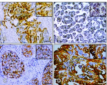
(4) 228 Jian Huang et al. Table 2. COX-2, CD44, Oct-3/4, and EGFR expression correlated with different histological types* Staining scores COX-2 (p = 0.001) Negative Low expression High expression CD44 (p = 0.079) Negative Low expression High expression Oct-3/4 (p = 0.016) Negative Low expression High expression EGFR (p = 0.103) Negative Low expression High expression. NMGs [n, (%)]. MHs [n, (%)]. MMTs [n, (%)]. Total. 4 (100.0) 0 (0.0) 0 (0.0). 2 (28.6) 5 (71.4) 0 (0.0). 18 (22.8) 25 (31.6) 36 (45.6). 24 30 36. 0 (0.0) 4 (100.0) 0 (0.0). 0 (0.0) 2 (28.6) 5 (71.4). 10 (12.7) 28 (35.4) 41 (51.9). 10 34 46. 1 (25.0) 3 (75.0) 0 (0.0). 4 (57.1) 3 (42.9) 0 (0.0). 49 (62.0) 12 (15.2) 18 (22.9). 54 18 18. 0 (0.0) 4 (100.0) 0 (0.0). 0 (0.0) 5 (71.4) 2 (28.6). 8 (10.1) 32 (40.5) 39 (49.4). 8 41 41. *p values indicating statistical differences between NMGs vs. MHs. vs. MMTs. p values < 0.05 were considered significant and were calculated using a chi-square test. COX: cyclooxygenase, EGFR: epidermal growth factor receptor, NMG: normal mammary gland, MH: mammary hyperplasia, MMT: malignant mammary tumor.. Fig. 1. Immunostaining for COX-2, Oct-3/4, CD44, and EGFR in canine mammary carcinoma. (A) Strong cytoplasmic COX-2 expression in the invasive components of ductal carcinoma with sporadic nucleic labeling. (B) Homogeneous nucleic expression of Oct-3/4 in well-differentiated simple carcinoma. (C) Predominant membrane expression of CD44 in ductal carcinoma. (D) Strong membrane and cytoplasmic expression of EGFR in tubulopapillary carcinoma with apical labeling in luminal cells. Scale bar = 50 m.. IHC phenotype of TSs Cell blocks from 24 CMTs with different histological grades. Journal of Veterinary Science. (seven WD tumors including one simple carcinoma and six simple carcinomas, nine MD tumors including three ductal carcinomas and six solid carcinomas, and eight PD tumors consisting of four solid carcinomas, three complex carcinomas, and one carcinosarcomas) were analyzed to evaluate COX-2, CD44, Oct-3/4, and EGFR immunolabeling. Representative TSs derived from tubular carcinomas (WDs), tubulopapillary carcinomas (MDs), and solid carcinomas (PDs) were assessed. The TSs had inter-sphere heterogeneity and a compact morphology. Enhanced COX-2 expression was consistently observed in the cytoplasm with sporadic nuclear staining in three TSs. COX-2 expression was stronger in TSs derived from more malignant (more poorly differentiated) tumors. CD44 staining was enriched in cellular membranes. Nuclear localization of Oct-3/4 was extensive in solid carcinomagenerated spheres compared to the other two samples. EGFR was located in the cytoplasm with negative or weak immunoreactivity. Comparative analysis showed that there was a significant difference in COX-2 (PD 10.0 ± 2.1, MD 7.6 ± 1.9, WD 10.0 ± 2.1; p = 0.006), CD44 (PD 10.9 ± 1.9, MD 8.9 ± 2.5, WD 6.5 ± 1.4; p = 0.002), and Oct-3/4 (PD 9.4 ± 2.5, MD 6.0 ± 1.4, WD 5.5 ± 1.4; p = 0.001) expression among the three histological grades (p < 0.01). This was not observed for EGFR expression (PD 1.7 ± 1.4, MD 2.2 ± 2.1, WD 2.0 ± 1.4; p = 0.856). Staining specific for CD44 and Oct-3/4 had a similar level of intensity along with elevated COX-2 expression in the TSs (Fig. 2)..
(5) Role of COX-2 in canine mammary tumor initiation 229. Table 3. Correlation of COX-2 with CD44, Oct-3/4, and EGFR expression in MMTs* Histological type. COX-2. WD (n = 17). – + – + – +. MD (n = 46) PD (n = 16). CD44 N. L. H. 2 1 3 1 3 0. 2 3 2 15 4 2. 1 8 1 24 0 7. p value 0.155 0.000 0.005. Oct-3/4 N. L. H. 3 9 2 28 5 2. 2 1 2 5 1 2. 0 2 2 7 1 5. p value 0.229 0.199 0.096. EGFR N. L. H. 0 0 2 5 1 0. 3 3 4 13 6 3. 3 8 0 22 0 6. p value 0.349 0.040 0.020. *p values denoting statistical differences between different groups. p values < 0.05 were considered significant and were calculated with a Chi-square test. N: negative expression, L: low expression, H: high expression.. Fig. 2. Immunolabeling of COX-2, CD44, Oct-3/4, and EGFR in tumorspheres (TSs). TSs generated from primary tumor tissues were compactly aggregated with a regular or irregular appearance. Heterogeneous patterns of COX-2 expression in the cytoplasm and nucleus were observed in all three TSs. Intensity of COX-2 expression in the TSs was elevated in a manner corresponding to primary tumor malignancy (histological grade) along with similar intensities of membrane CD44 and nucleic Oct-3/4 staining. Additionally, EGFR was absent or weakly expressed in all samples. Asterisks indicate a significant difference in IHC scores between PD (n = 8) vs. MD (n = 9) vs. WD (n = 7) primary tumor-derived TSs (p < 0.01). Scale bar = 50 m (TS) or 20 m (EGFR).. Discussion CSCs have the ability to self-renew and generate the diverse types of cells that form tumors. This makes CSCs a potential therapeutic target in human and veterinary oncology. Oncoprotein COX-2 is generally characterized as a pivotal regulator of growth and anti-apoptosis signals used by malignant tumor cells [7,24], and may also have an impact on CSCs [16,18,26]. In the current study, the possible role of COX-2 in tumor initiation was explored by assessing the expression patterns of COX-2 and CSC-related markers. COX-2 plays different roles in tumor initiation. According to our data, enhanced COX-2 expression and a heterogeneous pattern were commonly observed in pre-malignant and malignant. tumor tissues, and were strongly associated with malignant features such as large tumor size, complex histological type, poor differentiation, and an aggressive phenotype as previously reported in the literature [5,8,21]. Absent or low levels of COX-2 expression were found in normal and hyperplastic mammary tissues while expression was dramatically increased in tumor tissues. These results also imply that inducible COX-2 expression may promote canine mammary tumorigenesis. Moreover, differential stromal COX-2 staining in fibroblasts, endothelial, and infiltrating inflammatory cells may also suggest that stroma-derived COX-2 contributes to the acquired proliferative and invasive properties of tumor cells through tumor-stromal interaction during mammary tumorigenesis [1]. For cases of human breast cancer, it has been reported that. www.vetsci.org.
(6) 230 Jian Huang et al. +. +. proliferative tumors (COX-2 or Ki-67 ) possess more unfavorable characteristics and have a poorer prognosis than non-proliferative tumors (COX-2+ or Ki-67+) [20]. In our study, + we found that MTs with rapid growth (Ki-67 ) as well as moderate and poorly differentiated ones showed elevated COX-2 expression, which may suggest a dynamic switch of functional COX-2 during tumor proliferation and differentiation. It has been reported that down-regulated COX-2 expression suppresses the malignant phenotype, delays the onset of tumors formed by poorly differentiated cancer cells [24], and reverses the epithelial-to-mesenchymal transition [18]. In order to better understand the function of COX-2 in CSC-mediated tumor initiation, we evaluated the distribution of CSCs in mammary tissues by examining CD44, Oct-3/4, and EGFR expression patterns, and their respective associations with clinicopathological parameters. Aberrant expression of these factors was frequently observed in aggressive components of malignant tumors characterized by poor differentiation and rapid proliferation. Nevertheless, normal and hyperplastic tissues also had lower levels of expression. These findings indicate that CSCs maintain their malignant properties and can form a recurrent tumor that is more malignant. In contrast to COX-2, no significant correlation between CSC marker expression and poor prognostic variables was found. We therefore propose that these markers are not suitable independent prognostic factors. However, we further examined the co-expression of COX-2, CD44, Oct-3/4, and EGFR in MMTs and found significant associations among COX-2, CD44, and EGFR. These results show that COX-2 activation in CSCs may be CD44- or EGFR-dependent [17,29]. Although we did not observe a relationship between COX-2 and Oct-3/4 in primary CMTs, it has been previously hypothesized that Oct-3/4 expression is associated with nuclear COX-2 activation high + [25] and COX-2 or Oct-3/4 cells may acquire stem cell-like features in cases of human cancer [22]. The present study has provided new insight into the potential role of COX-2 in CSC-mediated tumor initiation by evaluating the expression patterns of COX-2 and CSC markers in TSs, which consist primarily of partially differentiated progenitor cells with few CSCs. The robust expression of COX-2 in heterogeneous patterns and at higher levels in more aggressive tumor-derived TSs suggests that COX-2 may contribute to the self-renewing ability and maintenance of CSCs. We compared COX-2, CD44, Oct-3/4, and EGFR expression in TSs according to tumor differentiation. The results confirmed that COX-2 expression accompanied enhanced CD44 and Oct-3/4 expression to a similar degree, and COX-2 up-regulation was associated with CSC enrichment in in vitro models. Therefore, it is reasonable to believe that COX-2 expression is necessary for CSC-mediated tumor initiation. Taken together, our data highlight the potential role of COX-2 in CSC-mediated tumor initiation. These findings suggest that Journal of Veterinary Science. CSCs may be more sensitive to direct COX-2 inhibition than the general cell population. Further studies should be conducted to explore COX-2-dependant elimination of CSCs as well as the development of novel therapeutic approaches for CMTs.. Acknowledgments The authors thank Dr. Dong for technical assistance. This work was supported by research grant no.31372489 from the National Natural Science Foundation of China.. Conflict of Interest There is no conflict of interest.. References 1. Arnoletti JP, Upson J, Babb JS, Bellacosa A, Watson JC. Differential stromal and epithelial localization of cyclooxygenase-2 (COX-2) during colorectal tumorigenesis. J Exp Clin Cancer Re 2005, 24, 279-287. 2. Borena BM, Bussche L, Burvenich C, Duchateau L, Van de Walle GR. Mammary stem cell research in veterinary science: an update. Stem Cells Dev 2013, 22, 1743-1751. 3. Clemente M, Pérez-Alenza MD, Illera JC, Peña L. Histological, immunohistological, and ultrastructural description of vasculogenic mimicry in canine mammary cancer. Vet Pathol 2010, 47, 265-274. 4. Cocola C, Anastasi P, Astigiano S, Piscitelli E, Pelucchi P, Vilardo L, Bertoli G, Beccaglia M, Veronesi MC, Sanzone S, Barbieri O, Reinbold RA, Luvoni GC, Zucchi I. Isolation of canine mammary cells with stem cell properties and tumour-initiating potential. Reprod Domest Anim 2009, 44 (Suppl 2), 214-217. 5. Doré M, Lanthier I, Sirois J. Cyclooxygenase-2 expression in canine mammary tumors. Vet Pathol 2003, 40, 207-212. 6. Goldschmidt M, Peña L, Rasotto R, Zappulli V. Classification and grading of canine mammary tumors. Vet Pathol 2011, 48, 117-131. 7. Greenhough A, Smartt HJ, Moore AE, Roberts HR, Williams AC, Paraskeva C, Kaidi A. The COX-2/PGE2 pathway: key roles in the hallmarks of cancer and adaptation to the tumour microenvironment. Carcinogenesis 2009, 30, 377-386. 8. Heller DA, Clifford CA, Goldschmidt MH, Holt DE, Shofer FS, Smith A, Sorenmo KU. Cyclooxygenase-2 expression is associated with histologic tumor type in canine mammary carcinoma. Vet Pathol 2005, 42, 776-780. 9. Hiraga T, Ito S, Nakamura H. Cancer stem-like cell marker CD44 promotes bone metastases by enhancing tumorigenicity, cell motility, and hyaluronan production. Cancer Res 2013, 73, 4112-4122. 10. Kumar SM, Liu S, Lu H, Zhang H, Zhang PJ, Gimotty PA, Guerra M, Guo W, Xu X. Acquired cancer stem cell phenotypes through Oct4-mediated dedifferentiation. Oncogene, 2012, 31, 4898-4911..
(7) Role of COX-2 in canine mammary tumor initiation 231. 11. Li HJ, Reinhardt F, Herschman HR, Weinberg RA. Cancer-stimulated mesenchymal stem cells create a carcinoma stem cell niche via prostaglandin E2 signaling. Cancer Discov 2012, 2, 840-855. 12. Liou JY, Ellent DP, Lee S, Goldsby J, Ko BS, Matijevic N, Huang JC, Wu KK. Cyclooxygenase-2-derived prostaglandin E2 protects mouse embryonic stem cells from apoptosis. Stem Cells 2007, 25, 1096-1103. 13. Magalhães GM, Terra EM, de Oliveira Vasconcelos R, de Barros Bandarra M, Moreira PRR, Rosolem MC, Alessi AC. Immunodetection of cells with a CD44+/CD24− phenotype in canine mammary neoplasms. BMC Vet Res 2013, 9, 205. 14. Michishita M, Akiyoshi R, Suemizu H, Nakagawa T, Sasaki N, Takemitsu H, Arai T, Takahashi K. Aldehyde dehydrogenase activity in cancer stem cells from canine mammary carcinoma cell lines. Vet J 2012, 193, 508-513. 15. Michishita M, Ezaki S, Ogihara K, Naya Y, Azakami D, Nakagawa T, Sasaki N, Arai T, Shida T, Takahashi K. Identification of tumor-initiating cells in a canine hepatocellular carcinoma cell line. Res Vet Sci 2014, 96, 315-322. 16. Moon CM, Kwon JH, Kim JS, Oh SH, Lee KJ, Park JJ, Hong SP, Cheon JH, Kim TI, Kim WH. Nonsteroidal anti-inflammatory drugs suppress cancer stem cells via inhibiting PTGS2 (cyclooxygenase 2) and NOTCH/HES1 and activating PPARG in colorectal cancer. Int J Cancer 2014, 134, 519-529. 17. Murphy JF, Lennon F, Steele C, Kelleher D, Fitzgerald D, Long AC. Engagement of CD44 modulates cyclooxygenase induction, VEGF generation, and proliferation in human vascular endothelial cells. FASEB J 2005, 19, 446-448. 18. Neil JR, Johnson KM, Nemenoff RA, Schiemann WP. Cox-2 inactivates Smad signaling and enhances EMT stimulated by TGF- through a PGE2-dependent mechanisms. Carcinogenesis 2008, 29, 2227-2235. 19. Pang LY, Argyle D. Cancer stem cells and telomerase as potential biomarkers in veterinary oncology. Vet J 2010, 185, 15-22.. 20. Park BW, Park S, Park HS, Koo JS, Yang WI, Lee JS, Hwang H, Kim SI, Lee KS. Cyclooxygenase-2 expression in proliferative Ki-67-positive breast cancers is associated with poor outcomes. Breast Cancer Res Treat 2012, 133, 741-751. 21. Queiroga FL, Pires I, Lobo L, Lopes CS. The role of Cox-2 expression in the prognosis of dogs with malignant mammary tumours. Res Vet Sci 2010, 88, 441-445. 22. Singh B, Cook KR, Vincent L, Hall CS, Martin C, Lucci A. Role of COX-2 in tumorospheres derived from a breast cancer cell line. J Surg Res 2011, 168, e39-49. 23. Sleeckx N, de Rooster H, Veldhuis Kroeze EJB, Van Ginneken C, Van Brantegem L. Canine mammary tumours, an overview. Reprod Domest Anim 2011, 46, 1112-1131. 24. Stasinopoulos I, Mori N, Bhujwalla ZM. The malignant phenotype of breast cancer cells is reduced by COX-2 silencing. Neoplasia 2008, 10, 1163-1169. 25. Thanan R, Murata M, Ma N, Hammam O, Wishahi M, El Leithy T, Hiraku Y, Oikawa S, Kawanishi S. Nuclear localization of COX-2 in relation to the expression of stemness markers in urinary bladder cancer. Mediators Inflamm 2012, 2012, 165879. 26. Trosko JE. Cancer stem cells and cancer nonstem cells: from adult stem cells or from reprogramming of differentiated somatic cells. Vet Pathol 2009, 46, 176-193. 27. Webster JD, Yuzbasiyan-Gurkan V, Trosko JE, Chang CC, Kiupel M. Expression of the embryonic transcription factor Oct4 in canine neoplasms: a potential marker for stem cell subpopulations in neoplasia. Vet Pathol 2007, 44, 893-900. 28. Wilson H, Huelsmeyer M, Chun R, Young KM, Friedrichs K, Argyle DJ. Isolation and characterisation of cancer stem cells from canine osteosarcoma. Vet J 2008, 175, 69-75. 29. Xu K, Shu HK. EGFR activation results in enhanced cyclooxygenase-2 expression through p38 mitogen-activated protein kinase−dependent activation of the Sp1/Sp3 transcription factors in human gliomas. Cancer Res 2007, 67, 6121-6129.. www.vetsci.org.
(8)
수치
관련 문서
