https://doi.org/10.5468/ogs.2020.63.3.225 pISSN 2287-8572 · eISSN 2287-8580
Introduction
Pregnancy-associated plasma protein-A (PAPP-A) is a zinc- bound metalloproteinase from the super family of metallo- proteases [1]. PAPP-A is a large glycoprotein made up of the placenta, decidua, and fetuses; however, the main source of this protein in pregnancy is placental syncytiotrophoblast [2-7]. Various functions of PAPP-A have been described. Pri- marily, PAPP-A releases insulin-like growth factor (IGF) via the breakdown of IGF-binding protein-4 [8-12]. Thus, PAPP-A facilitates the function of IGF of accelerating and augment- ing placental growth. Local IGF secretion plays an important role in increasing mitosis and cellular differentiation as well as in controlling trophoblastic invasion of the decidua [13].
The evaluating of pregnancy-associated plasma
protein-A with the likelihood of small for gestational age
Maryam Sadat Hoseini, MD
1, Samaneh Sheibani, MD
1, Mehrdad Sheikhvatan, MD, PhD
2,31Department of Obstetrics and Gynecology, Preventative Gynecology Research Center, Imam Hossein Hospital, Shahid Beheshti University of Medical Sciences, 2Pars Advanced and Minimally Invasive Medical Manners Research Center, Pars Hospital, Iran University of Medical Sciences, Tehran, Iran;
3Heidelberg University Hospital, University of Heidelberg, Heidelberg, Germany
Objective
Recently, strong evidences were obtained on the association between low pregnancy-associated plasma protein-A (PAPP-A) levels in the first trimester and poor outcomes of pregnancy.
Methods
This cross-sectional study was conducted on all pregnant women who were referred to the Obstetrics and Gynecology Clinic at Imam Hossein Hospital in Tehran, Iran in 2014. Women were asked to attend clinical examinations and screening at 11–14 weeks of gestation.
Results
Based on the definition, 14.5% of neonates found to be small for gestational age (SGA). There was a strong association between PAPP-A levels and birth weight. The mean PAPP-A level in the mothers of neonates who were SGA was significantly lower than those without this poor outcome. Based on the receiver operating characteristic curve analysis, serum PAPP-A level was a main determinant in the prediction of SGA neonates.
Conclusion
The serum PAPP-A level at 11–13 weeks of gestation can effectively predict the increased risk for fetal growth retardation. In patients in this study, the best cutoff value for PAPP-A was 0.75 MOM, which signifies that lower levels of this marker can predict fetal growth restriction with high sensitivity and specificity.
Keywords: Pregnancy; Plasma protein-A; Gestational age
Received: 2018.12.30. Revised: 2019.05.09. Accepted: 2019.05.28.
Corresponding author: Samaneh Sheibani, MD Department of Obstetrics and Gynecology, Preventative Gynecology Research Center, Imam Hossein Hospital, Shahid Beheshti University of Medical Sciences, Shahid Madani Street, Tehran 1617763141, Iran
E-mail: [email protected] https://orcid.org/0000-0002-8658-1867
Articles published in Obstet Gynecol Sci are open-access, distributed under the terms of the Creative Commons Attribution Non-Commercial License (http://creativecommons.
org/licenses/by-nc/3.0/) which permits unrestricted non-commercial use, distribution, and reproduction in any medium, provided the original work is properly cited.
Copyright © 2020 Korean Society of Obstetrics and Gynecology
The role of IGFs in controlling fetal growth is important as they regulate the absorption of amino acids and glucose in the trophoblast. Currently, it is hypothesized that low PAPP- A levels in the maternal serum represent a decrease in the regional levels of the protein, which results in a reduction in active IGF levels. Thus, inadequate free IGF levels, which affect the development of the embryo, may lead to other complications of pregnancy, such as stillbirth, risk of small for gestational age (SGA) neonates, preeclampsia, and prema- ture delivery [14,15]. In addition, other functions of PAPP-A include prevention of fetal targeting by the mother’s immune system, uterine mineralization, and angiogenesis [16]. Preg- nant women with PAPP-A levels less than 0.45 multiples of the median (MoM) have been significantly associated with increased risk of intrauterine growth restriction, SGA, pre- mature labor, preeclampsia, and stillbirth [17]. In total, low PAPP-A levels in the first trimester of pregnancy have been strongly associated with poor outcomes [18]. In other words, low PAPP-A levels are indicative of poor primary placenta, leading to serious complications such as embryonic growth restriction, fetal death, preterm labor, and preeclampsia in the third trimester. Considering the heavy economic and so- cial impact of chromosomal abnormalities and complications of pregnancy and childbirth for the community and given the fact that many of these complications are preventable, we attempted to examine the association between serum PAPP- A levels in the first trimester of pregnancy and the likelihood of SGA neonates.
Materials and methods
This cross-sectional study was conducted on all pregnant women who were referred to the Obstetrics and Gynecol- ogy Clinic at Imam Hossein Hospital in Tehran, Iran in 2014.
At first, all participants gave written/verbal informed con- sent before commencement of the study, following which a complete biography of the mothers was obtained. Data on maternal age, gestational age, parity, and history of pregnan- cies with complications were also recorded. The participants were asked to attend clinical examinations and screening at 11–14 weeks of gestation during which ultrasonography per- formed and blood samples were taken to measure common biochemical indices and markers such as β-HCG. In addition to the usual tests, we measured PAPP-A levels in the blood
samples of these individuals. The cases with fetal crown- rump length ranging from 45 to 84 mm were asked to pro- vide written informed consent if they wished to participate in the study. CRL values were converted to gestational age (GA) using Z score, and PAPP-A levels less than 5% in each GA were considered as low. Mothers were then followed up until delivery. During this period, all routine healthcare mea- sures were performed for these mothers and any abnormali- ties were treated. In this study, the results were determined based on outcome of the delivery. In cases of presence of a disorder other than SGA fetuses/neonates during pregnancy or at the time of delivery, the mother was excluded from the study. Finally, the data were statistically analyzed. The institutional ethics committee of the Preventative Gynecol- ogy Research Center (PGRC) at Shahid Beheshti University of Medical Sciences approved the study protocol. For analyses, results were presented as mean ± standard deviation (SD) for quantitative variables and as absolute frequencies and percentages for categorical variables. Normality of the data was analyzed using the Kolmogorov-Smirnoff test. Categori- cal variables were compared using chi-square test or Fisher’s exact test when more than 20% of cells with expected count of less than 5 were observed. The quantitative variables were also compared with t-test or Mann-Whitney U test. The value of PAPP-A to predict SGA neonates was assessed using the receiver operating characteristic (ROC) curve analysis. Multi- variable logistic regression model was also used to determine the value of low PAPP-A levels in predicting SGA neonates using baseline characteristics. For statistical analyses, SPSS version 16.0 for Windows (SPSS Inc., Chicago, IL, USA) was used. P-values of 0.05 or less were considered significant.
Results
In total, 715 pregnant women were assessed. The mean age of participants was 27.88±5.97 years (range, 17–38 years).
Regarding gestational age at the time of screening, 34.5%
were in the 11th week, 49.1% in the 12th week, and 16.4%
in the 13th week of pregnancy. The mean body weight of the participants was 68.90±12.55 kg (range, 47–105 kg).
Average birth weight of neonates was 3,100.91±553.82 g (range, 1,700–4,000 g). The mean PAPP-A level at the time of screening was 1.21±0.66 (range, 0.28–3.37) (Table 1).
Based on the definition of weight less than 10th percentile
based on gestational age, 14.5% of neonates were found to be SGA. There was no difference in the mean weight of mothers with and without SGA neonates (30.08±8.63 years vs. 27.51±5.43 years, P=0.439). No difference was also found in the mean weight of mothers with and without neonates who were SGA (73.81±18.21 kg vs. 68.06±11.39 kg, P=0.413). The overall prevalence of fetuses who were SGA at 11, 12, and 13 weeks of pregnancy was 26.3%, 3.7%, and 22.2%, respectively with no significant difference (P=0.078). There was a strong association between PAPP-A levels and birth weight (r=0.442, P=0.001) (Fig. 1). The mean PAPP-A level in mothers of neonates who were SGA was significantly lower than that in women with normal delivery
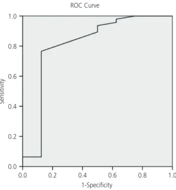
children (0.76±0.61 vs. 1.29±0.64, P=0.035). Based on the ROC curve analysis (Fig. 2), the serum PAPP-A level was a main determinant in the prediction of SGA neonates (Area under the curve=0.811, P=0.005). The best cutoff value for PAPP-A to discriminate between SGA neonates and those with normal weight was 0.75 yielding a sensitivity of 80.9%
and a specificity of 85.0%. According to the multivariable logistic regression model and with the presence of baseline variables including maternal age, weight, and gestational age, decreased PAPP-A levels could effectively predict the increased likelihood of SGA neonates (odds ratio, 12.347;
95% confidence interval, 0.012–165.917; P=0.048).
Discussion
Two methods of clinical examination and arterial Doppler are not very sensitive and precise for predicting fetal growth restriction (FGR) and may lead to over-estimation of cases that are SGA. In this regard, the use of biochemical markers (such as cystatin C, macrophage migration inhibitory factor, Table 1. Baseline characteristics of the study population
Characteristics Values
Mean age (yr) 27.88±5.97
Gestational age at screening (wk)
Eleventh week 34.5%
Twelfth week 49.1%
Thirteenth week 16.4%
Mean birth weight of neonates 3,100.91±553.82 The mean level of PAPP-A on screening
time 1.21±0.66
The prevalence of SGA 14.5%
PAPP-A, pregnancy-associated plasma protein-A; SGA, small for ges- tational age.
Fig. 1. The association between pregnancy-associated plasma protein-A (PAPP-A) level and birth weight (BW).
0.0000 1.0000 2.0000 3.0000 4.0000 PAPP-A
R Sq Linear=0.196
BW
4,000 3,500 3,000 2,500 2,000 1,500
Fig. 2. The receiver operating characteristic (ROC) curve analysis to determine the value of pregnancy-associated plasma protein-A (PAPP-A) to predict small for gestational age (SGA).
ROC Curve
0.0 0.2 0.4 0.6 0.8 1.0 1-Specificity
Diagonal segments are produced by ties.
Sensitivity
1.0
0.8
0.6
0.4
0.2
0.0
plasminogen activator inhibitor 2, and PAPP-A), maternal medical history (history of smoking, parity, body mass index, and increased systolic blood pressure), and some dynamic vascular parameters such as uterine artery pulsatility dur- ing the first or second trimester have been considered as predictors of FGR. Accordingly, in recent years, experts have considered the assessment of serum free β-HCG and PAPP- A levels in the prediction of FGR. Some studies have reported that increased β-HCG levels and decreased PAPP-A levels are critical in predicting FGR. However, a few important points are still questionable. First, the power and diagnostic capabil- ity of these biomarkers have not been tangibly considered in the prediction of FGR. Second, the best cut-off point for these biomarkers in predicting FGR in different communi- ties may vary and should, therefore, be considered in future studies. The present study aimed to determine the capability and value of PAPP-A in predicting FGR in a sample of Iranian population. First, our study showed that the assessment of serum PAPP-A levels was effective to predict FGR with high sensitivity and specificity. Second, based on the results of the ROC curve analysis, the best cutoff point for PAPP-A was 0.75;
thus, lower levels of this marker strongly correlated with high risk of FGR. However, the obtained cutoff value in our study was notably higher than that previously reported in some studies (0.75 vs. 0.30). As reported by Agarwal et al. [19] in 2017, with a cutoff value of 0.45, the specificity and positive predictive value of PAPP-A for FGR were 92.6% and 56.2%, respectively. However, in another study by Sung et al. [20] in 2017, the best cut-off value of PAPP-A for predicting a SGA infant was 1.06 MoM, which was significantly higher than that reported in the present study. This discrepancy might be caused by several factors such as the difference in the tools used for measurement of PAPP-A, the effect of genomic factors, and different gestational ages. In general, there is consensus among all studies with regard to the capability of PAPP-A in the prediction of FGR. In a study by Hansen et al.
[21], low PAPP-A levels were associated with the risk of SGA neonates. In a study by D’Antonio et al. [22], serum PAPP-A levels were significantly lower in women with SGA children than in those with normal delivery children. Another study by Loncar et al. [23] reported similar findings. In a survey by Kirkegaard et al. [24], PAPP-A levels less than 0.4 with a growth index less than 10 percentile were accompanied with an increased risk of SGA neonates up to 5.8-fold. Salvig et al.
[25] reported that the growth rate of infants from the first to
second trimester significantly correlated with PAPP-A levels.
In a study by Fox and Chasen [26], PAPP-A levels below the fifth percentile was associated with an increased rate of FGR in the third trimester, preterm birth, neonatal intensive care unit admission, intrauterine or neonatal death, smaller me- dian birth weight, and earlier median gestational age at de- livery. They also showed that PAPP-A values below the 10th percentile and the 25th percentile were associated with poor outcomes. Finally, Carbone et al. [27] indicated that PAPP- A below the 5th percentile had the highest sensitivity with a specificity of 82.1% for screening of SGA neonates.
In conclusion, the measurement of serum PAPP-A levels at 11–13 weeks of gestation can effectively predict the increased risk of SGA neonates. In patients in the present study, the best cutoff value for PAPP-A was 0.75, which sig- nifies that lower levels of this marker can predict SGA with high sensitivity and specificity.
Conflict of interest
No potential conflict of interest relevant to this article was reported.
Ethical approval
The study was approved by the Institutional Review Board of Shahid Beheshti University of Medical Sciences and per- formed in accordance with the principles of the Declaration of Helsinki.
Patient consent
The patients provided written informed consent for the pub- lication and the use of their images.
References
1. Kagan KO, Wright D, Valencia C, Maiz N, Nicolaides KH.
Screening for trisomies 21, 18 and 13 by maternal age, fetal nuchal translucency, fetal heart rate, free beta-hCG and pregnancy-associated plasma protein-A. Hum Re-
prod 2008;23:1968-75.
2. Kagan KO, Wright D, Spencer K, Molina FS, Nicolaides KH. First-trimester screening for trisomy 21 by free beta- human chorionic gonadotropin and pregnancy-associat- ed plasma protein-A: impact of maternal and pregnancy characteristics. Ultrasound Obstet Gynecol 2008;31:493- 502.
3. Ong CY, Liao AW, Spencer K, Munim S, Nicolaides KH.
First trimester maternal serum free β human chorionic gonadotrophin and pregnancy associated plasma pro- tein A as predictors of pregnancy complications. BJOG 2000;107:1265-70.
4. Yaron Y, Heifetz S, Ochshorn Y, Lehavi O, Orr-Urtreger A.
Decreased first trimester PAPP-A is a predictor of adverse pregnancy outcome. Prenat Diagn 2002;22:778-82.
5. Smith GC, Stenhouse EJ, Crossley JA, Aitken DA, Camer- on AD, Connor JM. Early pregnancy levels of pregnancy- associated plasma protein a and the risk of intrauterine growth restriction, premature birth, preeclampsia, and stillbirth. J Clin Endocrinol Metab 2002;87:1762-7.
6. Dugoff L, Hobbins JC, Malone FD, Porter TF, Luthy D, Comstock CH, et al. First-trimester maternal serum PAPP-A and free-beta subunit human chorionic gonado- tropin concentrations and nuchal translucency are asso- ciated with obstetric complications: a population-based screening study (the FASTER Trial). Am J Obstet Gynecol 2004;191:1446-51.
7. Spencer K, Yu CK, Cowans NJ, Otigbah C, Nicolaides KH. Prediction of pregnancy complications by first-tri- mester maternal serum PAPP-A and free β-hCG and with second-trimester uterine artery Doppler. Prenat Diagn 2005;25:949-53.
8. Pilalis A, Souka AP, Antsaklis P, Daskalakis G, Papantoni- ou N, Mesogitis S, et al. Screening for pre-eclampsia and fetal growth restriction by uterine artery Doppler and PAPP-A at 11–14 weeks’ gestation. Ultrasound Obstet Gynecol 2007;29:135-40.
9. Spencer K, Cowans NJ, Chefetz I, Tal J, Meiri H. First- trimester maternal serum PP-13, PAPP-A and second- trimester uterine artery Doppler pulsatility index as markers of pre-eclampsia. Ultrasound Obstet Gynecol 2007;29:128-34.
10. Plasencia W, Maiz N, Bonino S, Kaihura C, Nicolaides KH. Uterine artery Doppler at 11 + 0 to 13 + 6 weeks in the prediction of pre-eclampsia. Ultrasound Obstet Gy-
necol 2007;30:742-9.
11. von Dadelszen P, Magee LA, Roberts JM. Subclassifica- tion of preeclampsia. Hypertens Pregnancy 2003;22:143- 8.
12. Witlin GA, Saade GR, Mattar FM, Sibai BM. Predictors of neonatal outcome in women with severe pre-eclampsia or eclampsia between 24 and 33 weeks’ gestation. Am J Obstet Gynecol 1999;1:S19.
13. Irgens HU, Reisaeter L, Irgens LM, Lie RT. Long term mortality of mothers and fathers after pre-eclampsia:
population based cohort study. BMJ 2001;323:1213-7.
14. Snijders RJ, Noble P, Sebire N, Souka A, Nicolaides KH. UK multicentre project on assessment of risk of trisomy 21 by maternal age and fetal nuchal-translu- cency thickness at 10–14 weeks of gestation. Lancet 1998;352:343-6.
15. Spencer K, Spencer CE, Power M, Dawson C, Nicolaides KH. Screening for chromosomal abnormalities in the first trimester using ultrasound and maternal serum biochemistry in a one-stop clinic: a review of three years prospective experience. BJOG 2003;110:281-6.
16. Nicolaides KH, Spencer K, Avgidou K, Faiola S, Falcon O.
Multicenter study of first-trimester screening for trisomy 21 in 75 821 pregnancies: results and estimation of the potential impact of individual risk-orientated two-stage first-trimester screening. Ultrasound Obstet Gynecol 2005;25:221-6.
17. Ciobanu A, Rouvali A, Syngelaki A, Akolekar R, Nico- laides KH. Prediction of small for gestational age neo- nates: screening by maternal factors, fetal biometry, and biomarkers at 35–37 weeks’ gestation. Am J Obstet Gynecol 2019;220:486.e1-486.e11.
18. McCowan LM, Thompson JM, Taylor RS, Baker PN, North RA, Poston L, et al. Prediction of small for gesta- tional age infants in healthy nulliparous women using clinical and ultrasound risk factors combined with early pregnancy biomarkers. PLoS One 2017;12:e0169311.
19. Agarwal R, Kumari R, Mehndiratta M, Radhakrishnan G, Faridi MM, Chandra N. Pregnancy-associated plasma protein A levels in late first trimester pregnancies with small-for-gestational age neonates: a prospective case- control study. J Obstet Gynaecol India 2017;67:247-52.
20. Sung KU, Roh JA, Eoh KJ, Kim EH. Maternal serum pla- cental growth factor and pregnancy-associated plasma protein A measured in the first trimester as parameters
of subsequent pre-eclampsia and small-for-gestational- age infants: a prospective observational study. Obstet Gynecol Sci 2017;60:154-62.
21. Hansen YB, Myrhøj V, Jørgensen FS, Oxvig C, Sørensen S. First trimester PAPP-A2, PAPP-A and hCGβ in small- for-gestational-age pregnancies. Clin Chem Lab Med 2016;54:117-23.
22. D’Antonio F, Rijo C, Thilaganathan C, Akolekar B, Khalil R, Papageourgiou A, et al. Association between first- trimester maternal serum pregnancy-associated plasma protein-A and obstetric complications. Prenat Diagn 2013;33:839-47.
23. Loncar D, Varjacić M, Arsenijević S. Significance of preg- nancy-associated plasma protein A (PAPP-A) concentra- tion determination in the assessment of final outcome of pregnancy. Vojnosanit Pregl 2013;70:46-50.
24. Kirkegaard I, Henriksen TB, Uldbjerg N. Early fetal
growth, PAPP-A and free β-hCG in relation to risk of delivering a small-for-gestational age infant. Ultrasound Obstet Gynecol 2011;37:341-7.
25. Salvig JD, Kirkegaard I, Winding TN, Henriksen TB, Tør- ring N, Uldbjerg N. Low PAPP-A in the first trimester is associated with reduced fetal growth rate prior to gesta- tional week 20. Prenat Diagn 2010;30:503-8.
26. Fox NS, Chasen ST. First trimester pregnancy associated plasma protein-A as a marker for poor pregnancy out- come in patients with early-onset fetal growth restric- tion. Prenat Diagn 2009;29:1244-8.
27. Carbone JF, Tuuli MG, Bradshaw R, Liebsch J, Odibo AO. Efficiency of first-trimester growth restriction and low pregnancy-associated plasma protein-A in predict- ing small for gestational age at delivery. Prenat Diagn 2012;32:724-9.