circumferential cortical bone defects
Seung-Yun Shin1, Seung-Il Shin1, Seung-Beom Kye2, Seok-Woo Chang3, Jongrak Hong4, Jun-Young Paeng5, Seung-Min Yang2,*
1Department of Periodontology, Institute of Oral Biology, Kyung Hee University School of Dentistry, Seoul, Korea
2Department of Periodontology, The Institute of Oral Health Science, Samsung Medical Center, Sungkyunkwan University School of Medicine, Seoul, Korea
3Department of Conservative Dentistry, Institute of Oral Biology, Kyung Hee University School of Dentistry, Seoul, Korea
4Department of Oral Maxillofacial Surgery, The Institute of Oral Health Science, Samsung Medical Center, Sungkyunkwan University School of Medicine, Seoul, Korea
5Department of Oral and Maxillofacial Surgery, Kyungpook National University Hospital, Daegu, Korea
Research Article
J Periodontal Implant Sci 2015;45:30-35 http://dx.doi.org/10.5051/jpis.2015.45.1.30
Purpose: Implant beds with an insufficient amount of cortical bone or a loss of cortical bone can result in the initial instability of a dental implant. Thus, the objective of this study was to evaluate the effect of bone cement grafting on implant initial stability in areas with insufficient cortical bone.
Methods: Two different circumferential defect depths (2.5 mm and 5 mm) and a control (no defect) were prepared in six bovine rib bones. Fourteen implants of the same type and size (4 mm×10 mm) were placed in each group. The thickness of the cortical bone was measured for each defect. After the implant stability quotient (ISQ) values were measured three times in four different directions, bone cement was grafted to increase the primary stability of the otherwise unstable implant. After grafting, the ISQ values were measured again.
Results: As defect depth increased, the ISQ value decreased. In the controls, the ISQ value was 85.45±3.36 (mean±standard deviation). In circumferential 2.5-mm and 5-mm defect groups, the ISQ values were 69.42±7.06 and 57.43±6.87, respectively, before grafting.
These three values were significantly different (P<0.001). After grafting the bone cement, the ISQ values significantly increased to 73.72±8.00 and 67.88±10.09 in the 2.5-mm and 5.0-mm defect groups, respectively (P <0.05 and P <0.001). The ISQ value increased to more than double that before grafting in the circumferential 5-mm defect group. The ISQ values did not significantly differ when measured in any of the four directions.
Conclusions: The use of bone cement remarkably increased the stability of the implant that otherwise had an insufficient level of stability at placement, which was caused by in- sufficient cortical bone volume.
Keywords: Alveolar bone loss, Bone cements, Dental implants.
Received: Nov. 20, 2014 Accepted: Dec. 24, 2014
*Correspondence:
Seung-Min Yang
Department of Periodontology, The Institute of Oral Health Science, Samsung Medical Center, Sungkyunkwan University School of Medicine, 81 Irwon-ro, Gangnam-gu, Seoul 135-710, Korea E-mail: [email protected]
Tel: +82-2-792-6114 Fax: +82-2-792-6117
INTRODUCTION
Primary implant stability is typically defined as the stability of an implant immediately af- ter its placement. Primary implant stability is a prerequisite for obtaining adequate osseoin- tegration of dental implants and is considered an important predictor for successful osseo- integration [1]. Without primary stability, the implant can become surrounded by fibrous encapsulation [2]. A lack of primary stability seems to be one of the leading causes of early implant failure.
The degree of primary stability achieved is influenced by numerous factors, including the
This is an Open Access article distributed under the terms of the Creative Commons Attribution Non-Commercial License (http://creativecommons.org/licenses/by-nc/3.0/).
bone quality and quantity, implant geometry, implant surface characteristics, surgical technique, and operator skills [3-9]. Corti- cal bone is the main structure that provides implant stability dur- ing surgery and resists the axial loading of an osseointegrated im- plant [10]. One study reported the use of dental implant bone ce- ment to provide early implant stability in a loosely prepared bony socket. The mechanical properties and secondary stability of the bone-cement-implant interfaces of implants were found to be su- perior to those of the self-threaded implants throughout one to 12 weeks of healing. Roze et al. [7] showed a significant correlation between implant stability quotient (ISQ) values and the cortical bone thickness measured with a computed tomography scan.
Moreover, our previous study showed that thin cortical bone thick- ness was associated with a low ISQ value [11]. The ISQ measuring direction also influenced the ISQ values.
Bone cement is composed of 100% pure synthetic β-tricalcium phosphate (β-TCP) bone filler or bone substitute that contains a polyphosphate, which is an osteogenesis stimulating factor. It has a macroporous structure that enables blood vessels and/or bones to grow inside it. After approximately seven weeks, the commence- ment of absorption can be identified. Bone cement is commonly employed in orthopedic surgery and neurosurgery [12-14]. The ideal hardening time of bone cement is less than 5 minutes, and this re- action is not an exothermic reaction. Furthermore, multilayer aug- mentation is possible in most cases.
For dental implants, the initial stability of the implant is depen- dent on the presence of a sufficient amount of cortical bone in the implant bed. Therefore, to improve the initial stability of implant, especially in patients with insufficient cortical bone, bone cement can be used as the bone graft material. The objective of the present study was to evaluate the effect of bone cement grafting on the initial stability of a bone implant in an area with insufficient cortical bone.
MATERIALS AND METHODS
Drilling procedure and defect preparation
The defect was prepared following our previously described pro- cedure [11]. Six frozen bovine rib bones from a Korean bull or cow were used in this study. For thawing, the frozen bovine rib bones were left at room temperature for 3 hours before performing the experiment. To allow for the creation of an 8-mm diameter defect, bovine rib bones with minimum width of 8 mm were used. Seven implant beds were prepared in each rib bone, and the centers of the implant beds were 15 mm apart. The drilling sequence recom- mended according to the manufacturer’s protocol (Shinhung Co., Seoul, Korea) was modified to accommodate the thick cortical bone in bovine rib bones; our drilling sequence was a 2.0-mm drill, 2.4-/2.8-mm drill, 3.2-/3.6-mm twist drill, and 4.0-mm tap drill.
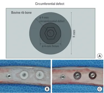
Two kinds of circumferential defects were made: the first was 8 mm in diameter and had a depth of 2.5 mm, and the second was 8 mm diameter and had a depth of 5.0 mm. The control area had no
defect (Fig. 1A and B). For each of the two defect groups and the control group, 14 samples were prepared for each group. The cir- cumferential defect was made around the prepared drilling hole using a trephine bur 8 mm in diameter. After drilling with the tre- phine bur, bone was removed using a #770 bur to create the 2.5- mm or 5-mm depth in the circumferential defect (Fig. 1B). After preparation of the bone defects, the cortical bone thickness was measured using a UNC-15 probe. In this bovine bone model, we considered the area outside of the rib arch as the buccal side and the area inside of the rib arch as the lingual side. In addition, the left side was considered the mesial side, and the right side was con- sidered the distal side.
Implant placement and ISQ measurement
In the six bovine rib bones used in our study, seven implants were placed in each bone. These implants were external connection-type implants (φ 4 mm×10 mm) (Sola, Shinhung Co.) and were placed in each prepared hole. Each implant fixture had a platform diameter of 4.1 mm, with a height of 0.7 mm (the hexagonal connection), a pitch length of 6 mm, and a self-tapping fixture design. ISQ values were measured three times from the four directions (mesial, distal, buccal, and lingual sides) with an Osstell instrument (Integration Di- agnostics AB, Göteborg, Sweden). After measuring the ISQ values, the bone cement (Polybone, Hanmi Medicare Inc., Seoul, Korea) was mixed for 30 seconds, allowed to react for 10–30 seconds, and then grafted into each prepared circumferential bone defect (Fig. 1C), which took approximately 4 minutes. After grafting, the cement was allowed to harden for 3 minutes. Last, the ISQ values were measured again.
Circumferential defect Bovine rib bone
ϕ 4-mm
circumferential defect
ϕ 4-mm fixture
8 mm
A
B C
Figure 1. Schematic drawings of the circumferential defect (occlusal view) and photos of a sample bovine rib bone before and after bone cement graft- ing. (A) Schematic drawing of the circumferential defect (occlusal view). (B) Before bone cement grafting. (C) After bone cement grafting.
Statistical analysis
Differences in the thickness of the cortical bone and the ISQ val- ues between all three groups were analyzed using one-way analysis of variance with the Tukey test. The differences in the ISQ values be- fore and after bone cement placement were analyzed using paired t-tests. The significance level of these tests was determined at P<
0.05. All statistical calculations were performed using PASW Statis- tics ver. 18.0 (SPSS Inc., Chicago, IL, USA).
RESULTS
The thickness of the cortical bone before drilling in the control, circumferential 2.5-mm defect, and circumferential 5-mm defect groups were 2.75 mm, 2.68 mm, and 2.86 mm, respectively (P>0.05) (Table 1). After preparation of the bone defect, no cortical bone re-
mained in the 5-mm defect group, and only a thin layer of cortical bone remained in the 2.5-mm depth group. The ISQ values before and after bone cement grafting are shown in Table 1. Before bone cement grafting, the ISQ values varied largely across the defect depth groups. As the defect depth increased, the ISQ value decreased. In the controls, the ISQ value was 85.45±3.36 (mean±standard deviation).
In the 2.5-mm and 5-mm defect groups, the ISQ values were 69.42±
7.06 and 57.43±6.87, respectively, before bone cement grafting. These three values were significantly different from one another (P<0.001).
After bone cement grafting, the ISQ values increased to 73.72±8.00 and 67.88±10.09 in the 2.5-mm (P<0.05) and 5-mm defect groups (P<0.001), respectively. Although the ISQ value in the 5-mm defect group was lower than that in 2.5-mm defect group, the difference between the ISQ values before and after grafting were greater in 5-mm defect group than that in the 2.5-mm defect group was.
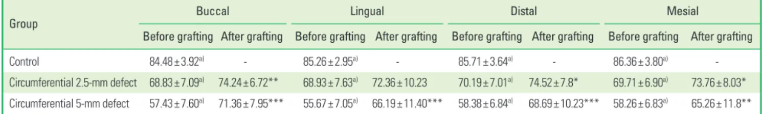
The ISQ values were measured from four directions to evaluate whether these values differed depending upon the measuring di- rection. For all measuring directions, the ISQ values decreased as the defect depth increased (Table 2). After grafting the bone ce- ment in the 5-mm defect group, the ISQ value in buccal direction was the highest, and then decreased in the order of the distal, lin- gual, and mesial direction (P<0.05). Moreover, all of the ISQ values before and after grafting were statistically significant except for those in the lingual direction. After grafting, none of the ISQ values in the 2.5-mm and 5-mm defect groups was statistically significant except for those in the mesial direction.
The ISQ values were then categorized as being measured perpen- dicular to the rib bone or parallel to the rib bone (Table 3). The buc- cal and lingual directions were considered as perpendicular to the rib bone axis, and the mesial and distal directions were considered Table 1. The thickness of initial cortical bone (mm) and implant stability quo-
tient (ISQ) values for the three experimental groups.
Group Thickness of the
initial cortical bone (mm)
values before ISQ graftinga)
values after ISQ grafting
Control (n=14) 2.75±0.43 85.45±3.36 -
Circumferential 2.5-mm defect (n=14)
2.68±0.42 69.42±7.06 73.72±8.00*
Circumferential 5-mm defect (n=14)
2.86±0.60 57.43±6.87 67.88±10.09***
Values are presented as mean±standard deviation.
A significant difference was found before and after grafting: *P<0.05 and
***P<0.001.
a)The ISQ values significantly differed among the three groups before grafting.
Table 2. The implant stability quotient values recorded in each measurement direction.
Group Buccal Lingual Distal Mesial
Before grafting After grafting Before grafting After grafting Before grafting After grafting Before grafting After grafting
Control 84.48±3.92a) - 85.26±2.95a) - 85.71±3.64a) - 86.36±3.80a) -
Circumferential 2.5-mm defect 68.83±7.09a) 74.24±6.72** 68.93±7.63a) 72.36±10.23 70.19±7.01a) 74.52±7.8* 69.71±6.90a) 73.76±8.03*
Circumferential 5-mm defect 57.43±7.60a) 71.36±7.95*** 55.67±7.05a) 66.19±11.40*** 58.38±6.84a) 68.69±10.23*** 58.26±6.83a) 65.26±11.8**
Values are presented as mean±standard deviation.
A significant difference was found before and after grafting: *P<0.05, **P<0.01, and ***P<0.001.
a)The implant stability quotient values significantly differed among the three groups before grafting.
Table 3. The implant stability quotient values for the perpendicular and parallel measurements.
Group Buccal and lingual sides (perpendicular to the rib axis) Mesial and distal side (parallel to the rib direction)
Before grafting After grafting Before grafting After grafting
Control 84.87±3.37 - 86.04±3.69 -
Circumferential 2.5-mm defect 68.88±7.32 73.30±8.34* 69.95±6.94 74.14±7.87*
Circumferential 5-mm defect 56.55±7.03 68.77±9.38*** 58.32±6.80 66.98±10.95***
Values are presented as mean±standard deviation.
A significant difference was found before and after grafting: *P<0.05 and ***P<0.001.
as parallel to the rib bone axis. The ISQ values did not significantly differ between these two different directions. When the ISQ values were compared before and after grafting, they were found to be significantly greater after bone cement grafting. In the 2.5-mm defect group, the ISQ values measured in the parallel and perpen- dicular directions increased from 68.88 and 69.95 before grafting to 73.30 and 74.14 after grafting, respectively. Likewise, in the 5.0- mm defect group, the ISO values measured in perpendicular and parallel directions increased from 56.55 mm and 58.32 mm before grafting to 68.77 mm and 66.98 mm after grafting, respectively.
DISCUSSION
β-TCP has been a used widely graft material in dental, cranial, and spinal surgery [15-20]. In addition, cement type bone graft material, which is composed of β-TCP and polyphosphate, has been used to induce bone regeneration after cranial or spinal surgery [12-14]. It has also been used as a scaffold because of its high porosity that provides sufficient spaces for newly formed bone to grow after spi- nal surgery. Moreover, it has been used as a grafting material to fill defects or sinuses in cranial surgery.
The main determinants of implant stability are the mechanical properties of the bone tissue at the implant site and the ability to obtain firm implant engagement with the bone tissue [1,21]. In a previous study that tested the use three different kinds of defects [11], the ISQ value decreased as the amount of cortical bone around the implant decreased. Moreover, a loss of cortical bone reduced the stability of these implants and the ISQ values. The ISQ values of the three-wall defect group (81.74±3.32) did not significantly dif- fer from the no defect (control) group (84.70±5.52). However, the ISQ values for the circumferential and one-wall defect groups were significantly lower than that of the other groups [11]. Another study found the initial implant mobility within the cortical layer to result in a significantly less amount of bone around the implants than that around the stable controls was [22].
Risk factors for the unstable placement of implants include poor bone quality, maxillary posterior jaw locations, and short implants.
Implant diameter is not related to primary stability [23]. Herr [24]
summarized several methods that increase implant success when the initial stability at the implant placement site is insufficient. First, if the alveolar ridge width and height are sufficient, a longer or wider implant fixture placement should be considered. Second, if the alveolar ridge width and height are insufficient and it is not possible to install a longer and wider implant, extending the time between the first and second surgery or delaying the prosthodontic treatment should be considered to provide more time for osseoin- tegration. Third, the use of a self-tapping implant was recommend- ed to overcome any insufficient initial stability. Fourth, inserting a grafting bone substitute into the preparation hole before implant placement was suggested to improve the primary stability of the implant. Last, the use of a bone cement graft around the prepared implant beds was also recommended as a method to overcome any
insufficient initial stability of the dental implant.
In 2011, Seong et al. [10] tested the experimental placement of dental implants with a smaller diameter than the drill used for the preparation of the bone in rabbit tibia. Thereafter, bone cement was grafted into the space between the prepared bone and the placed implants. The bone cement used around the initially unstable dental implants in the rabbit model was found to result in early implant stability and improved mechanical properties of the implant.
In the present study, the ISQ values increased four to 10 points more after bone cement application, which served to increase the initial stability. The ISQ value increased the most in the 5-mm depth defect group. Although the ISQ values after grafting were not as high as that of the control group, a significant increase in the ISQ values of the defect groups was observed. After bone cement graft- ing, the manipulation of the implant by inserting a cover screw and/or healing abutment connection might be easier than that be- fore grafting is. In addition, measurement of the ISQ level has been found to be influenced by the measurement orientation along the axis of the alveolar ridge [25-27]. In a hard jawbone model, the ini- tial measurement of the ISQ values recorded in the parallel orienta- tion was greater than that in the perpendicular orientation. How- ever, these two values did not show any significant difference in the present study; a slight difference was found between the perpen- dicular ISQ values and that of the parallel ISQ values, but it was not significant.
According to a study by Morris et al. [23], initial implant mobility (the mobility observed at implant placement) was observed in 2.8%
of 1,554 placed implants. Implant mobility was the mostly found in the maxillary posterior area that has poor bone density (6.3%). Of the implants placed in D4 bone, 12.2% were found to be mobile at place- ment, which was higher than the percentage of implants placed in denser bone. Moreover, 97.7% of these mobile implants were stabi- lized by stage II, the uncovering, was carried out. This promising find- ing might result from the sufficient healing period. Before abutment connection, implants were allowed to heal for 4–6 months in the mandible and 6.5–8 months in the maxilla. In this study, the direction that the measurement was made did not result in different findings.
In our previous study, the direction of the measurement was signifi- cantly different in the one- and three-wall defect groups, but did not significantly differ in circumferential defect group.
This study showed that the use of bone cement remarkably in- creased the stability of implants that otherwise had insufficient stabilities at placement, which was caused by an insufficient vol- ume of cortical bone. The main limitations of this study were the limited number of samples and the potential variations between the rib bone samples. These results might not be applicable to clin- ical situations. Bone cement should become resorbed and replaced by newly formed bone if allowed the proper amount of time. Fur- ther studies are necessary to confirm the clinical performance of bone cement grafts in in vivo animal studies.
CONFLICT OF INTEREST
No potential conflict of interest relevant to this article was re- ported.
ACKNOWLEDGEMENTS
Authors would like to thank Dr. Sang-Ik Park, Dr. Jin-Hwan Jung, and Dr. Byung-In Han for assisting during the experiment.
ORCID
Seung-Yun Shin http://orcid.org/0000-0001-6980-7556 Seung-Il Shin http://orcid.org/0000-0001-8762-6169 Seung-Beom Kye http://orcid.org/0000-0002-9135-2591 Seok-Woo Chang http://orcid.org/0000-0003-4461-3274 Jongrak Hong http://orcid.org/0000-0003-3569-2046 Jun-Young Paeng http://orcid.org/0000-0002-0104-9338 Seung-Min Yang http://orcid.org/0000-0002-8002-5800
REFERENCES
1. Albrektsson T, Zarb GA. Current interpretations of the osseointe- grated response: clinical significance. Int J Prosthodont 1993;6:
95-105.
2. Lioubavina-Hack N, Lang NP, Karring T. Significance of primary stability for osseointegration of dental implants. Clin Oral Implants Res 2006;17:244-50.
3. Bayarchimeg D, Namgoong H, Kim BK, Kim MD, Kim S, Kim TI, et al. Evaluation of the correlation between insertion torque and pri- mary stability of dental implants using a block bone test. J Peri- odontal Implant Sci 2013;43:30-6.
4. Hong J, Lim YJ, Park SO. Quantitative biomechanical analysis of the influence of the cortical bone and implant length on primary stability. Clin Oral Implants Res 2012;23:1193-7.
5. Javed F, Almas K. Osseointegration of dental implants in patients undergoing bisphosphonate treatment: a literature review. J Peri- odontol 2010;81:479-84.
6. Javed F, Romanos GE. Impact of diabetes mellitus and glycemic control on the osseointegration of dental implants: a systematic literature review. J Periodontol 2009;80:1719-30.
7. Roze J, Babu S, Saffarzadeh A, Gayet-Delacroix M, Hoornaert A, Layrolle P. Correlating implant stability to bone structure. Clin Oral Implants Res 2009;20:1140-5.
8. Tabassum A, Meijer GJ, Wolke JG, Jansen JA. Influence of the surgical technique and surface roughness on the primary stabili- ty of an implant in artificial bone with a density equivalent to maxillary bone: a laboratory study. Clin Oral Implants Res 2009;
20:327-32.
9. Tabassum A, Meijer GJ, Wolke JG, Jansen JA. Influence of surgical technique and surface roughness on the primary stability of an implant in artificial bone with different cortical thickness: a lab-
oratory study. Clin Oral Implants Res 2010;21:213-20.
10. Seong WJ, Kim HC, Jeong S, DeVeau DL, Aparicio C, Li Y, et al. Ex vivo mechanical properties of dental implant bone cement used to rescue initially unstable dental implants: a rabbit study. Int J Oral Maxillofac Implants 2011;26:826-36.
11. Shin SY, Shin SI, Kye SB, Hong J, Paeng JY, Chang SW, et al. The effects of defect type and depth, and measurement direction on the implant stability quotient (ISQ) value. J Oral Implantol 2014 Jun 26 [Epub]. http://dx.doi.org/10.1563/aaid-joi-D-13-00331 12. Park JH, Choi CG, Jeon SR, Rhim SC, Kim CJ, Roh SW. Radiographic
analysis of instrumented posterolateral fusion mass using mixture of local autologous bone and b-TCP (PolyBone(R)) in a lumbar spinal fusion surgery. J Korean Neurosurg Soc 2011;49:267-72.
13. Park JH, Roh SW. Anterior cervical interbody fusion using poly- etheretherketone cage filled with autologous and synthetic bone graft substrates for cervical spondylosis: comparative analysis be- tween PolyBone® and iliac bone. Neurol Med Chir (Tokyo) 2013;
53:85-90.
14. Park YH, Kim SG, Lee JW, Yoon YH. Obliteration of temporal dorsal bulla in guinea pigs using different types of calcium phosphate.
Int J Pediatr Otorhinolaryngol 2011;75:1176-80.
15. Abbah SA, Lam CX, Hutmacher DW, Goh JC, Wong HK. Biological performance of a polycaprolactone-based scaffold used as fu- sion cage device in a large animal model of spinal reconstructive surgery. Biomaterials 2009;30:5086-93.
16. Byun HY, Wang HL. Sandwich bone augmentation using recombi- nant human platelet-derived growth factor and beta-tricalcium phosphate alloplast: case report. Int J Periodontics Restorative Dent 2008;28:83-7.
17. Cha JK, Park JC, Jung UW, Kim CS, Cho KS, Choi SH. Case series of maxillary sinus augmentation with biphasic calcium phosphate: a clinical and radiographic study. J Periodontal Implant Sci 2011;41:
98-104.
18. Elahi MM, Vanduzer S, Spears J, Gibson J, Mitra A. Frontal sinus obliteration with beta-tricalcium phosphate. J Craniofac Surg 2004;15:967-70.
19. Kishimoto M, Kanemaru S, Yamashita M, Nakamura T, Tamura Y, Tamaki H, et al. Cranial bone regeneration using a composite scaf- fold of Beta-tricalcium phosphate, collagen, and autologous bone fragments. Laryngoscope 2006;116:212-6.
20. Suba Z, Takacs D, Matusovits D, Barabas J, Fazekas A, Szabo G.
Maxillary sinus floor grafting with beta-tricalcium phosphate in humans: density and microarchitecture of the newly formed bone.
Clin Oral Implants Res 2006;17:102-8.
21. Sennerby L, Meredith N. Implant stability measurements using resonance frequency analysis: biological and biomechanical as- pects and clinical implications. Periodontol 2000 2008;47:51-66.
22. Ivanoff CJ, Sennerby L, Lekholm U. Influence of initial implant mobility on the integration of titanium implants. An experimen- tal study in rabbits. Clin Oral Implants Res 1996;7:120-7.
23. Morris HF, Ochi S, Orenstein IH, Petrazzuolo V. AICRG, Part V: Fac- tors influencing implant stability at placement and their influ-
ence on survival of Ankylos implants. J Oral Implantol 2004;30:
162-70.
24. Herr Y. Atlas of periodontology-based implantology I. Seoul: Yenang Inc.; 2012.
25. Fischer K, Stenberg T, Hedin M, Sennerby L. Five-year results from a randomized, controlled trial on early and delayed loading of im- plants supporting full-arch prosthesis in the edentulous maxilla.
Clin Oral Implants Res 2008;19:433-41.
26. Valderrama P, Oates TW, Jones AA, Simpson J, Schoolfield JD, Co- chran DL. Evaluation of two different resonance frequency devic- es to detect implant stability: a clinical trial. J Periodontol 2007;
78:262-72.
27. Veltri M, Balleri P, Ferrari M. Influence of transducer orientation on Osstell stability measurements of osseointegrated implants.
Clin Implant Dent Relat Res 2007;9:60-4.