Korean J Gastroenterol Vol. 65 No. 5, 326-329 http://dx.doi.org/10.4166/kjg.2015.65.5.326 pISSN 1598-9992 eISSN 2233-6869
IMAGE OF THE MONTH
Korean J Gastroenterol, Vol. 65 No. 5, May 2015 www.kjg.or.kr
급속하게 진행된 중간 대장암
김재현, 박무인
고신대학교 의과대학 내과학교실
Rapidly Growing Interval Colon Cancer
Jae Hyun Kim and Moo In Park
Department of Internal Medicine, Kosin University College of Medicine, Busan, Korea
CC This is an open access article distributed under the terms of the Creative Commons Attribution Non-Commercial License (http://creativecommons.org/licenses/
by-nc/4.0) which permits unrestricted non-commercial use, distribution, and reproduction in any medium, provided the original work is properly cited.
Copyright © 2015. Korean Society of Gastroenterology.
교신저자: 박무인, 602-702, 부산시 서구 감천로 262, 고신대학교복음병원 소화기내과
Correspondence to: Moo In Park, Division of Gastroenterology, Department of Internal Medicine, Kosin University College of Medicine, 262 Gamcheon-ro, Seo-gu, Busan 602-702, Korea. Tel: +82-51-990-5061, Fax: +82-51-990-5055, E-mail: [email protected]
Financial support: None. Conflict of interest: None.
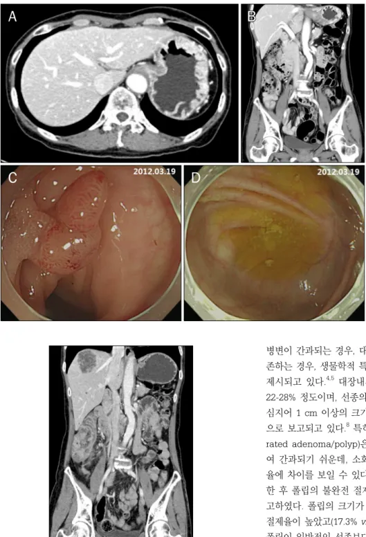
증례: 58세 여자 환자가 한 달 전부터 발생한 간헐적인 메 스꺼움과 복통을 주소로 내원하였다. 공복 시에 메스꺼움이 있었으며, 복통은 하복부에 쥐어짜는 듯한 양상을 보였고 주 기적으로 호전과 악화를 반복하였으며 그 강도가 점점 심해지 는 양상이었다. 그러나 발열, 혈변, 설사 등은 없었다. 당뇨, 간염, 폐결핵의 병력은 없었고, 5년 전에 고혈압으로 진단되 었다고 하나 약을 복용하지는 않았다. 평소 일주일에 2회, 소 주 1병의 음주력이 있었으며, 약 10년 동안 하루 반 갑의 흡연 력이 있었다. 암의 가족력은 없는 환자로, 내원 2년 전 검진 목적으로 위내시경, 대장내시경, 복부 및 흉부 전산화단층촬 영을 시행하였고(Fig. 1A, B), S자 결장과 직장에서 크기가 10 mm 이상인 4개의 폴립이 관찰되어(Fig. 1C), 대장의 폴립 제거를 위해 검진 한 달 후 대장내시경 점막하 절제술을 시행 받았다. 조직검사 결과는 저도 이형성 선종이었으며, 두 차례 의 대장내시경검사에서 상행 결장 및 맹장에서는 특이 소견이 보이지 않았다(Fig. 1D). 내원 당시 활력 징후는 혈압 110/80 mmHg, 맥박수 70회/분, 체온 36.5oC로 측정되었다. 신체검 사에서 결막이 창백하였고, 복부 촉진에서 경미한 우하복부 압통이 관찰되었으나 반발 압통은 없었고, 간비종대 및 촉지 되는 종괴는 없었다.
위내시경검사에서 특이 소견은 보이지 않고 증상이 지속되 어 복부 전산화단층촬영 검사를 시행하였다. 맹장 및 상행결 장의 벽이 비후되어 관강이 좁아져 있었으며, 다발성 간 전이
소견이 관찰되었다(Fig. 2). 말초혈액검사에서 백혈구 9,260/
μL, 혈색소 9.4 g/dL, 혈소판 198,000/μL로 측정되었고, 생화 학검사에서 AST/ALT 18/8 IU/L, 총 빌리루빈 0.78 mg/dL, ALP 97 IU/L로 측정되었다. CEA와 CA 19-9는 각각 85.37 ng/mL, 277.60 U/mL로 상승되어 있었다. 대장내시경검사에 서 상행 결장에 약 5 cm 크기의 Borrmann type 3 형태의 종양이 관찰되었고, 종양으로 인해 관강이 좁아져 있어 대장 내시경이 더 이상 진입하지 못하였다(Fig. 3A, B). 대장내시경 조직검사에서 고분화 선암종이 확인되었고, 간 조직검사에서 도 같은 소견이 확인되었다. 양전자 단층촬영 검사에서 폐 전 이 소견 또한 관찰되었다.
내원 2년 전의 검사에서 대장의 폴립 이외에 특이 소견을 보이지 않았으나, 불과 2년 만에 다발성 간 전이 및 폐 전이를 동반한 대장암으로 진행된 환자로, 현재 항암 약물 치료 (bevacizumab+FOLFIRI regimen) 중이다.
진단: 급속하게 진행된 중간 대장암
중간 대장암(interval colon cancer)은 대장내시경 검사 후 권고되는 추적검사 기간 이내에 발생한 대장암을 말하며, 통 상적으로 대장내시경검사 후 5년 이내 또는 폴립 절제를 받은 환자에서는 3-5년 이내에 발생한 암을 의미한다. Cooper 등1 의 연구에서는 대장내시경검사를 시행한 57,839명을 추적 관 찰하였고 7.2%에서 중간 대장암이 진단되었다. Baxter 등2의
Kim JH and Park MI. Rapidly Growing Interval Colon Cancer
327
Vol. 65 No. 5, May 2015
Fig. 1. Initial abdomen-pelvis CT and endoscopic findings. (A, B) No ab- normal findings are noted on abdo- men-pelvis CT (A, transverse view; B, coronal view). (C) About 10 mm sized, pedunculated polyp is observed on sigmoid colon and removed by endo- scopic mucosal resection. (D) No ab- normal endoscopic findings are pre- sent on cecum.
Fig. 2. Follow-up abdomen-pelvis CT findings. Irregular wall thickening and luminal narrowing of the cecum and ascending colon is seen along with multiple liver metastasis.
연구에서는 14,064명의 대장암 환자 중 근위부 대장암의 12.4%, 원위부 대장암의 6.8%가 중간암이었다. 국내의 연구 에서도 중간 대장암은 6.2%로 발생하였고 근위부 대장에서 호발하는 경향을 보였다.3 이러한 중간 대장암이 발생하는 원 인으로 불량한 장정결이나 불완전한 관찰 수기 등으로 인해
병변이 간과되는 경우, 대장 폴립의 불완전한 절제로 인해 잔 존하는 경우, 생물학적 특성으로 인해 성장이 빠른 경우 등이 제시되고 있다.4,5 대장내시경검사에서 폴립의 간과율은 대략 22-28% 정도이며, 선종의 간과율은 20-24%로 알려져 있다.6,7 심지어 1 cm 이상의 크기가 큰 폴립조차도 종종 간과되는 것 으로 보고되고 있다.8 특히 무경성 톱니선종/폴립(sessile ser- rated adenoma/polyp)은 근위부 대장에서 호발하고 편평하 여 간과되기 쉬운데, 소화기내과 의사 간에도 1-18%로 발견 율에 차이를 보일 수 있다.9 Pohl 등10은 346개의 폴립을 제거 한 후 폴립의 불완전 절제 여부를 조사하여 10.1%였다고 보 고하였다. 폴립의 크기가 큰 경우가 작은 경우에 비해 불완전 절제율이 높았고(17.3% vs. 6.8%; RR=2.1), 무경성 톱니선종/
폴립이 일반적인 선종보다 불완전 절제율이 높았다(31.0% vs.
7.2%; RR=3.7). 무경성 톱니선종/폴립은 BRAF 돌연변이가 많고 현미부수체 불완전성(microsatellite instability)과 CpG island methylator phenotype과 연관된 대장암이 많아 비교 적 빠르게 악성화가 진행될 수 있을 것으로 생각되어 중간암 의 주요한 원인으로 고려되고 있다.11,12 하지만 대부분의 연구 에서 중간암과 일반 대장암의 생존율에는 차이가 없어, 무경 성 톱니선종/폴립이 분자 생물학적 차이로 인해 더 빠른 성장 속도 때문에 중간암의 원인이 되는지, 아니면 간과되기 쉽고 편평하면서 우측에 호발하고 불완전 절제율이 높아서 중간암
328
김재현, 박무인. 급속하게 진행된 중간 대장암The Korean Journal of Gastroenterology
Fig. 3. Follow-up endoscopic findings. (A) About 5 cm sized, Borrmann type 3 tumor is observed on ascending colon. (B) Because of the luminal narrowing, the scope could not be passed into the cecum.
의 원인이 되는지에 대해서는 좀더 연구가 필요하다.
2012년에 발표된 대장 폴립 진료 가이드라인에 따르면,13 일정한 자격을 갖춘 대장내시경 의사가 양호한 대장정결상태 에서 양질의 기준 대장내시경검사를 시행하였음을 전제로, 기 준 대장내시경검사 소견이 폴립 절제 후 진행 신생물의 발생 고위험군에 해당되지 않는 경우 추적 대장내시경검사를 폴립 절제 후 5년에 시행할 것을 권고하였고, 고위험군에 해당되는 경우에는 폴립 절제 후 3년에 시행할 것을 권고하였다. 폴립 절제 후 진행 신생물의 발생 고위험군에는 (1) 선종의 개수가 3개 이상, (2) 선종의 크기가 10 mm 이상, (3) 관융모 또는 융모 선종(tubulovillous or villous adenoma), (4) 고도 이형 성을 동반한 선종(adenoma with high-grade dysplasia), (5) 10 mm 이상 크기의 톱니모양폴립이 진단된 경우가 포함된 다. 이번 증례의 경우 2년 전 대장내시경검사에서 10 mm 이 상 크기의 선종이 발견되어 고위험군에 해당되므로 권고안에 따르면 폴립 절제 후 3년에 추적 대장내시경을 시행하는 것이 바람직하나, 일정한 자격을 갖춘 대장내시경 의사가 양호한 대장정결상태에서 양질의 기준 대장내시경검사를 2회에 걸쳐 시행하였음에도 불과 2년만에 다발성 간 전이 및 폐 전이를 동반한 대장암으로 진행된 경우로, 문헌 고찰에서 이번 증례 처럼 급속하게 진행된 중간 대장암의 보고는 없었다. 따라서 대장 폴립 절제 후 고위험군에 해당되는 경우에는 이번 증례 처럼 권고안의 범주를 벗어나는 급속하게 진행되는 중간 대장 암의 발생 가능성도 염두에 두고, 추적 대장내시경검사 간격 을 좀 더 짧게 하는 것을 고려해 보아야 할 것으로 생각된다.
REFERENCES
1. Cooper GS, Xu F, Barnholtz Sloan JS, Schluchter MD, Koroukian SM. Prevalence and predictors of interval colorectal cancers in medicare beneficiaries. Cancer 2012;118:3044-3052.
2. Baxter NN, Sutradhar R, Forbes SS, Paszat LF, Saskin R, Rabeneck L. Analysis of administrative data finds endoscopist quality measures associated with postcolonoscopy colorectal cancer. Gastroenterology 2011;140:65-72.
3. Kim CJ, Jung YS, Park JH, et al. Prevalence, clinicopathologic characteristics, and predictors of interval colorectal cancers in Korean population. Intest Res 2013;11:178-183.
4. Bressler B, Paszat LF, Vinden C, Li C, He J, Rabeneck L.
Colonoscopic miss rates for right-sided colon cancer: a pop- ulation-based analysis. Gastroenterology 2004;127:452-456.
5. Bressler B, Paszat LF, Chen Z, Rothwell DM, Vinden C, Rabeneck L. Rates of new or missed colorectal cancers after colonoscopy and their risk factors: a population-based analysis. Gastroenterology 2007;132:96-102.
6. van Rijn JC, Reitsma JB, Stoker J, Bossuyt PM, van Deventer SJ, Dekker E. Polyp miss rate determined by tandem colonoscopy:
a systematic review. Am J Gastroenterol 2006;101:343-350.
7. Heresbach D, Barrioz T, Lapalus MG, et al. Miss rate for colorectal neoplastic polyps: a prospective multicenter study of back-to- back video colonoscopies. Endoscopy 2008;40:284-290.
8. Pickhardt PJ, Nugent PA, Mysliwiec PA, Choi JR, Schindler WR.
Location of adenomas missed by optical colonoscopy. Ann Intern Med 2004;141:352-359.
9. Kahi CJ, Hewett DG, Norton DL, Eckert GJ, Rex DK. Prevalence and variable detection of proximal colon serrated polyps during screening colonoscopy. Clin Gastroenterol Hepatol 2011;9:42- 46.
10. Pohl H, Srivastava A, Bensen SP, et al. Incomplete polyp re-
Kim JH and Park MI. Rapidly Growing Interval Colon Cancer
329
Vol. 65 No. 5, May 2015 section during colonoscopy-results of the complete adenoma
resection (CARE) study. Gastroenterology 2013;144:74-80.e1.
11. Sawhney MS, Farrar WD, Gudiseva S, et al. Microsatellite in- stability in interval colon cancers. Gastroenterology 2006;131:
1700-1705.
12. Arain MA, Sawhney M, Sheikh S, et al. CIMP status of interval co- lon cancers: another piece to the puzzle. Am J Gastroenterol
2010;105:1189-1195.
13. Hong SN, Yang DH, Kim YH, et al; Multi-Society Task Force for- Development of Guidelines for Colorectal Polyp Screening, Surveillance and Management. Korean guidelines for post-poly- pectomy colonoscopic surveillance. Korean J Gastroenterol 2012;59:99-117.