Anterior cruciate ligament (ACL) ruptures are commonly associated with meniscus tears. The prevalence of associ- ated meniscus injuries in patients with ACL ruptures has been found to be 65% in acute injuries and 90% in chronic injury of ACL.1,2) Meniscus repair performed simultane- ously with ACL reconstruction can give additional stability to the knee joint.3) The healing rate of repaired meniscus associated with ACL reconstruction is 92%, compared to only 63% in meniscus tears not associated with ACL
The Results of All-Inside Meniscus Repair Using the Viper Repair System Simultaneously with
Anterior Cruciate Ligament Reconstruction
Hong Je Kang, MD, Churl Hong Chun, MD, Kwang Mee Kim, PhD*, Hang Hwan Cho, MD, Johnsel C. Espinosa, MD
†Department of Orthopedic Surgery, Wonkwang University School of Medicine, Iksan,
*Department of Nursing, Chodang University, Muan, Korea
†Department of Orthopedics Surgery, Philippine Orthopedic Center, Manila, Philippines
Background: Meniscus tears are commonly associated with anterior cruciate ligament (ACL) ruptures. It is essential to repair meniscal tears as much as possible to prevent early osteoarthritis and to gain additional stability in the knee joint. We evaluated the results of arthroscopic all-inside repair using the Meniscal Viper Repair System (Arthrex) on meniscus tears simultaneously with ACL reconstruction.
Methods: Nineteen out of 22 patients who were treated with arthroscopic all-inside repair using the Meniscal Viper Repair System for meniscus tear associated with ACL rupture were evaluated. ACL reconstructions were performed at the same period.
The mean follow-up period was 16.5 months (range, 12 to 24 months). The clinical results of the meniscus repair were evaluated by symptoms (such as catching or locking), tenderness, effusion, range of motion limitation, and the McMurray test. Clinical suc- cess was defined by negative results in all five categories. The Hospital for Special Surgery (HSS) score was evaluated. Objective results were evaluated with secondary look arthroscopy or magnetic resonance imaging (MRI). The MRI results were categorized as completely repaired, incompletely repaired, and failure by Henning’s classification. The results of second-look arthroscopy were evaluated with the criteria of meniscal healing.
Results: The clinical success rate was 95.4% and the HSS scores were 93.9 ± 5.4 at the final follow-up. According to Henning’s classification, 15 out of 18 cases showed complete healing (83.3%) and two cases (11.1%) showed incomplete healing. Seventeen out of 18 cases that underwent second-look arthroscopy showed complete healing (94.4%) according to the criteria of meniscal healing. Only one case showed failure and the failure was due to a re-rupture at the sutured area. Complications of ACL recon- struction or meniscus repair were not present.
Conclusions: The results demonstrate that arthroscopic all-inside repair using the Meniscal Viper Repair System is an effective treatment method when it is performed simultaneously with ACL reconstruction.
Keywords: Anterior cruciate ligament, Meniscus, Reapir, Viper, All inside
Copyright © 2015 by The Korean Orthopaedic Association
This is an Open Access article distributed under the terms of the Creative Commons Attribution Non-Commercial License (http://creativecommons.org/licenses/by-nc/4.0) which permits unrestricted non-commercial use, distribution, and reproduction in any medium, provided the original work is properly cited.
Clinics in Orthopedic Surgery • pISSN 2005-291X eISSN 2005-4408 Received June 19, 2014; Accepted November 10, 2014
Correspondence to: Churl Hong Chun, MD
Department of Orthopedic Surgery, Wonkwang University School of Medicine, 460 Iksan-daero, Iksan 570-974, Korea
Tel: +82-63-859-1360, Fax: +82-63-852-9329 E-mail: [email protected]
reconstruction.4-7) Generally, the treatment of a meniscus tear combined with an ACL rupture is repair rather than a meniscectomy because the bleeding and debris from the ACL reconstruction starts an inflammatory reaction that helps the meniscus’ healing phase.7,8) The other reason that repair is preferred, is that the healed meniscus can help add stability to the knee joint.9)
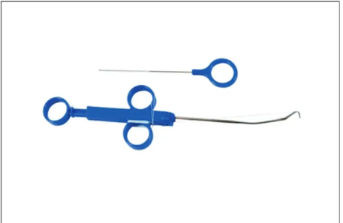
The arthroscopic meniscus repair method can be categorized as all-inside, outside-in, or inside-out. Inside- out or outside-in repair techniques require additional skin incisions and can cause neurovascular injury and soft tissue irritation.10-13) All-inside repair with meniscal implants and devices has been popularly used because of its advantages, such as its ease of use and shorter repair time.14,15) However, it has some disadvantages such as im- plant breakage, failure of repair, infection, and chondral injury.16,17) The Meniscal Viper Repair System (Arthrex, Naples, FL, USA) is a new device for the all-inside suture technique, allowing surgeons to avoid the disadvantages of
the outside-in or inside-suture techniques (Fig. 1).14) There have been some reports regarding the results of meniscus repair using the Meniscal Viper Repair System simultane- ously with ACL reconstruction.18,19)
The purpose of this study was to evaluate the results of arthroscopic all-inside repair using the Meniscal Viper Repair System on meniscus tears with ACL reconstruction through clinical outcomes and second-look arthroscopy or magnetic resonance imaging (MRI).
METHODS
Study Group Collection
From January 2010 to April 2012, there were 92 cases that underwent ACL reconstruction at Wonkwang University Hospital. Among these cases, 37 cases had meniscal tear.
Out of the 37 cases, 22 cases underwent arthroscopic all- inside meniscus repair using the Meniscal Viper Repair System with simultaneous ACL reconstruction. Out of the 22 cases, the 19 cases that underwent second arthroscopy or MRI were selected for the study (Fig. 2).
The mean age at operation time was 35.4 years (range, 18 to 45 years) with 15 men and 4 women consti- tuting the patients. Among the 19 patients there were four cases of medial meniscus tears, 14 cases of lateral meniscus tears, and one case where both were torn (Table 1).
The indications for meniscal repair in this study were: meniscal tears that were reparable using the Menis- cal Viper Repair System, including tears of the middle body of the meniscus, tears approximately one-third of a posterior medial meniscal tear, and tears of the posterior half of the lateral meniscus.
We excluded isolated meniscal repair or combined knee injuries other than ACL. White-white zone tears Fig. 1. The Meniscal Viper Repair System.
92 Cases ACL reconstruction
37 Cases
ACL reconstruction + meniscus injury
22 Cases
ACL reconstruction + meniscus repair with the Meniscal Viper Repair System
15 Cases
ACL reconstruction + meniscus repair with other repair technique/meniscectomy
19 Cases
Follow-up with MRI and 2nd look arthroscopy
Fig. 2. Patient selection process. ACL:
anterior cruciate ligament, MRI: magnetic resonance imaging,
were treated with meniscectomy. Also, we excluded those patients who had anterior horn tears or root of meniscus tears that were irreparable using the Meniscal Viper Repair System.
Operation Methods and Rehabilitation
The Meniscal Viper Repair System is a device that can tie a suture at the tip of a needle which passes through the meniscus. After thorough inspection of the knee joint with arthroscopy, the Viper was positioned at the posterior site of the torn meniscus and the needle was passed through the torn site. A non-absorbable suture material followed the needle and made the natural loop. The medial menis- cus repair was performed prior to the ACL reconstruction.
Some cases of posterior horn tear of the medial meniscus with lax joint space were repaired using the Viper System. However, not all posterior horn tears were repaired because the Viper System could not reach area:
only the posterior one-third of the medial meniscal tear was repaired with the system.
The key elements of the arthroscopic all-inside menis- cus repair using the Meniscal Viper Repair System include an insertion angle parallel to the meniscus with a clear field of view. The position of the knee should be fixed during the meniscal repair. In some cases, only the torn meniscus was sutured due to incorrect positioning.
ACL reconstructions were performed using fresh frozen Achilles allograft and an all-inside meniscus repair Table 1. Patients’ Data
Patient
no. Gender/
age (yr) Tear
type Medial/
lateral Tear
location All-inside
suture Technique for
femoral tunnel Follow-up
MRI Second-look arthroscopy
Clinical result
Meniscal healing*
HSS at last
follow-up ROM at last follow-up
1 Male/27 O M RR 3 AM O O 97 125 CH
2 Male/39 LO L RR 3 AM O O 98 127 CH
3 Male/41 LO L RR 3 TT O O 91 123 CH
4 Male/39 C ML RW 4 AM O O 93 129 CH
5 Male/16 LO L RW 4 AM O O 92 121 CH
6 Female/15 LO M RR 3 TT O O 97 124 CH
7 Male/36 C M RR 4 AM O O 95 125 CH
8 Female/18 LO L RW 3 TT O O 94 123 CH
9 Female/53 LO L RR 2 TT O O 97 128 CH
10 Male/24 O L RR 4 AM O O 92 122 CH
11 Male/32 H L RR 3 AM X O 93 122 CH
12 Female/42 C L RW 3 TT O O 82 112 Failure
13 Female/50 LO L RR 2 TT O O 98 127 CH
14 Male/54 LO L RR 3 TT O O 97 128 CH
15 Female/55 LO M RR 4 TT O X 91 123 CH
16 Male/17 LO L RR 4 AM O O 93 121 CH
17 Male/19 LO L RR 4 AM O O 98 127 CH
18 Female/42 H L RR 2 TT O O 94 129 CH
19 Male/22 O L RR 4 AM O O 92 128 CH
MRI: magnetic resonance imaging, HSS: Hospital for Special Surgery, ROM: range of motion, O: oblique tear, M: medial meniscus, RR: red-red zone, AM:
accessory medial portal, CH: complete healing, LO: longitudinal tear, L: lateral meniscus, TT: transtibial portal, C: complex tear, ML: medial and lateral meniscus, RW: red-white zone, H: horizontal tear.
*MRI and second-look result.
using the Meniscal Viper Repair System by a single sur- geon. All ACLs were reconstructed using the single bundle technique. A femoral tunnel was positioned in the 10:30 direction using the accessory medial portal in 10 cases, with patients who were younger, male, who played a lot of sports, and in those with a large anatomical femoral con- dyle. The other nine cases featured a femoral tunnel in the 11 o’clock direction using the transtibial approach.
Rehabilitation after the operation was performed for four weeks with brace protection that limited the flexion up to 60° to protect the reconstructed ACL and repaired meniscus. After 4 weeks, the range of motion (ROM) was increased every week up to 120° flexion with brace pro- tection six weeks after the operation. Crutch walking was
recommended for partial weight-bearing during the reha- bilitation process.
Clinical Evaluation
During the last follow-up consultation prior to the second- look arthroscopy, physical exams were conducted to evalu- ate the patients’ symptoms (catching, locking, and giving way), including tenderness, effusion, ROM limitations, and the McMurray test. Clinical success was considered if all five categories were negative. The Hospital for Special Surgery (HSS) score was added for the analysis of func- tional evaluation.
Fig. 3. Healing rates by segment accor- ding to Henning’s criteria: (A) complete healing, (B) partial healing > 50%, and (C)
< 50% healing.
A B C
D E F
Fig. 4. An 18-year-old woman with a complete rupture of the anterior cruciate ligament and longitudinal tear of the medial meniscus of her left knee.
(A) Arthroscopic findings of longitudinal tear of medial meniscus. (B–D) Arthroscopic findings during meniscus repair using the Meniscal Viper Repair System. (E) Arthroscopic finding immediately after suture. (F) Second-look arthroscopic findings after meniscus repair show complete healing.
Complete healing Partial healing > 50% < 50% Healing
Magnetic Resonance Imaging and Second-Look Arthroscopic Evaluation
The mean follow-up period was 16.5 months (range, 12 to 24 months), and the second-look arthroscopy was per- formed at 17.3 months on average (range, 12 to 23 months;
18 patients) and the MRIs were taken at about 15.4 months (range, 12 to 24 months; 19 patients) after the first opera- tion.
The results of MRI were evaluated with Henning’s classification,20) which categorizes healing states as com- plete, incomplete (more than 50%), or failure (less than 50%) (Fig. 3). The same surgeon who performed the meniscus repair examined the meniscus with a probe at the time of the second-look arthroscopy. The results of second-look arthroscopy were evaluated with the criteria of meniscal healting,9,21) which are classified by a residual cleft at the repair site of the meniscus; complete healing (less than 10% of residual cleft), incomplete healing (less than 50%), and failure (more than 50%).
RESULTS
Clinical Results
In 19 cases, the clinical cure rate was 94.7% with a mean HSS score of 93.9 at the last follow-up. There was one case of stiffness 12 weeks after the operation, with a ROM less than 90° on the physical examination. Passive manipula- tion under spinal anesthesia was performed and the ROM was increased to more than 120° at the last follow-up. There were no cases of deep infection or neurovascular injuries.
Magnetic Resonance Imaging and Second-Look Arthroscopy Results
According to Henning’s classification, in 17 out of the 18 cases performed, MRI showed complete healing (83.3%), and in 2 cases (11.1%), incomplete healing without any clinical symptoms (Fig. 4). According to criteria of menis- cal healing, in 17 out of 18 cases performed, second-look arthroscopy showed complete healing (94.4%). Only one case showed failure according to Henning’s classification and the criteria of meniscal healing. Authors performed the repair with 2 notes of all-inside suture and 2 notes of
A B
E D
C
Fig. 5. A 41-year-old man with complete rupture of the anterior cruciate ligament and longitudinal tear of the lateral meniscus of his right knee. (A) Arthroscopic findings of longitudinal tear of lateral meniscus. (B, C) Arthroscopic findings during meniscus repair using the Meniscal Viper Repair System. (D) Arthroscopic findings immediately after suture. (E) Second-look arthroscopic findings after meniscus repair show complete healing.
inside-out suture during second-look arthroscopy (Figs. 5 and 6).
DISCUSSION
In ACL ruptures, the meniscus tear is a common associ- ated injury. It can be associated in 65% of acute injuries and 90% in chronic injury of ACL.1,2) Higher incidence rates of lateral meniscus injuries have been reported by numerous authors.22,23) Cerabona et al.24) insisted that there is no difference in either type of meniscus tear occurrence.
However, some authors reported that the medial menis- cus is a much more common site of tears than the lateral meniscus.23,25) In our study, the lateral meniscus (17 cases) outnumbered the medial (4 cases) and both (1 case) me- niscus tears.
A recent systematic review showed that the menis- cectomy procedure is performed 2–3 times more frequent- ly than a meniscus repair during ACL reconstruction.26)
However, long-term studies have shown significantly better clinical outcomes when the menisci are repaired at the time of the ACL reconstruction.27) Bellabarba et al.28) compared the healing rate of meniscus repair with ACL reconstruction to that of meniscus repair alone and found the rates were similar.
The meniscus repair method can be categorized as all-inside, outside-in, and inside-out.29) These methods have different indications due to the site, type, and size of the meniscus tear. Each study reported various results regarding meniscus tear repair with ACL reconstruction.
Gill and Diduch23) reported that in 32 patients who under- went meniscal repair using the Meniscus Arrow (Bionx, BlueBell, PA, USA) with concurrent ACL reconstruction, there was a 90.6% success rate. However, this success rate was clinically evaluated without objective methods, such as second arthroscopy or postoperative MRI. Ahn et al.9) reported on 39 patients undergoing arthroscopic all-inside suture using a suture hook for medial meniscus poste- rior horn (MMPH) tears with ACL reconstruction. They found that 32 (82.1%) of the knees showed complete heal- ing and 6 (15.4%) showed incomplete healing without any positive findings of the clinical symptoms by second-look arthroscopy. In our study, the clinical cure rate was 94.7%.
The MRI healing rate and second-look arthroscopic heal- ing rate were 83.3% and 94.4%, respectively. This healing rate is comparable to those previously reported with the other techniques when the ACL is simultaneously recon- structed.9,23) The objective healing rate does not always correlate with clinical results, however. In our study, the clinical cure rate is better than the objective healing rate because a few asymptomatic patients still showed a failure of meniscal healing. In our study, one case showed failure and underwent revision repair during the second-look ar- throscopy. Two cases showed incomplete healing on MRI, although they showed complete healing on second-look arthroscopy. We suggest the cause of the discrepancy is the intrameniscal tear on the MRI, which cannot be seen with second-look arthroscopy. However, there were no clinical symptoms and complete healing was seen on the second-look arthroscopy. So, the patients were not treated and could be symptomatic during the long term follow-up period.
We compared our MRI results with those of Lee et al.,30) who found that an MRI showed 25 (89%) and 24 (86%) healed menisci in the sagittal and coronal views, respectively. They also obtained similar results to our re- sults with respect to the percentage of complete healing seen on MRI.27) Only a few reports on the Viper Repair System have been published. Chang et al.19) reported their C
A
B
Fig. 6. (A) The preoperative coronal magnetic resonance imaging (MRI) scan revealed a longitudinal tear of the medial meniscus. (B) The follow- up MRI obtained 15 months after the operation shows partial healing at the previous tear of medial meniscus. (C) The MRI at the 24-month follow-up appointment shows almost complete healing at the medial meniscus lesion.
in vitro study which found that the Viper Repair System is an appropriate device for meniscus tears running about 1–2 mm. Gunes et al.14) reported that the all-inside vertical suture technique using a Viper Repair System is similar to the strength with outside-in vertical suture technique due to the high primary fixation strength compared to all-in- side meniscus implants in a biomechanical study. Hagino et al.16) studied 57 patients with average of 19 months of follow-up and showed a 86% of cure rate in lateral menis- cus tears. However, unlike the inside-out or outside-in, the Viper Repair System has low fixation power, and narrow joint spaces such as the MMPH space are difficult to repair with the system. It is also relatively more expensive than the other methods and the device is fragile. These flaws re- quire consideration before undertaking a procedure with the equipment.
There was a case of failure in our study, which was apparent on both the 6-month follow-up MRI and the second-look arthoscopy. In this case, we repaired the re- torn meniscus using the inside-out technique during the second-look arthroscopy, and planned tear site capsule penetration by needle. In the relatively older woman, we could see degenerating changes on some meniscuses that were located in the red-white zone, which is a relatively disadvantageous zone for healing using the second-look
arthroscopy.
There were several weaknesses in our study, includ- ing the number of patients. Our study examined 19 pa- tients, and it is suggested that a large patient group should be included in future studies. A longer follow-up period may also be advantageous; our follow-up period was, on average, 17.3 months. Lastly, this study was not a com- parative study with other repair methods, although it did compare previous literature.
In conclusion, arthroscopic all-inside repair using the Meniscal Viper Repair System is an effective treatment method and can be performed simultaneously with ACL reconstruction on patients.
CONFLICT OF INTEREST
No potential conflict of interest relevant to this article was reported.
ACKNOWLEDGEMENTS
This study was financially supported by Wonkwang In- stitute of Clinical Medicine, Iksan, Republic of Korea, in 2015.
REFERENCES
1. DeHaven KE. Diagnosis of acute knee injuries with hemar- throsis. Am J Sports Med. 1980;8(1):9-14.
2. Warren RF, Marshall JL. Injuries of the anterior cruciate and medial collateral ligaments of the knee: a long-term follow- up of 86 cases: part II. Clin Orthop Relat Res. 1978;(136):
198-211.
3. de Girolamo L, Galliera E, Volpi P, et al. Why menisci show higher healing rate when repaired during ACL reconstruc- tion? Growth factors release can be the explanation. Knee Surg Sports Traumatol Arthrosc. 2015;23(1):90-6.
4. Horibe S, Shino K, Nakata K, Maeda A, Nakamura N, Mat- sumoto N. Second-look arthroscopy after meniscal repair:
review of 132 menisci repaired by an arthroscopic inside- out technique. J Bone Joint Surg Br. 1995;77(2):245-9.
5. Kocabey Y, Tetik O, Isbell WM, Atay OA, Johnson DL. The value of clinical examination versus magnetic resonance imaging in the diagnosis of meniscal tears and anterior cru- ciate ligament rupture. Arthroscopy. 2004;20(7):696-700.
6. Murrell GA, Maddali S, Horovitz L, Oakley SP, Warren RF.
The effects of time course after anterior cruciate ligament
injury in correlation with meniscal and cartilage loss. Am J Sports Med. 2001;29(1):9-14.
7. Shelbourne KD, Gray T. Results of anterior cruciate liga- ment reconstruction based on meniscus and articular carti- lage status at the time of surgery: five- to fifteen-year evalu- ations. Am J Sports Med. 2000;28(4):446-52.
8. Warren RF. Meniscectomy and repair in the anterior cru- ciate ligament-deficient patient. Clin Orthop Relat Res.
1990;(252):55-63.
9. Ahn JH, Wang JH, Yoo JC. Arthroscopic all-inside suture repair of medial meniscus lesion in anterior cruciate liga- ment: deficient knees: results of second-look arthroscopies in 39 cases. Arthroscopy. 2004;20(9):936-45.
10. Rodeo SA. Arthroscopic meniscal repair with use of the outside-in technique. Instr Course Lect. 2000;49:195-206.
11. Johnson D, Weiss B. Meniscal repair using the inside-out suture technique. Sports Med Arthrosc. 2012;20(2):68-76.
12. Vinyard TR, Wolf BR. Meniscal repair: outside-in repair.
Clin Sports Med. 2012;31(1):33-48.
13. Kang HJ, Chun CH, Kim SH, Kim KM. A ganglion cyst generated by non-absorbable meniscal repair suture mate- rial. Orthop Traumatol Surg Res. 2012;98(5):608-12.
14. Gunes T, Bostan B, Erdem M, Asci M, Sen C, Kelestemur MH. Biomechanical evaluation of arthroscopic all-inside meniscus repairs. Knee Surg Sports Traumatol Arthrosc.
2009;17(11):1347-53.
15. Pujol N, Tardy N, Boisrenoult P, Beaufils P. Long-term out- comes of all-inside meniscal repair. Knee Surg Sports Trau- matol Arthrosc. 2015;23(1):219-24.
16. Hagino T, Ochiai S, Watanabe Y, et al. Clinical results of arthroscopic all-inside lateral meniscal repair using the Meniscal Viper Repair System. Eur J Orthop Surg Trauma- tol. 2014;24(1):99-104.
17. Kurzweil PR, Tifford CD, Ignacio EM. Unsatisfactory clini- cal results of meniscal repair using the meniscus arrow. Ar- throscopy. 2005;21(8):905.
18. Starke C, Kopf S, Petersen W, Becker R. Meniscal repair. Ar- throscopy. 2009;25(9):1033-44.
19. Chang HC, Caborn DN, Nyland J, Burden R. Effect of lesion location on fixation strength of the meniscal viper repair system: an in vitro study using porcine menisci. Arthros- copy. 2006;22(4):394-9.
20. Henning CE, Clark JR, Lynch MA, Stallbaumer R, Yearout KM, Vequist SW. Arthroscopic meniscus repair with a pos- terior incision. Instr Course Lect. 1988;37:209-21.
21. Scott GA, Jolly BL, Henning CE. Combined posterior inci- sion and arthroscopic intra-articular repair of the meniscus:
an examination of factors affecting healing. J Bone Joint Surg Am. 1986;68(6):847-61.
22. Shelbourne KD, Nitz PA. The O'Donoghue triad revisited:
combined knee injuries involving anterior cruciate and
medial collateral ligament tears. Am J Sports Med. 1991;
19(5):474-7.
23. Gill SS, Diduch DR. Outcomes after meniscal repair using the meniscus arrow in knees undergoing concurrent ante- rior cruciate ligament reconstruction. Arthroscopy. 2002;
18(6):569-77.
24. Cerabona F, Sherman MF, Bonamo JR, Sklar J. Patterns of meniscal injury with acute anterior cruciate ligament tears.
Am J Sports Med. 1988;16(6):603-9.
25. Sherman MF, Lieber L, Bonamo JR, Podesta L, Reiter I. The long-term followup of primary anterior cruciate ligament repair: defining a rationale for augmentation. Am J Sports Med. 1991;19(3):243-55.
26. Noyes FR, Barber-Westin SD. Treatment of meniscus tears during anterior cruciate ligament reconstruction. Arthros- copy. 2012;28(1):123-30.
27. Shelbourne KD, Carr DR. Meniscal repair compared with meniscectomy for bucket-handle medial meniscal tears in anterior cruciate ligament-reconstructed knees. Am J Sports Med. 2003;31(5):718-23.
28. Bellabarba C, Bush-Joseph CA, Bach BR Jr. Patterns of meniscal injury in the anterior cruciate-deficient knee: a review of the literature. Am J Orthop (Belle Mead NJ). 1997;
26(1):18-23.
29. Belzer JP, Cannon WD Jr. Meniscus tears: treatment in the stable and unstable knee. J Am Acad Orthop Surg. 1993;
1(1):41-7.
30. Lee DW, Jang HW, Lee SR, Park JH, Ha JK, Kim JG. Clini- cal, radiological, and morphological evaluations of posterior horn tears of the lateral meniscus left in situ during ante- rior cruciate ligament reconstruction. Am J Sports Med.
2014;42(2):327-35.