55
A Case of Asymmetric Bilateral Discoid Medial Menisci
Ju-Hong Lee, M.D., Seong-Il Wang, M.D., Jong-Hyuk Park, M.D. and Young-Jin Lim, M.D.2 Department of Orthopedic Surgery, Chonbuk National University Medical School, Jeonju,
2Saint Carollo Hospital, Sunchon, Korea
Received: December 15, 2010 Revised: January 24, 2011 Accepted: January 29, 2011
Corresponding author: Seong-Il Wang, M.D.
Department of Orthopedic Surgery, Chonbuk National University Hospital, 634-18, Keum-am dong, Jeonju 561-712, Korea
TEL: 82-63-250-1760, FAX: 82-63-271-6538 E-mail: [email protected]
Discoid medial meniscus is a very rare condition of the knee. Even less frequent is the presence of bilateral medial discoid menisci and in fact only 18 cases have been described in the medical literature. We present here one case of asymmetric bilateral discoid medial meniscus. One knee had an incomplete type of discoid medial meniscus with a horizontal cleavage tear confirmed by both magnetic resonance imaging (MRI) and arthroscopy. The other knee showed a complete type of discoid medial meniscus with posterior cyst formation on MRI.
Key Words: Knee, Bilateral discoid medial meniscus, Magnetic resonance imaging
The first medial discoid meniscus was reported by Cave and Staples in 1941. Fewer than 80 cases have been reported in literature but rarely several authors have published about this anomaly. The reported in- cidence rates range from 0.06% to 0.3%1). Some of the discoid meniscus of the literature might have asympto- matic discoid meniscus on the opposite side because there is high probability of symmetric appearance of meniscus in both knees9). Bilateral discoid medial me- nisci are even more rare. Since Murdoch reported the first case of bilateral discoid medial menisci in 1956, only fewer than 20 cases mentioned in literature. The types of the discoid medial menisci have been the same in both knees in bilateral cases. Only Kim and Seo5) re- ported an asymmetric bilateral case, one knee had an incomplete type of discoid medial meniscus with hori-
zontal cleavage and longitudinal tears and the other had a complete type of discoid medial meniscus. We experi- enced an asymmetric bilateral discoid medial meniscus, one knee had an incomplete type of discoid medial me- niscus with horizontal cleavage tear and the other had a subclinical complete type of discoid medial meniscus with posterior cyst formation.
CASE REPORT
A 22-year-old male presented with a one-month his- tory of medial knee pain in his right knee. He had a history of a specific acute injury during basketball. The pain was aggravated by climbing stairs and long distance walking. Because of pain he had to quit his sports activities.
Examination of the knee showed a range of motion from 10o to 140o, with mild effusion. The patient had medial joint line tenderness. A McMurray test revealed a click on the medial side and pain with external rotation. No abnormality was detected on radiographs.
There were no ligamentous instability, patellofemoral abnormality on his right knee. MRI of the right knee
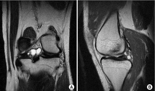
Fig. 1. MRI of the right knee showing a incomplete discoid medial meniscus with horizon- tal cleavage. (A) Coronal view and (B) Sagittal view.
Fig. 2. MRI of the left knee demonstrating the complete discoid medial meniscus with grade II intrasubstance tear.
(A) Coronal view and (B) sagittal view.
showed a discoid medial meniscus with a horizontal cleavage tear in mid-body to posterior horn and lateral meniscus was normal (Fig. 1).
Even if he had no complaints on his left knee and physical examination didn't demonstrate abnormal signs, we evaluated MRI of the opposite knee due to possi- bility of combined anomalies on the left knee. This showed a complete discoid medial meniscus with posteri- or cyst formation, which was apparent on coronal and
sagittal T2 weighted image (Figs. 2, 3).
The patient underwent arthroscopy of the his right knee, which revealed a horizontal cleavage tear in mid- dle and posterior segment of the incomplete discoid me- dial meniscus (Fig. 4) and a pathologic medial plica syndrome. Saucerization with partial meniscectomy was performed, involving removal of the central portion of the meniscus (Fig. 4) and medial patella plica was parti- ally excised. Upon finding a pathologic thick medial pa-
Fig. 3. MRI of the left knee demonstrating the complete discoid medial meniscus with posterior cyst formation. (A) Coronal view and (B) sagittal view.
Fig. 4. Arthroscopic views showing. (A, B) The incom- plete discoid medial meniscus of the right knee. F, femoral condyle; D, discoid medial me- niscus; T, tibial condyle. (C) Horizontal cleavage on probing was suspected. (D) Horizon- tally torn meniscus was res- haped by partial meniscec- tomy and saucerization.
tella plica that were impinging between patella and me- dial femoral condyle during extension, we proceeded to remove it. The patient returned to a productive level of
functions, recovering full extension of the knee and no limitations in ADL. The patient had no symptoms 2 year postoperatively.
DISCUSSION
The discoid shaped meniscus occurs almost exclusively on the lateral side of the knee. Smillie8) reported 467 had a discoid lateral meniscus and only 7 had a discoid medial meniscus in 10,000 meniscectomies. In fact, dis- coid meniscus is a relatively rare condition of the knee more frequently found in the lateral meniscus. Discoid medial menisci are even rare and bilateral discoid medial meniscus was extremely rare. 18 cases are only de- scribed in literature4,6).
The types of the discoid medial meniscus were the same in both knees in bilateral cases3). Over 99% of the medial meniscus have symmetric appearance in both knees9) and reported cases of bilateral discoid medial meniscus were of the same types except one case re- ported by Kim and Seo5). To the our knowledge, this is the 2nd case of bilateral medial discoid meniscus with asymmetric appearance.
The abnormal radiographic findings associated with medial discoid meniscus have been reported, as cupping of the medial tibial plateau on anteroposterior radio- graphs3), proximal mediotibial physis collapse and wid- ening of the medial joint margin. However, our case did not have abnormal radiographic findings. Several ano- malies related to the medial discoid meniscus have been reported, including anomalous insertion of the anterior horn of the medial meniscus into the anterior cruciate ligament7), discoid lateral meniscus in the same knee9,10), meniscal cyst and pathologic medial patella plica7). In our case, there was not only pathological medial patella plica on right knee, but also posterior cyst formation of medial meniscus on his left knee.
For the purpose of confirming bilaterality of this anomaly and subclinical pathology of opposite knee, the unaffected knee should be evaluated with MRI even if asymptomatic whenever a discoid medial meniscus is found. We believed that as Tachibana, et al.9) stated
"the reported prevalence of bilateral discoid medial me-
nisci will probably increase, because when a discoid me- dial meniscus is encountered currently, an MRI is used to find knee disorders, including in the contralateral knee". Therefore bilateral discoid medial meniscus may be much more than our prediction.
Surgical treatment of discoid menisci should be con- sidered only if the patient is symptomatic. Our patient complained of right pain associated with swelling with lack of full extension of knee but no complaint on left knee. Although stable discoid menisci without associated tears are usually asymptomatic and can be identified on- ly as incidental findings on MRI or arthroscopy2), the patients may be recommended about activity mod- ification to prevent possible injury if there are sub- clinical meniscal pathologies in the unaffected knee.
MRI can show discoid meniscus and associated con- ditions better than any other radiological methods, but arthroscopy is mandatory for confirming the diagnosis and for the treatment. In our case, arthroscopy allowed to confirm diagnosis and treat meniscal abnormalities, reshaping medial discoid on right knee. The use of MRI was helpful for the diagnosis of the unaffected knee as well as the affected knee.
CONFLICT OF INTEREST
No potential conflict of interest relevant to this article was reported.
REFERENCES
1. Dickason JM, Del Pizzo W, Blazina ME, Fox JM, Friedman MJ, Snyder SJ: A series of ten discoid medial menisci. Clin Orthop Relat Res, (168); 75-79:
1982.
2. Franceschi F, Longo UG, Ruzzini L, Simoni P, Zobel BB, Denaro V: Bilateral complete discoid medial meni- scus combined with posterior cyst formation. Knee Surg Sports Traumatol Arthrosc, 15; 266-268: 2007.
3. Fujikawa K, Tomatsu T, Matsu K: Morphological analysis of meniscus and articular cartilage in the knee
비대칭적 양측성 원판형 연골
전북대학교 의학전문대학원 정형외과학교실, 2성가롤로병원 정형외과
이주홍ㆍ왕성일ㆍ박종혁ㆍ임영진
2슬관절의 내측 원판형 연골은 매우 드물며, 특히 양측성 내측 원판형 연골은 더욱 드물어 지금까지 세계 적으로 18예가 보고되고 있다. 저자들은 자기공명영상 및 관절경 검사에서 우측 슬관절의 횡파열을 동반 한 불완전 원판형 연골과 반대측 슬관절에 대한 자기공명영상에서 후방 연골낭을 동반한 완전형 원판형 연골의 비대칭적 양측성 내측 원판형 연골 1예를 경험하였기에 보고하는 바이다.
색인 단어: 슬관절, 양측성 내측 원판형 연골, 자기공명영상 joint by means of arthrogram. J Jpn Orthop Assoc, 52;
203-215: 1978.
4. Heybeli N: Bilateral complete discoid medial meniscus:
how many cases? Associated pathologies? Knee Surg Sports Traumatol Arthrosc, 15; 1062: 2007.
5. Kim SJ, Seo YJ: Bilateral discoid medial menisci:
Incomplete type in one knee and complete type in opposite knee. Knee, 13; 255-257: 2006.
6. Lee BI, Lee YS, Kwon SW, Choi SW, Cho KH, Kwon YJ: Bilateral symptomatic discoid medial meniscus:
report of three cases. Knee Surg Sports Traumatol
Arthrosc, 15; 739-743: 2007.
7. Pinar H, Akseki D, Karaoğlan O, Ozkan M, Uluç E:
Bilateral discoid medial menisci. Arthroscopy, 16; 96- 101: 2000.
8. Smille IS: The congenital discoid meniscus. J Bone Joint Surg Br, 30; 671-682: 1948.
9. Tachibana Y, Yamazaki Y, Ninomiya S: Discoid medial meniscus. Arthroscopy, 19; E12-E18: 2003.
10. Yáñez-Acevedo A: Bilateral discoid lateral menisci and unilateral discoid medial menisci. Arthroscopy, 17; 772- 775: 2001.