120 Ann Dermatol Letter to the Editor
Received May 11, 2012, Revised May 29, 2012, Accepted for publication June 3, 2012
Corresponding author: Kwang Hyun Cho, Department of Dermatology, Seoul National University College of Medicine, 101 Daehak-ro, Jongno-gu, Seoul 110-744, Korea. Tel: 82-2-2072-2412, Fax: 82-2-742-7344, E-mail: [email protected]
This is an Open Access article distributed under the terms of the Creative Commons Attribution Non-Commercial License (http://
creativecommons.org/licenses/by-nc/3.0) which permits unrestricted non-commercial use, distribution, and reproduction in any medium, provided the original work is properly cited.
Schiefferdecker R, et al. Vasculitis and retinoids. Lancet 1989;2:494-496.
4. Reynolds P, Fawcett H, Waldram R, Prouse P. Delayed onset of vasculitis following isotretinoin. Lancet 1989;2:1216.
5. Aractingi S, Lassoued K, Dubertret L. Post-isotretinoin vasculitis. Lancet 1990;335:362.
6. Epstein EH Jr, McNutt NS, Beallo R, Thyberg W, Brody R,
Hirsch A, et al. Severe vasculitis during isotretinoin therapy.
Arch Dermatol 1987;123:1123-1125.
7. Chochrad D, Langhendries JP, Stolear JC, Godin J. Isotre- tinoin-induced vasculitis imitating polyarteritis nodosa, with perinuclear antineutrophil cytoplasmic antibody in titers correlated with clinical symptoms. Rev Rhum Engl Ed 1997;
64:129-131.
http://dx.doi.org/10.5021/ad.2013.25.1.120
Eosinophilic Pustular Folliculitis Involving Labial Mucosa, Which Improved with Naproxen
Seon-Pil Jin, Song Youn Park, Kkot Bora Yeom, You Chan Kim
1, Kwang Hyun Cho
Department of Dermatology, Seoul National University School of Medicine, Seoul,
1Department of Dermatology, Ajou University College of Medicine, Suwon, Korea
Dear Editor:
Eosinophilic pustular folliculitis (EPF) is a sterile eosino- philic infiltration of hair follicles. There are three variants, including the classic, immunosuppression-associated, and infantile type1. Although they are histologically indis- tinguishable from each other, some propose that the three types be regarded as different disease entities due to their different responses to treatment.
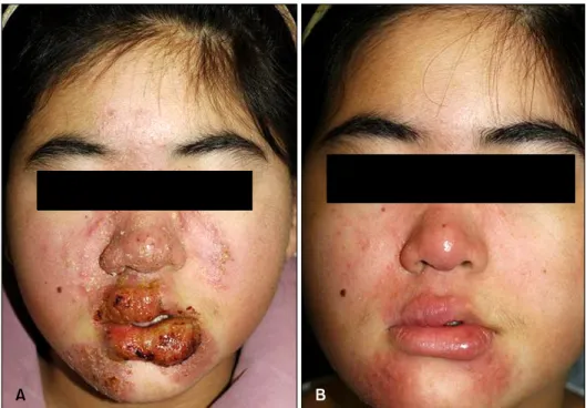
Histologically, the most diagnostic feature of EPF is the infiltration of eosinophils in hair follicles and perifollicular areas1. However, the term ‘folliculitis’ is being challenged2, on account of the fact that approximately 20% of patients with classic EPF have the disease on palms and soles1. Here, we report a case of EPF involving the labial mucosa, which improved with naproxen. An 11 year-old girl pre- sented with itching erythematous plaques and clustering pustules on the lateral side of the nose and perioral area, with erosive lesions on the external lips (Fig. 1A). A biopsy was taken from the lateral side of the nose at a district hospital. The lesion at nose showed the same feature and connected with labial lesion. It demonstrated follicular and perifollicular infiltration by eosinophils and
other inflammatory cells (Fig. 2). The diagnosis of classic EPF was made at the hospital, and she was treated with systemic prednisolone, cyclosporine, dapsone, and/or topical corticosteroid, pimecrolimus. However, her symp- toms waxed and waned over 6 months and showed improvement only with the systemic prednisolone.
Routine blood test was within normal limits without eosinophilia. Because pruritus was the prominent symp- tom rather than pain and tenderness, infectious condition was less suspected. Suspecting EPF, we started her on naproxen, 250 mg twice a day; and after three months, her symptoms greatly improved (Fig. 1B).
The etiology and pathogenesis of EPF have still yet to be fully elucidated, and there are multiple treatment options.
The utilization and efficacy of therapies seem to depend on the type of EPF2. Topical corticosteroids tend to be the first choice in all EPF variants1. In children, topical calcineurin inhibitors and oral antihistamines are also usually effective and are viewed as the first-line agents3. However, our case was recalcitrant to various treatments, including topical corticosteroids and topical calcineurin inhibitor. Only the naproxen had a remarkable effect.
Vol. 25, No. 1, 2013 121
Letter to the Editor
Fig. 1. (A) Erythematous plaques and clustering pustules on both lateral side of the nose with central clearing and peripheral extension, involving lips. (B) Greatly im- proved state after treatment with naproxen.
Fig. 2. (A) Inflammatory cell infiltration into hair follicle and around perifollicular area (H&E, ×100), (B) perifollicular eosinophilic and neutrophilic infiltration (H&E, ×400).
Nonsteroidal anti-inflammatory drugs (NSAID), particularly indomethacin, have been suggested as the treatment of choice in classic EPF2. There are few reports dealing with naproxen, a type of NSAID widely being used, on EPF4,5. We have reported the effects of naproxen as a first-line option on classic EPF in a Korean literature4. It showed 69% (11/16) of complete response rate in adult cases. This report implies that naproxen may also be safely used in children.
There has been only one case in a German literature, which involved oral mucosa in classic EPF. Our case had the labial lesions mainly on the external surface, which is
a keratinized stratified squamous epithelium rather than a true mucosa. But as there are no follicles in the external lip, as in the palms and soles, it is more likely that this disease may not be a real follicular disorder at all.
REFERENCES
1. Wolff K, Goldsmith LA, Katz SI, Gilchrest BA, Paller AS, Leffell DJ, editors. Fitzpatrick's dermatology in general medicine. 7th ed. New York: McGraw-Hill, 2008:314-315.
2. Ellis E, Scheinfeld N. Eosinophilic pustular folliculitis: a comprehensive review of treatment options. Am J Clin Dermatol 2004;5:189-197.
122 Ann Dermatol Letter to the Editor
Received November 29, 2011, Revised May 3, 2012, Accepted for publication June 9, 2012
Corresponding author: Gyong Moon Kim, Department of Dermatology, St. Vincent’s Hospital, College of Medicine, The Catholic University of Korea, 93 Jungbu-daero, Paldal-gu, Suwon 442-723, Korea. Tel:
82-31-249-7460, Fax: 82-31-242-8927, E-mail: gyongmoonkim@
catholic.ac.kr
This is an Open Access article distributed under the terms of the Creative Commons Attribution Non-Commercial License (http://
creativecommons.org/licenses/by-nc/3.0) which permits unrestricted non-commercial use, distribution, and reproduction in any medium,
provided the original work is properly cited. Fig. 1. A 0.5×0.7 cm sized, skin-colored, subcutaneous nodule of the right cheek.
3. James WD, Berger TG, Elston DM, editors. Andrew's diseases of the skin: clinical dermatology. 10th ed.
Philadelphia: WB Saunders, 2005:69-90.
4. Yeon JH, Youn SW, Kim KH, Cho KH. Clinical effects of oral
naproxen for the treatment of eosinophilic pustular folliculitis. Korean J Dermatol 2008;46:1609-1614.
5. Youn CS, Cho KH. Eosinophilic pustular folliculitis treated with naproxen. Br J Dermatol 2001;145:514-515.
http://dx.doi.org/10.5021/ad.2013.25.1.122
Myopericytoma of the Facial Cheek
Eun Kyung Kim, Ji Hyun Lee, Si Yong Kim, Gyong Moon Kim
Department of Dermatology, St. Vincent’s Hospital, College of Medicine, The Catholic University of Korea, Seoul, Korea
Dear Editor:
A 44-year-old woman presented with a painful, solitary nodule on her right cheek. She noticed the flesh-colored nodule several years prior, and the nodule expanded slowly in size. There was no history of trauma. The patient’s past medical history and family history were un- remarkable. Upon physical examination, a 0.5×0.7 cm- sized, skin-colored, firm nodule on the right cheek was seen (Fig. 1). The biopsy specimen revealed a concentric perivascular proliferation of blank, spindle-shaped myoid- appearing cells (Fig. 2A, B). Immunohistochemical stain showed diffuse immunoreactivity on smooth muscle actin and was negative for desmin (Fig. 2C, D). The CD34 stain highlighted only the endothelium of the vessel, but the perivascular concentric myoid tumor cells were not immu- noreactive (Fig. 2E). From the clinicopathological fin- dings, the diagnosis of myopericytoma (MPC) was made.
MPC is a rare, recently delineated tumor that originates from the perivascular myoid cells1. It was described by Granter et al.2 in 1998 and newly entered into a subgroup of perivascular tumors in the World Health Organization classification of soft tissue tumors3. MPCs are well-circum-
scribed and composed of a mixture of solid cellular areas intermixed with variable numbers of vascular channels.
The latter are often elongated and display prominent bran- ching, resulting in a stag-horn appearance. The cells in the solid areas are round, or short and spindle shaped with eosinophilic cytoplasm and vesicular nuclei. The presence of concentric layers of tumor cells around vascular chann- els resulting in a typical onion ring appearance is a hall mark of this tumor2. However, MPC tumors exhibits a broad spectrum of growth patterns: solid classic, heman- giopericytoma-like, angioleiomyoma-like, hypocellular fib- roma-like, solitary fibrous tumor-like, glomus tumor-like, cellular immature, intravascular and malignant subtypes.
The present case was classified as angioleiomyoma-like pattern. By immunohistochemistry, the neoplastic cells