Original Article: Biochemistry/Molecular Biology
Micronucleus Test for the Classification of Chemical Mutagenicity according to Globally Harmonized System
Kyung-Taek Rim* · Hyeon-Yeong Kim · Yong-Hyun Chung
Received: 23 May 2013 / Accepted: 11 June 2013 / Published Online: 31 December 2013
© The Korean Society for Applied Biological Chemistry 2013
Abstract To classify the chemical hazard according to globally harmonized system of classification and labeling of chemicals (GHS), we investigated the genotoxicity of three chemicals, methyl myristate, 2-ethylhexanoic acid zinc salt, N,N,N',N'- tetrakis(2-hydroxyethyl) ethylenediamine, using male ICR mice bone marrow cells for the screening of micronucleus induction.
Although these three chemicals have already been tested numerous times, a micronucleus test has not been conducted. The seven week-old male ICR mice were tested at three dosages for the three chemicals, respectively. After 24 h of oral administration with the three chemicals, the mice were sacrificed and their bone marrow cells were prepared for smearing slides. As a result of counting the micronucleated polychromatic erythrocyte (MNPCE) of 2,000 polychromatic erythrocytes, all treated groups expressed no statistically significant increase of MNPCE compared to the negative control group. There were no clinical signs related with the oral exposure of these three chemicals. It was concluded that these three chemicals did not induce micronucleus in the bone marrow cells of ICR mice, and there was no direct proportion with dosage. These results indicate that the three chemicals have no mutagenic potential under each test condition, and it is not classified these chemicals as mutagens by GHS.
Keywords globally harmonized system of classification and labeling of chemicals · methyl myristate · micronucleus · mutagenicity · N,N,N',N'-tetrakis(2-hydroxyethyl) ethylenediamine
· 2-ethylhexanoic acid zinc salt
Introduction
Chemical substances, whether single or combined in mixtures, may have various hazardous effects on human health or environment. Some of them may be carcinogenic, mutagenic or reproductive toxic, which is categorized as “CMRs”. In view of the hazards they present, these classified substances and mixtures are subject to restrictive regulations, particularly in the workplace.
The necessity for a chemical hazard assessment has increased because the number of workers exposed to chemicals has risen with the development of many industries, and it is also necessary to discover what these substances are and how they are regulated.
Carcinogens are substances and preparations which, if they are inhaled, ingested or penetrated the skin, may induce cancer or increase its incidence. Mutagens are substances may induce heritable genetic defects or increase their incidence. The mutagens cause permanent changes in the amount or structure of the genetic material in a cell, and these chemicals in accordance with UN GHS shall be considered mutagens for purposes of this article.
Cellular mutagens/genotoxins are substances that cause heritable changes in the genetic material in cells (ECVAM, 2002; UNECE, 2004). The term mutagen refers to a substance that induces transmissible changes in DNA structure involving a single gene or a group of genes. Genotoxins are a broader category of substances that induce changes to the structure or number of genes via chemical interaction with DNA and/or non-DNA targets (Maurici et al., 2005). At the early testing stages, the genotoxicity assays for predicting potential heritable cellular damage are the same as used for predicting carcinogenicity because the endpoints measured in genotoxicity tests are common precursors for both of these adverse health outcomes. The classification of cellular mutagenicity is hazarding based, taking into account a chemical’s intrinsic ability to induce genotoxicity in cells, and is not meant for quantitative risk assessment. Category 1 chemicals are “known to induce heritable mutations or to be regarded as if they induce heritable mutations in the cells of humans,” and Category 2 chemicals are those that “cause concern for humans owing to the possibility that K. -T. Rim · H. -Y. Kim · Y. -H. Chung
Center for Chemical Safety and Health, Occupational Safety and Health Research Institute, Korea Occupational Safety and Health Agency, Daejeon, Republic of Korea
*Corresponding author (K. -T. Rim: [email protected])
they may induce heritable mutations in the cells of humans”
(UNECE, 2004). Substances that induce heritable damage in animal germ cells are regarded as suspect human cellular mutagens as well as possibly having implications for carcinogenicity.
The globally harmonized system of classification and labeling of chemicals (GHS) is an internationally agreed-upon system, created by the United Nations (UK government, 2012). This classification system is a complex with data obtained from tests, literature, and practical experience. It is designed to replace the various classification and labeling standards used in different countries by using consistent criteria on a global level. Its development began at the UN Rio Conference in 1992, when the International Labor Organization, the Organization for Economic Co-operation and Development (OECD), various governments and other stakeholders met at a UN conference. It supersedes the relevant European Union (EU) and US standards. Classification of chemicals with repeated dose, mutagenicity and much other toxicity testing according to GHS are used for the purpose of classification for all substances, which are placed on the market.
Genotoxicity/mutagenicity testing is conducted for pharmaceuticals, industrial chemicals, consumer products and the results are used to classify chemicals for heritable cellular mutagenicity as well as carcinogenicity.
Most regulatory agencies and international authorities recommend a test scheme consisting of in vitro and in vivo methods to identify genotoxic/mutagenic substances. A tiered test scheme would likely start with computer-based prediction using (quantitative) structure-activity relationships and in vitro testing. The GHS recommends use of OECD Test Guidelines for genotoxicity testing and states that “if new, well validated, tests arise these may also be used in the total weight of evidence to be considered”
(UNECE, 2004). There is a need to evaluate diverse types of biological alterations in order to thoroughly assess the genotoxic/
mutagenic potential of a substance; this requires the use of a battery of tests. This article focuses on the role of mutagenicity testing in the prediction of cellular damage, and classifies the mutagenicity according to GHS. The OECD TGs available for in vivo and in vitro genotoxicity and mutagenicity testing are listed in OECD Guidelines for the Testing of Chemicals, Section 4 Health Effects (OECD, 2011).
To classify the chemical mutagenicity according to GHS, the in vivo micronucleus test was performed on mammalian bone marrow cells treated with three chemicals of methyl myristate (CAS No. 124-10-7), 2-ethylhexanoic acid zinc salt (CAS No.
136-53-8), N,N,N',N'-tetrakis(2-hydroxyethyl) ethylenediamine (CAS No. 140-07-8) for which the definitive information is insufficient. Although many toxicological studies have been conducted other than the micronucleus test, the available genotoxic data on these three chemicals are still controversial with and without mammalian metabolic activation (S9). So it was necessary for further study according to Good Laboratory Practice guideline to secure quality assurance of the test. The purpose of this micronucleus test is to screen the cytogenetic damage that results in the formation of micronuclei containing lagging
chromosome fragments or whole chromosomes. Micronuclei were first used to quantify chromosomal damage and are now recognized as one of the most successful and reliable assays for genotoxic carcinogens (Scott and Evans, 1967).
The toxicological information on these three chemicals as gained in this study will be used to promote workers’ rights to know and to prepare or update the material safety data sheet with GHS. This study will also contribute to improving the testing of chemicals by generally used genotoxicity testing methods, as well as investigations on the underlying mechanism and the interpretation of genotoxicity data on hazardous chemicals.
The physico-chemical and toxicological information of these three chemicals are shown in Table 1.
Materials and Methods
Chemicals and animal feeding conditions. Methyl myristate (Acros Organics, Belgium), 2-ethylhexanoic acid zinc salt (Alfa Aesar, USA) and N,N,N',N'-tetrakis(2-hydroxyethyl) ethylenediamine (Acros Organics, Belgium) were used as the test chemicals. Corn oil (Sigma, USA) was used as a solvent in case of the two former, and water for injection (Choongwae Pharma Corp., Korea) of the other one according to the results of the solubility test. The positive control used mitomycin C (MMC) (Sigma, USA).
These entire three test substances are liquid forms that have molecular weight 242.4, 351.8 and 236.3, respectively.
Animals and experimental design. The mouse (Mus musculus) bone marrow micronucleus test was carried out according to OECD guidelines, TG 474 (OECD, 1997). Groups of specific pathogen free male ICR mice were treated with the test substance at three dosage levels, the highest dosage level being the estimated maximum tolerated dose or the standard limit dose for the micronucleus test, whichever is least. Concurrent negative and positive control groups were also treated.
According to the OECD test guideline (OECD, 2011), the dose- setting criteria should be evaluated on a case-by-case basis. A range finding study was performed because there are no suitable data available, it should be performed in the same conditions and settings to be used in the main study. The highest dose is defined as the dose producing signs of toxicity such that higher dose levels would be expected to produce lethality, may also be defined as a dose that produces some indication of toxicity of the bone marrow. In this study, It was performed using 7 week-old male ICR mice at 500, 1,000, and 2,000 mg/kg with two (the first and third chemicals) and at 187.5, 375, 750 mg/kg with second chemical, respectively. At 24 h after treatment with these chemicals administered orally there were normally 6 male animals per group. The experimental animal room was maintained at a temperature of 22.7–23.9 and relative humidity of 44.6–47.9%.
The animal studies were approved by an animal ethics committee to ensure that appropriate animal care before the animals was obtained for research (Approval No. 06138, 06109, 06141).
Bone marrow preparation and micronucleus test. Bone
marrow cells were obtained from the femurs immediately following sacrifice. Immature erythrocytes could be differentiated using a variety of staining techniques that rely on their relatively high content of residual DNA. 5% Giemsa was used for mouse bone marrow/peripheral blood and stained immature erythrocytes blue, while the mature erythrocytes with low nucleic acid content appeared pinkish orange. Based on the cell cycle and maturation times of the erythrocytes, the bone marrow was harvested after 24 h. The bone marrow was flushed from the femurs and spread onto slides. The slides were air-dried, fixed, and stained with a fluorescent DNA specific stain that easily illuminates any micronuclei that may be present. The 2000 polychromatic erythrocytes (PCEs, reticulocytes; immature erythrocytes) were scored per animal for the frequency of micronucleated cells in each of the 6 animals per dosage group. In addition, the percentage of PCEs among the 500 erythrocytes in the bone marrow was scored for each dosage group as an indicator of chemical-induced toxicity.
The presence of micronucleated polychromatic erythrocytes was visually scored (at least 2000 cells per mouse) by optical microscopy using a fluorescence microscope (BX51, Olympus, Japan). Cells were considered to be micronucleated when they neatly contained defined chromatin corpuscles with a diameter of less than one-third the diameter of the cell nucleus and stained equal or lighter than the nucleus of the cell from which the micronucleated cell was developed.
Evaluation and data analysis. Data were presented as the mean
number of micronucleated cells per 2000 cells for each treatment group. The final conclusion for a micronucleus test was determined in consideration of the results of the statistical analyses.
The experimental and control micronucleus frequency data were tested statistically by the Kastenbaum-Bowman method (5%) (Kastenbaum and Bowman, 1970), t-test (5%) and the ANOVA test (5%) using the SigmaStat v. 3.11.
Results
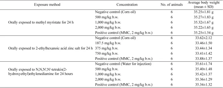
Animal body weights with oral exposure to 3 chemicals. There were no specific symptoms among animals orally exposed to methyl myristate, 2-ethylhexanoic acid zinc salt and N,N,N',N'- tetrakis(2-hydroxyethyl) ethylenediamine. The ranges of body weights of animals exposed to these three chemicals were 35.22 to 35.32 g, 33.38 to 33.62 g, and 35.34 to 35.61 g, respectively (Table 2).
Frequencies of micronucleus induction and cytotoxicity. The preliminary tests were performed as a limit test to determine the maximum dosage. The inhibition of proliferation in the bone marrow cells was not observed in these tests for the two chemicals.
The ratios of erythrocytes with micronucleus induction (MNPCE) were 0.7±0.8, 0.5±0.5, 0.5±0.5 and 0.3±0.5 counts/2,000 PCE cells in the negative control group, 500, 1,000 and 2,000 mg/kg Table 1 Physico-chemical and toxicological information of three chemicals be tested
Methyl myristate (CAS No. 124-10-7) 2-Ethylhexanoic acid zinc salt (CAS No.
136-53-8)
N,N,N',N'-Tetrakis(2-hydroxyethyl) ethylenediamine (CAS No. 140-07-8)
Uses
Intermediate for myristic acid detergents, emulsifiers, wetting agents, stabilizers, resins, lubricants, plasticizers, textiles, animal feeds, standard for gas chromatography, flavouring
Drying agent for paints, inks, enamels and varnishes, Wood protectant, Engine coolant anti-corrosive, stabilizer in plastic, emulsifier for cosmetics
Laboratory chemicals, Manufacture of substances
Physico-chemical properties
m.p. 19oC b.p. 295oC f.p. >112oC
water solubility - insoluble log Kow 6.41
M.W. 242.42
m.p. 115oC b.p. 219oC solubility 0.92 mg/L log Kow 4.47 M.W. 351.81
m.p. 155.76oC b.p. 414.75oC f.p. 99oC log Kow -3.75 M.W. 184.31
Toxicological Properties
Acute oral LD50 >2,000 mg/kg (Rat) Severe Skin irritation (Rabbit) Mild Eye irritation (Rabbit)
Ames test (S. typhimurium) - negative
No data Severe Skin irritation (Rabbit)
Environmental Properties
Fish LC50 1700 mg/L (96 hr, Danio rerio) Daphnia Magna LC50 0.025 mg/L (48 h) Algae EC50 0.018 mg/L (96 h)
B.C.F. 1100
B.C.F. 17.43 (estimate) Non-degradable Koc 869
B.C.F. 3.162
GHS classification
Skin irritation cat. 2; Respiratory sensitization cat. 1; Skin sensitization cat.
1; Germ cell mutagenicity cat. 2; Chronic marine environmental hazard cat. 4
Chronic marine environmental hazard cat. 4
Eye irritation cat. 2; Chronic marine environmental hazard cat. 4
m.p. melting point; b.p. boiling point; f.p. flash point; log Kow octanol-water partition coefficient; M.W. molecular weight; LD50 half-lethal dose; LC50 half-lethal concentration; B.C.F. bioconcentration factor
*Mostly referred from material safety data sheets information in KOSHANET (http://www.kosha.or.kr/bridge?menuID=69). Searches were conducted using keywords chemical name AND/OR CAS number.
methyl myristate treated group, respectively. Positive control was 168.7±4.0 counts/2,000 PCE cells. The ratios of PCEs (polychromatic erythrocytes) within total erythrocytes were 0.474±0.049, 0.467±
0.057, 0.457±0.043 and 0.458±0.028 counts in the negative control group, 500, 1,000 and 2,000 mg/kg methyl myristate treated group, respectively. Statistically significant changes were not observed compared with the negative control group (Table 3).
The ratios of erythrocytes with micronucleus induction were 1.3±0.8, 0.7±0.5, 0.8±1.0 and 1.0±0.9 counts/2,000 PCE cells in the negative control group, 187.5, 375 and 750 mg/kg 2- ethylhexanoic acid zinc salt treated group, respectively. Positive control was 156.8±9.3 counts/2,000 PCE cells. The ratio of PCEs within total erythrocytes were 0.487±0.031, 0.482±0.052, 0.496±
0.027 and 0.512±0.035 counts in the negative control group, 187.5, 375 and 750 mg/kg 2-ethylhexanoic acid zinc salt treated group, respectively. There were also no statistically significant changes observed compared with the negative control group (Table 4).
The ratios of erythrocytes with micronucleus induction (MNPCE) were 0.8±0.4, 1.5±0.5, 0.8±0.8 and 0.7±0.8 counts/2,000 PCE cells in the negative control group, 500, 1,000 and 2,000 mg/kg N,N,N',N'-tetrakis(2-hydroxyethyl)ethylenediamine treated group, respectively. Positive control was 158.2±13.2 counts/2,000 PCE cells. The ratios of PCEs (polychromatic erythrocytes) within total erythrocytes were 0.506±0.042, 0.501±0.042, 0.516±0.045 and 0.499±0.035 counts in the negative control group, 500, 1,000 and Table 2 Animal body weight in micronucleus tests with oral exposure to three chemicals
Exposure method Concentration No. of animals Average body weight
(mean ± SD)
Negative control (Corn oil) 6 35.25±1.81 g
500 mg/kg b.w. 6 35.27±1.83 g
Orally exposed to methyl myristate for 24 h 1,000 mg/kg b.w. 6 35.32±1.67 g
2,000 mg/kg b.w. 6 35.22±1.65 g
Positive control (MMC, 2 mg/kg b.w.) 6 35.25±1.54 g
Negative control (Corn oil) 6 33.62±2.12
187.5 mg/kg b.w. 6 33.46±1.50
Orally exposed to 2-ethylhexanoic acid zinc salt for 24 h 375 mg/kg b.w. 6 33.44±1.34
750 mg/kg b.w. 6 33.41±1.42
Positive control (MMC, 2 mg/kg b.w.) 6 33.88±1.37 Negative control (Water for injection) 6 35.61±1.74 Orally exposed to N,N,N',N'-tetrakis(2-
hydroxyethyl)ethylenediamine for 24 hours
500 mg/kg b.w. 6 35.40±1.48
1,000 mg/kg b.w. 6 35.42±1.37
2,000 mg/kg b.w. 6 35.36±1.29
Positive control (MMC, 2 mg/kg b.w.) 6 35.34±1.32 MMC: mitomycin C. b.w: body weight. All values are expressed as mean ± SD.
Table 3 Results of the main micronucleus test with methyl myristate (for 24 h)
Groups Counted *PCE Counted ‡NCE PCE/(PCE+NCE) †MNPCE/PCE
Negative control 237.2±24.5 262.8±24.5 0.474±0.049 0.7±0.8
500 mg/kg b.w. 233.67±28.71 266.33±28.71 0.467±0.057 0.5±0.5
1,000 mg/kg b.w. 0228.5±21.27 0271.5±21.27 0.457±0.043 0.5±0.5
2,000 mg/kg b.w. 00.229±13.80 0.0271±13.80 0.458±0.028 0.3±0.5
Positive control 0215.5±11.15 0284.5±11.15 0.431±0.022 168.7±4.000
*PCE: polychromatic erythrocyte. ‡NCE: normochromatic erythrocyte. †MNPCE: micronucleated polychromatic erythrocyte.
All values are expressed as mean ± SD.
b.w: body weight.
Table 4 Results of the main micronucleus test with 2-ethylhexanoic acid zinc salt (for 24 h)
Groups Counted *PCE Counted ‡NCE PCE/(PCE+NCE) †MNPCE/PCE
Negative control 243.67±15.71 256.33±15.71 0.487±0.031 1.3±0.8
187.5 mg/kg b.w. 240.83±26.25 259.17±26.25 0.482±0.052 0.7±0.5
375 mg/kg b.w. 247.83±13.30 252.17±13.30 0.496±0.027 0.8±1.0
750 mg/kg b.w. 255.83±17.49 244.17±17.49 0.512±0.035 1.0±0.9
Positive control 00.255±16.35 00.245±16.35 0.510±0.033 156.8±9.300
*PCE: polychromatic erythrocyte. ‡NCE: normochromatic erythrocyte. †MNPCE: micronucleated polychromatic erythrocyte.
All values are expressed as mean ± SD.
b.w: body weight.
2,000 mg/kg N,N,N',N'-tetrakis(2-hydroxyethyl)ethylenediamine treated group, respectively. Statistically significant changes were also not observed compared with the negative control group (Table 5).
These three chemicals did not inhibit the bone marrow cell proliferation in all treated groups, and did not make the micronucleus induction. Based on the results of these tests it is concluded that these three chemicals are not mutagenic in the mammalian bone marrow micronucleus test, and it cannot be classified any categories in mutagenicity of GHS.
Discussion
The GHS defines many hazards. There are three main categories:
physical hazards, and health & environmental hazards (United Nations, 2011). The physical hazards are related to damages that can occur due to heat, over-pressurization, or radiation exposure.
Physical hazards include explosives, flammables, oxidizers, gases under pressure, self-reactive substances, pyrophoric substances, and corrosives. The other category is health & environmental hazards. These hazards deal with damage to a physical body from contact or chemical reaction. These include acute toxicity, skin irritation, eye damage, respiratory or skin sensitization, germ cell mutagenicity, carcinogenicity, reproductive toxicity, and hazards to aquatic life (Willey, 2012). In 2012, the Ministry of Employment and Labor in Korea were classified and reported the industrial mutagens as governmental notification (MoEL, 2012).
In vivo tests for assessing potential heritable genotoxicity include that heritable cell mutagenicity tests that include a component that measures damage passed onto progeny are that the mouse heritable translocation test (OECD Test Guideline (TG) 485), the mouse specific locus test, and the rodent dominant lethal test (OECD TG 478). Assays for measuring genotoxicity induction in germ cells that are used to predict chemicals that might induce heritable damage include the mammalian spermatogonial chromosome aberration test (OECD TG 483), the spermatid micronucleus assay, the mammalian oocyte chromosome aberration/aneuploidy test, and unscheduled DNA synthesis test in testicular cells.
Assays for measuring genotoxicity induction in somatic cells that are used to predict whether a chemical has the potential to induce cell genotoxicity include the mammalian erythrocyte micronucleus test (OECD TG 474), the mammalian bone marrow chromosome
aberration test (OECD TG 475), the liver unscheduled DNA synthesis (UDS) (OECD TG 486), and the mouse spot test (OECD TG 484) which measures genotoxicity in fetal somatic cells.
In addition to the standard in vitro mammalian cell genotoxicity tests, there are in vitro assays for measurement of genotoxicity in primary germ cells but these are not standardized or validated;
there are no ongoing coordinated activities to address this at this time. The European Centre for the Validation of Alternative Methods (ECVAM) panels estimated that total replacement of animal testing for genotoxicity/mutagenicity at the EU level would take at least 12 years and require models for evaluating toxicokinetics and metabolism (Maurici et al., 2005). The Comet assay, both in vitro and in vivo, is being developed and validated for regulatory purposes. Recommendations for standardizing protocols were published (Tice et al., 2000), and an ECVAM panel provided additional recommendations for completing validation (Maurici et al., 2005). The Japanese Center for the Validation of Alternative Methods (JaCVAM) is leading the validation of the Comet assay. The Comet assay involves an electrophoretic method to detect DNA strand breaks and alkaline-labile DNA lesions in cells. It is commonly used for eukaryotic cells owing to its versatility (Yu and Dashwood, 2007).
Other in vitro methods for genotoxicity/mutagenicity testing are in various stages of development and validation, yet assays specifically to address effects in germ cells are not a current focus.
An integrated testing scheme for genotoxicity and carcinogenicity has been proposed to address the anticipated increase in testing requirements under the EU Registration, Evaluation, Authorization and Restriction of Chemicals (Combes et al., 2007). An ECVAM panel proposed that total replacement of animal testing for genotoxicity/mutagenicity would require models for evaluating toxicokinetics and metabolism (Maurici et al., 2005). In vitro genotoxicity tests also need to be modified to use cell lines relevant to the target organs of interest, which for the purposes of predicting heritable germ cell damage would require standardization and validation of in vitro assays in mammalian germ cells.
Although there are several chemical classifications for regulating these substances and their use in terms of their mutagenic properties, only one of these classifications is included in a regulation. It is the european classification provided for under what is referred to as the CLP regulation for classification, labeling and packaging (European Union, 2008). Genetic toxicologists as Table 5 Results of the main micronucleus test with N,N,N',N'-tetrakis(2-hydroxyethyl)ethylenediamine (for 24 h)
Groups Counted *PCE Counted ‡NCE PCE/(PCE+NCE) †MNPCE/PCE
Negative control 252.83±21.08 247.17±21.08 0.506±0.042 0.8±0.4
500 mg/kg b.w. 250.33±20.97 249.67±20.97 0.501±0.042 1.5±0.5
1,000 mg/kg b.w. 00.258±22.57 00.242±22.57 0.516±0.045 0.8±0.8
2,000 mg/kg b.w. 249.67±17.55 250.33±17.55 0.499±0.035 0.7±0.8
Positive control 245.17±20.07 254.83±20.07 0.490±0.040 158.2±13.20
*PCE: polychromatic erythrocyte. ‡NCE: normochromatic erythrocyte. †MNPCE: micronucleated polychromatic erythrocyte.
All values are expressed as mean ± SD.
b.w: body weight.
experts should consider data quality and reliability, and give a critical review of all available information for support of classification. A weight of evidence approach is also required to assess mutagenic potential of chemicals (Morita et al., 2009).
Another use of tiered testing at EPA, and within other governmental regulatory programs globally is found in the evaluation of chemical mutagenicity (Brusick et al., 2008). Here initial testing typically focuses on in vitro tests of mutagenicity and progresses to more sophisticated in vivo testing, with the types of tests performed chosen based on results from the in vitro studies (Plunkett et al., 2010).
There are three tests typically used to classify the mutagenicity with GHS category using in industrial chemicals, reverse mutation (Ames) test, in vitro chromosomal aberration test and in vivo micronucleus test. The objective of in vivo micronucleus test are to evaluate the test article for in vivo clastogenic activity and/or disruption of the mitotic apparatus by detecting micronuclei in PCE in mouse bone marrow (Heddle et al., 1983; 1991; Schmid, 1975). The micronucleus assay is now recognized as one of the most successful and reliable assays for genotoxic carcinogens because the micronucleus (MN) formation results either from chromosome breakage (clastogenicity) or aneuploidy. By using pancentromeric probes, it is possible to draw conclusions if MN is formed as a consequence of chromosomal breakage (clastogenicity) or aneuploidy (Kim et al., 2010).
In the dose range-finding assay, the test article is formulated in vehicle and administered once by oral gavages to three males and three females per dose level. The animals are usually dosed at 500, 1,000, or 2,000 mg/kg and observed for up to 2 days after dosing for toxic signs and/or mortality. Data analysis is performed using an analysis of variance (Winer, 1971) on untransformed proportions of cells with micronuclei per animal on untransformed PCE: normochromatic erythrocytes (NCE) ratios when the variances were homogeneous. Ranked proportions are used for heterogeneous variances. If the analysis of variance was statistically significant, Dunnett’s t-test (Dunnett, 1955; 1964) are used to determine which dose groups, if any, are statistically significantly different from the control. Analyses are performed separately for each sampling time.
In this paper, we performed these in vivo micronucleus tests based on the results of the dose range-finding assay; the maximum dose was estimated to be 2,000 mg/kg, the limit dose based on regulatory guidelines. Bone marrow was extracted and at least 2000 PCEs per animal were analyzed for the frequency of micronuclei. Cytotoxicity was assessed by scoring the number of PCEs and NCEs in at least the first 500 total erythrocytes for each animal. The test article did not induce signs of clinical toxicity in the animals treated at the highest dose level (the limit dose based on regulatory guidelines). The test article did not induce statistically significant increases in micronucleated PCEs at any test article doses. In addition, the test article was not cytotoxic to the bone marrow (i.e., did not produce statistically significant decreases in the PCE:NCE ratio) at any dose of the test article.
These results indicate that these test chemicals have no mutagenic
potential under each test condition, and it does not classify them as mutagens by GHS categories. Although it is not mutagen, the tested compounds pose a risk for irritation or sensitization of the eye or skin, it should be kept in mind that dermal or ocular uptake, acute and chronic toxicity by dermal contact have not been tested so far.
These compounds should be further evaluated in integrated testing approaches for dermal or optical exposure. Additional studies focusing on lung exposure and long-term effects of these low-level contaminants are also needed to improve assessment of hazardous effects to predict risks for human health.
Acknowledgments The in vivo micronucleus tests in this article were performed by Biotoxtech (Korea) with supported by the Korea Occupational Safety and Health Agency (KOSHA), the Ministry of Employment and Labor, Republic of Korea and Grant-in-Aid for chemical hazard assessment.
References
Brusick DJ, Fields WR, Myhr BC, and Doolittle DJ (2008) Chapter 23:
Genetic toxicology. In Principles and Methods of Toxicology , Wallace Hayes A. (5th ed ), pp. 1195–6. Informa Healthcare USA Inc., USA.
Combes R, Grindon C, Cronin MT, Roberts DW, and Garrod J (2007) Proposed integrated decision-tree testing strategies for mutagenicity and carcinogenicity in relation to the EU REACH legislation. Altern Lab Anim 35, 267–87.
Dunnett CW (1955) A multiple comparisons procedure several treatments with a control. J Am Stat Assoc 50, 1096–121.
Dunnett CW (1964) New tables for multiple comparisons with a control.
Biometrics 20, 482–91.
ECVAM (2002) Genotoxicity and carcinogenicityAltern Lab Anim 30, Suppl.
1, 83–93.
European Union, Regulation (EC) (2008) No 1272/2008 of the European Parliament and of the councel of 16 December 2008 on classification, labelling and packaging of substances and mixtures, amending and repealing Directives 67/548/EEC and 1999/45/EC, and amending Regulation (EC) No 1907/2006., EU
Heddle JA, Cimino MC, Hayashi M, Romagna F, Shelby MD, Tucker JD et al. (1991) Micronuclei as an index of cytogenetic damage: past, present, and future. Environ Mol Mutagen 18, 277–91.
Heddle JA, Hite M, Kirkhart B, Larson K, MacGregor JT, Newell GW et al.
(1983) The induction of micronuclei as a measure of genotoxicity. A report of the US Environmental Protection Agency Gene-Tox Program.
Mutat Res 123, 61–118.
Kastenbaum MA and Bowman KO (1970) Tables for determining the statistical significance of mutation frequencies. Mutat Res 9, 527–49.
Kim SJ, Rim KT, Kang MG, Kim JK, Chung YH, and Yang JS (2010) A Study of Micronucleus Induction with Methyl Formate and 2- Methylbutane in Bone Marrow Cells of Male ICR Mice. Saf Health Work 1, 80–6.
Maurici D, Aardema M, Corvi R, Kleber M, Krul C, Laurent C et al. (2005) Genotoxicity and mutagenicity. Altern Lab Anim 33, Suppl. 1, 117–30.
Ministry of Employment and Labor (MoEL) (2012)Governmental Notification No. 2012–31. Exposure limits of chemicals and physical agents, Korea.
Morita T, Hayashi M, Nakajima M, Tanaka N, Tweats DJ, Morikawa K et al.
(2009) Practical issues on the application of the GHS classification criteria for germ cell Mutagens. Regul Toxicol Pharmacol 55, 52–68.
OECD (2011) OECD Guidelines for the Testing of Chemicals, Section 4 Health Effects. OECD iLibrary, France
Plunkett LM, Kaplan AM, and Becker RA (2010) An enhanced tiered toxicity testing framework with triggers for assessing hazards and risks of commodity chemicals. Regul Toxicol Pharmacol 58, 382–94.
Schmid W (1975) The micronucleus test. Mutat Res 31, 9–15.
Scott D and Evans HJ (1967) X-ray-induced chromosomal aberrations in vicia faba: changes in response during the cell cycle. Mutat Res 4, 579–99.
Tice RR, Agurell E, Anderson D, Burlinson B, Hartmann A, Kobayashi H et al. (2000) Single cell gel/comet assay: Guidelines for in vitro and in vivo genetic toxicology testing. Environ Mol Mutagen 35, 206–21.
UK government (2012) Health and Safety Executive. United Nations Globally Harmonised System of Classification and Labelling of Chemicals (GHS). HSE, UK.
United Nations Economic Commission for Europe (UNECE) (2004) Globally Harmonized System of classification and labeling of chemicals (GHS).
In Part 3. Health and environmental hazards. Chapter 3.5. Germ cell
mutagenicity. pp. 155–62, UNECE, USA.
United Nations (UN) (2011) Globally Harmonized System of Classification and Labeling of Chemicals (GHS). 4th revised edition., USA
Willey RJ (2012) International Symposium on Safety Science and Technology, Understanding a safety data sheet (SDS) in regards to process safety.
Procedia Engineering 45, 857–67.
Winer BJ (1971) In Statistical Principles in Experimental Design, (2nd ed.).
McGraw-Hill, USA.
Yu TW and Dashwood RH (2007) Measuring antigenotoxic effects using the Ames test and Comet assay. Am Biotech Lab 25, 22–3.