Background and Purpose Responses to repetitive nerve stimulation (RNS) in patients with muscle-specific tyrosine kinase (MuSK) antibody (Ab)-positive myasthenia gravis (MG) vary depending on the muscles tested. We analyzed the RNS responses of limb and facial muscles in MuSK-Ab-positive and acetylcholine receptor (AChR)-Ab-negative MG (MuSK MG) and MuSK-Ab-negative and AChR-Ab-negative [double-seronegative (DSN)] MG pa- tients.
Methods We retrospectively compared RNS responses between 45 MuSK MG and 29 DSN MG. RNS was applied to the abductor digiti minimi, flexor carpi ulnaris, trapezius, orbicu- laris oculi, and nasalis muscles.
Results Abnormal RNS responses in limb muscles were observed in 22.2 and 58.6% of MuSK MG and DSN MG patients, respectively, with abnormal facial responses observed in 77.8 and 65.5%, and abnormal responses observed in any of the five muscles in 86.7 and 72.4%. Abnormal RNS responses in the abductor digiti minimi or flexor carpi ulnaris were less frequent in MuSK MG (8.9 and 15.6%, respectively) than in DSN MG (37.9 and 55.2%), whereas the findings for other muscles were not significantly different between the groups.
Abnormal facial responses but normal limb responses were independently associated with MuSK MG (odds ratio=5.224, 95% confidence interval=1.300–20.990).
Conclusions Abnormal RNS responses primarily in facial muscles without involvement of limb muscles were more pronounced in MuSK MG than in DSN MG. RNS of both facial and limb muscles in AChR-Ab-negative MG can increase the test sensitivity and aid in early suspi- cion of MuSK MG.
Key Words MuSK, muscle-specific tyrosine kinase, myasthenia gravis, repetitive nerve stimulation, acetylcholine receptor.
Repetitive Nerve Stimulation in MuSK-Antibody-Positive Myasthenia Gravis
INTRODUCTION
Myasthenia gravis (MG) is a relatively rare autoimmune disorder characterized by fatiga- ble weakness of voluntary muscles.1,2 Since impaired neuromuscular transmission is the main pathomechanism of MG, identifying this is essential in an MG diagnosis. Therefore, electrodiagnostic studies including repetitive nerve stimulation (RNS) testing are impor- tant diagnostic tools, especially in patients without antibodies (Abs) to the acetylcholine receptor (AChR).
Typically 40–70% of generalized MG patients without AChR Abs have Abs to muscle- specific tyrosine kinase (MuSK).3-6 The few studies in which RNS was performed in patients with MuSK-Ab-positive MG produced wide ranges for the proportion of patients with ab- normal responses (12% to 86%).7-10 These previous studies were heterogeneous with respect to the nerve–muscle sets studied in the RNS: whereas some studies performed RNS only in Seung Woo Kima*
Mun Kyung Sunwoob* Seung Min Kima Ha Young Shina Il Nam Sunwooa
a Department of Neurology, Yonsei University
College of Medicine, Seoul, Korea
b Department of Neurology, Bundang Jesaeng General Hospital, Seongnam, Korea
pISSN 1738-6586 / eISSN 2005-5013 / J Clin Neurol 2017;13(3):287-292 / https://doi.org/10.3988/jcn.2017.13.3.287
Received January 9, 2017 Revised March 9, 2017 Accepted March 9, 2017 Correspondence Ha Young Shin, MD Department of Neurology,
Yonsei University College of Medicine, 50-1 Yonsei-ro, Seodaemun-gu, Seoul 03722, Korea
Tel +82-2-2228-1600 Fax +82-2-393-0705 E-mail [email protected]
*These authors contributed equally to this work.
cc This is an Open Access article distributed under the terms of the Creative Commons Attribution Non-Com- mercial License (http://creativecommons.org/licenses/by-nc/4.0) which permits unrestricted non-commercial use, distribution, and reproduction in any medium, provided the original work is properly cited.
JCN
Open Access ORIGINAL ARTICLERepetitive Nerve Stimulation in MuSK MG
JCN
limb muscles,3,7 others performed RNS in both limb and fa- cial muscles.5,10-12 Also, most previous studies had small pop- ulations.5,6,9
The aim of the present study was to determine the differ- ence in sensitivity of RNS according to the tested muscles and reveal any distinctive RNS features of MuSK-Ab-posi- tive MG patients among AChR-Ab-negative MG patients.
We analyzed the RNS responses of limb and facial muscles in MuSK-Ab-positive and AChR-Ab-negative MG (MuSK MG) patients and compared the results with those of MuSK- Ab-negative and AChR-Ab-negative [double-seronegative (DSN)] MG patients.
METHODS
Subjects
We retrospectively reviewed medical records of MG patients between January 1992 and December 2015 in Severance Hospital. The diagnosis of MG was based on clinical symp- toms and signs of muscle fatigue, decremental responses to low-frequency RNS, serum levels of AChR and MuSK Abs, and improvement of muscle fatigue after the intramuscular injection of neostigmine. The following inclusion criteria were used: 1) completed AChR Ab and MuSK Ab assay, 2) negative AChR Ab results, and 3) RNS performed in all of five muscles (abductor digiti minimi, flexor carpi ulnaris, nasalis, orbi- cularis oculi, and trapezius). In total, 81 AChR-Ab-negative MG patients met the inclusion criteria, of which 8 with pure ocular MG at the time of RNS and 1 with concomitant diag- nosis of amyotrophic lateral sclerosis were excluded. All of the pure ocular cases were DSN MG patients. Finally, data from 45 MuSK MG patients (i.e., negative for AChR Ab and positive for MuSK Ab) and 29 DSN MG patients (i.e., nega- tive for both AChR and MuSK Abs) were analyzed. The Uni- versity of Yonsei Institutional Review Board approved this research (Approval No. 4-2016-0775).
Diagnostic testing
RNS was performed using the method of Oh et al.13 in five muscles: the abductor digiti minimi and flexor carpi ulnaris after ulnar nerve stimulation, the orbicular oculi and nasa- lis after facial nerve stimulation, and the trapezius after spi- nal accessory nerve stimulation. Nerve stimulation was per- formed by delivering supramaximal stimulation for 0.2 ms.
The compound muscle action potential (CMAP) was record- ed in each tested muscle at baseline and immediately after 30 seconds of muscle exercise. Low-frequency RNS was per- formed at 1, 3, and 5 minutes after exercise, with five 3-Hz stim- ulations delivered at 3 minutes after exercise. The responses to RNS at 3 minutes after exercise were analyzed in this study.
The ratio between the first CMAP and the lowest CMAP among the first five waveforms was calculated. A decrement in CMAP of ≥10% was considered abnormal. In those patients taking anticholinesterase agents, RNS was conducted at least 12 hours after the last dose. RNS was conducted using the Neu- roscreen System (Toennies, Bavaria, Germany) or the Schwar- zer topas EMG system (Natus, Bavaria, Germany). The AChR Ab was analyzed using a commercially available assay (anti- AChR-binding Ab, Seoul Clinical Laboratories, Seoul, Korea), with the result considered negative when the Ab level was
≤0.2 nmol/L. An anti-MuSK Ab assay was also conducted us- ing a commercially available assay (anti-MuSK Ab, Athena Di- agnostics, Worcester, MA, USA), with the results categorized into negative (<10 titer units), borderline (≥10 titer units and
<20 titer units), or positive (≥20 titer units); those patients cat- egorized as positive were included in the MuSK MG group.
Data collection
Clinical features including age, sex, age at symptom onset, age at initial RNS, symptoms at disease onset, Myasthenia Gravis Foundation of America (MGFA) clinical classifica- tion, quantitative myasthenia gravis (QMG) score, state of immunosuppressive treatment, and presence of MG crisis were collected by reviewing medical records. RNS results at the time of initial visit to our clinic were also recorded based on electrodiagnostic reports.
Statistical analysis
Fisher’s exact test or the chi-square test was used to compare categorical variables, and the t-test or Mann-Whitney test was used to compare continuous variables. The associations between clinical and electrodiagnostic variables and MuSK MG were evaluated using multiple logistic regression analy- ses. Covariates with p value <0.1 in the univariate analyses were included in multivariate analyses. Because of the rela- tively small sample, the MGFA classification was included as a representative of clinical features in the multivariate analy- sis. Patients who were assigned a MGFA classification of “b”
were regarded as those with bulbar predominance. All statis- tical analyses were two-tailed, and a p value <0.05 was consid- ered to be statistically significant. Statistical analyses were performed using R software (version 3.2.2, R Foundation, Vienna, Austria).
RESULTS
Clinical characteristics
The clinical features in the MuSK MG and DSN MG groups are listed in Table 1. The proportion of females was greater in the MuSK MG group (91.1%) than in the DSN MG group
Kim SW et al.
JCN
(65.5%, p=0.006). Bulbar and respiratory symptoms were more frequent in the MuSK MG group (91.1 and 31.1%) than in the DSN MG group (65.5 and 6.9%, p=0.006 and 0.014, respectively). A MGFA classification of IV or V was more frequent in the MuSK MG group (33.3%) than in the DSN MG group (6.9%, p=0.008). Among the subgroup of patients whose QMG scores were recorded, the total QMG score did not differ between the two groups. However, the QMG score for bulbar and facial weakness was higher in MuSK MG (3.6±1.9, mean±standard deviation) than in DSN MG (1.3±2.1, p=0.001). The state of immunosuppressive treatment or the presence of an MG crisis before the initial RNS did not differ between the two groups.
Comparison of repetitive nerve stimulation between groups
The RNS results for the MuSK MG and DSN MG groups are presented in Table 2. Abnormal RNS responses in the ab- ductor digiti minimi or flexor carpi ulnaris occurred less fre- quently in the MuSK MG group (8.9 and 15.6%) than in the DSN MG group (37.9 and 55.2%, p=0.002 and p<0.001, respectively). By contrast, the proportions of abnormal RNS responses in the trapezius, orbicularis oculi, and nasalis mus- cles were not significantly different across the groups. The magnitude of the CMAP decrement showed similar patterns.
The CMAP decrement in the abductor digiti minimi or flex- or carpi ulnaris muscles was significantly greater in the DSN MG group (-15.03 and -24.35%, respectively) than in the MuSK MG group (-2.35 and -3.67%, p=0.002 and 0.001, re- spectively). In contrast, the CMAP decrement in the orbicu- Table 1. Clinical features of MuSK-Ab-positive and AChR-Ab-negative MG (MuSK MG) patients and MuSK-Ab-negative and AChR-Ab-negative (DSN) MG patients at the time of an initial RNS test
MuSK MG (n=45) DSN MG (n=29) p
Sex, female 41 (91.1) 19 (65.5) 0.006
Age at onset, years 37.0 (27.0–44.0) 35.0 (26.0–46.5) 0.982
Age at time of RNS, years 40.0 (29.5–46.0) 37.0 (28.5–47.0) 0.820
Symptoms at time of RNS
Ocular 34 (75.6) 22 (75.9) 0.976
Bulbar 41 (91.1) 19 (65.5) 0.006
Limb 23 (51.1) 17 (58.6) 0.527
Neck 22 (48.9) 16 (55.2) 0.598
Respiratory 14 (31.1) 2 (6.9) 0.014
MGFA classification at time of RNS 0.049
Class II 16 (35.6) 17 (58.6)
Class III 14 (31.1) 10 (34.5)
Class IV 5 (11.1) 0 (0)
Class V 10 (22.2) 2 (6.9)
Severity (MGFA II+III:IV+V) 30:15 27:2 0.008
MGFA “b” classification at time of RNS 36 (80.0) 9 (31.0) <0.001
QMG score at the time of RNS*
Total 11.8±4.9 9.7±6.8 0.263
Ocular 2.9±2.1 3.1±2.5 0.844
Bulbar and facial 3.6±1.9 1.3±2.1 0.001
Limb 3.4±2.8 4.2±3.1 0.361
Respiratory 0.8±1.1 0.4±0.7 0.119
Neck 1.1±1.2 0.8±1.0 0.376
Immunosuppressive treatment at time of RNS 0.333
RNS before treatment 26 (57.8) 20 (69.0)
RNS during treatment 19 (42.2) 9 (31.0)
Crisis before RNS 10 (22.2) 2 (6.9) 0.110
Data are n (%), mean±standard deviation, or median (interquartile range) values.
*QMG scores at the time of initial RNS were recorded in 22 MuSK MG and 19 DSN MG patients.
Ab: antibody, AChR: acetylcholine-receptor, DSN: double-seronegative, MG: myasthenia gravis, MGFA: Myasthenia Gravis Foundation of America, MuSK: muscle-specific tyrosine kinase, QMG: quantitative myasthenia gravis, RNS: repetitive nerve stimulation.
Repetitive Nerve Stimulation in MuSK MG
JCN
laris oculi, nasalis, and trapezius muscles did not differ be- tween the two groups.
The anatomical distribution of abnormal RNS responses differed between MuSK MG and DSN MG. An RNS pattern that was abnormal in facial muscles (abnormal in either the orbicularis oculi or nasalis) and normal in limb muscles (normal in both the abductor digiti minimi and flexor carpi ulnaris) occurred significantly more frequently in the MuSK MG group (60%) than in the DSN MG group (13.8%, p<0.001).
A pattern that was abnormal in both facial and limb muscles
was more frequent in DSN MG (51.7%) than in MuSK MG (17.8%, p=0.002).
Multivariate analysis
Multiple logistic regression analysis was performed to assess clinical and electrodiagnostic factors associated with MuSK MG. In univariate analysis, female sex, bulbar MGFA classi- fication, severity of disease, and RNS patterns that were ab- normal in the face but normal in limbs were related to MuSK MG. In multivariate analysis, bulbar MGFA classification [odds Table 2. Initial RNS results for MuSK MG and DSN MG patients
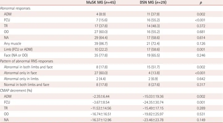
MuSK MG (n=45) DSN MG (n=29) p
Abnormal responses
ADM 4 (8.9) 11 (37.9) 0.002
FCU 7 (15.6) 16 (55.2) <0.001
TR 17 (37.8) 14 (48.3) 0.372
OO 27 (60.0) 16 (55.2) 0.681
NA 29 (64.4) 17 (58.6) 0.614
Any muscle 39 (86.7) 21 (72.4) 0.126
Limb (FCU or ADM) 10 (22.2) 17 (58.6) 0.001
Face (NA or OO) 35 (77.8) 19 (65.5) 0.246
Pattern of abnormal RNS responses
Abnormal in both limbs and face 8 (17.8) 15 (51.7) 0.002
Abnormal only in face 27 (60.0) 4 (13.8) <0.001
Abnormal only in limbs 2 (4.4) 2 (6.9) 0.642
Normal in both limbs and face 8 (17.8) 8 (27.6) 0.317
CMAP decrement (%)
ADM -2.35±6.44 -15.03±19.36 0.002
FCU -3.67±8.54 -24.35±30.74 0.001
TR -11.53±14.56 -15.49±17.15 0.289
OO -16.74±16.51 -19.82±25.97 0.531
NA -16.37±12.96 -23.46±23.78 0.149
Data are n (%) or mean±standard deviation values.
ADM: abductor digiti minimi, CMAP: compound muscle action potential, DSN: double-seronegative, FCU: flexor carpi ulnaris, MG: myasthenia gravis, MuSK: muscle-specific tyrosine kinase, NA: nasalis, OO: orbicularis oculi, RNS: repetitive nerve stimulation, TR: trapezius.
Table 3. Results of univariate and multivariate analyses evaluating the clinical and electrodiagnostic factors associated with MuSK MG compared to DSN MG
Variable Univariate logistic regression Multivariate logistic regression
OR 95% CI p OR 95% CI p
Age at onset 0.995 0.954–1.037 0.801
Sex, female 5.359 1.499–19.418 0.010 1.655 0.350–7.826 1.655
MGFA “b” at time of RNS 8.889 3.038–26.006 <0.001 6.410 1.892–21.718 0.003
Disease severity at time of RNS
MGFA II+III Reference Reference
MGFA IV+V 6.750 1.412–32.262 0.017 3.466 0.575–20.897 0.175
Abnormal RNS response in the face and
normal RNS response in limbs 9.375 2.789–31.512 <0.001 5.224 1.300–20.990 0.020
Immunosuppressive treatment before RNS 1.624 0.607–4.346 0.334
CI: confidence interval, DSN: double-seronegative, MG: myasthenia gravis, MGFA: Myasthenia Gravis Foundation of America, MuSK: muscle-specific tyrosine kinase, OR: odds ratio, RNS: repetitive nerve stimulation.
Kim SW et al.
JCN
ratio (OR)=6.410, 95% confidence interval (CI)=1.892–21.718]
and RNS patterns that were abnormal in the face but normal in limbs (OR=5.224, 95% CI=1.300–20.990) were found to be independently associated with MuSK MG (Table 3).
Change in sensitivity of repetitive nerve stimulation Overall, abnormal responses in any of the five muscles were observed in 86.7% of MuSK MG patients and 72.4% of DSN MG patients (Fig. 1). Abnormal responses in limb muscles were observed in 22.2% of MuSK MG patients and 58.6%
of DSN MG patients. When the trapezius muscle was addi- tionally evaluated, the proportion of abnormal responses increased to 48.9% in the MuSK MG group, whereas the sen- sitivity did not change in the DSN MG group. When con- sidering both the limb and facial muscles, the proportion of patients with abnormal RNS responses increased to 82.2% in the MuSK MG group and 72.4% in the DSN MG group.
DISCUSSION
Abnormal RNS responses of limb muscles occurred less frequently in MuSK MG patients than in DSN MG patients.
A pattern of abnormal facial muscle responses but normal limb muscle responses was more frequent in MuSK MG patients than in DSN MG patients. Whereas the proportion of MuSK MG patients with abnormal responses was 22.2%
when considering only limb muscles, this increased to 82.2%
when the orbicularis oculi and nasalis muscles were also evaluated. By contrast, the additional evaluation of facial muscles in DSN MG patients increased the sensitivity by only 13.8%. These findings suggest that abnormal RNS re- sponses primarily in facial muscles without involvement of distal limb muscles are more pronounced in MuSK MG than in DSN MG, and that performing RNS in both the face and limbs can increase the test sensitivity.
Our findings are consistent with those of previous stud- ies. Some of the previous studies found a low sensitivity of
RNS in MuSK-Ab-positive MG patients, but they only per- formed RNS in hand or shoulder muscles and not in facial muscles.6,7 Consistent with this, we found abnormal RNS responses in limb muscles in only 22.2% of MuSK MG pa- tients. Other studies that performed RNS in both limb and facial muscles found abnormal responses to be more com- mon in facial muscles (75–80%) than in limb muscles (25–
36%).8,11,14 In the present study, the sensitivities of RNS in the facial and limb muscles were 77.8 and 22.2%, respective- ly. In addition, the RNS responses tended to be abnormal only in the face, and rarely in both the face and limbs, which is consistent with a previous study suggesting focal muscle involvement in MuSK-Ab-positive MG.10
The proportion of abnormal RNS responses in the pres- ent DSN MG patients is also consistent with previous stud- ies. Abnormal RNS responses were reportedly observed in 30–74% of subjects in facial muscles and in 36–78% of sub- jects in limb muscles.3,9,11,12 Similarly, in the present study, the RNS responses of facial and limb muscles were abnor- mal in 65.5 and 58.6% of DSN MG patients, respectively.
The strengths of the present study include its relatively large population and the similarity of the methods used to perform RNS across patients. By contrast, previous studies have included relatively small populations or recruited pa- tients from different institutions, and did not perform RNS in the same muscles.8,9,14 Also, in the present study we were able to calculate the sensitivity of RNS in each tested mus- cle and demonstrate how the sensitivity changed as the num- ber of muscles tested increased. This revealed that addition- ally evaluating the orbicularis oculi and nasalis muscles increased the overall sensitivity in MuSK MG by 60%.
Early suspicion of MuSK-Ab-positive MG is important since this condition is often associated with rapid disease pro- gression, frequent respiratory crisis, poor response to cholin- esterase inhibitor, and early requirement of immunosuppres- sive treatment.15,16 Because bulbar and facial muscle weakness is prominent in MuSK-Ab-positive MG, this condition can
Fig. 1. Abnormal rate of repetitive nerve stimulation responses depending on the site of examination in (A) muscle-specific tyrosine kinase-anti- body-positive myasthenia gravis group and (B) double-seronegative myasthenia gravis group. NA: nasalis, OO: orbicularis oculi, TR: trapezius.
A
All muscles Limb
+OO+NA Limb
+NA Limb
+OO Limb
+TR Limb
100 80 60 40 20
0 Limb All muscles
+OO+NA Limb
+NA Limb
+OO Limb +TR Limb
B
100 80 60 40 20 0
Repetitive Nerve Stimulation in MuSK MG
JCN
be suspected based on clinical characteristics. However, a differential diagnosis is difficult based solely on clinical data.
In the present study, abnormal RNS responses primarily in facial muscles were shown to be independently associated with MuSK MG after adjusting for sex, disease severity, and clinical presentation. RNS is usually performed when MG is initially suspected, with the results being immediately avail- able to physicians after the test. Thus, identification of the typi- cal RNS pattern of MuSK MG can contribute to early diag- noses.
In conclusion, conducting RNS in multiple muscles in- cluding those of the face and limbs in AChR-Ab-negative MG patients increases the diagnostic sensitivity and may be help- ful in differentiating MuSK MG from DSN MG.
Conflicts of Interest
The authors have no financial conflicts of interest.
Acknowledgements
This work was supported by the National Research Foundation of Korea (NRF) grant funded by the Korean government (MSIP) (No. NRF- 2016R1C1B1010120).
REFERENCES
1. Gilhus NE. Myasthenia gravis. N Engl J Med 2016;375:2570-2581.
2. Lee HS, Lee HS, Shin HY, Choi YC, Kim SM. The epidemiology of myasthenia gravis in Korea. Yonsei Med J 2016;57:419-425.
3. Evoli A, Tonali PA, Padua L, Monaco ML, Scuderi F, Batocchi AP, et al. Clinical correlates with anti-MuSK antibodies in generalized sero- negative myasthenia gravis. Brain 2003;126(Pt 10):2304-2311.
4. Hoch W, McConville J, Helms S, Newsom-Davis J, Melms A, Vincent A. Auto-antibodies to the receptor tyrosine kinase MuSK in patients
with myasthenia gravis without acetylcholine receptor antibodies.
Nat Med 2001;7:365-368.
5. Lavrnic D, Losen M, Vujic A, De Baets M, Hajdukovic LJ, Stojanovic V, et al. The features of myasthenia gravis with autoantibodies to MuSK.
J Neurol Neurosurg Psychiatry 2005;76:1099-1102.
6. Sanders DB, El-Salem K, Massey JM, McConville J, Vincent A. Clini- cal aspects of MuSK antibody positive seronegative MG. Neurology 2003;60:1978-1980.
7. Padua L, Tonali P, Aprile I, Caliandro P, Bartoccioni E, Evoli A. Sero- negative myasthenia gravis: comparison of neurophysiological pic- ture in MuSK+ and MuSK- patients. Eur J Neurol 2006;13:273-276.
8. Pasnoor M, Wolfe GI, Nations S, Trivedi J, Barohn RJ, Herbelin L, et al. Clinical findings in MuSK-antibody positive myasthenia gravis: a U.S. experience. Muscle Nerve 2010;41:370-374.
9. Oh SJ, Hatanaka Y, Hemmi S, Young AM, Scheufele ML, Nations SP, et al. Repetitive nerve stimulation of facial muscles in MuSK anti- body-positive myasthenia gravis. Muscle Nerve 2006;33:500-504.
10. Nikolic A, Basta I, Stojanovic VR, Stevic Z, Lavrnic D. Electrophysi- ological profile of the patients with MuSK positive myasthenia gra- vis. Neurol Res 2014;36:945-949.
11. Oh SJ. Muscle-specific receptor tyrosine kinase antibody positive myasthenia gravis current status. J Clin Neurol 2009;5:53-64.
12. Wolfe GI, Oh SJ. Clinical phenotype of muscle-specific tyrosine ki- nase-antibody-positive myasthenia gravis. Ann N Y Acad Sci 2008;
1132:71-75.
13. Oh SJ, Eslami N, Nishihira T, Sarala PK, Kuba T, Elmore RS, et al.
Electrophysiological and clinical correlation in myasthenia gravis.
Ann Neurol 1982;12:348-354.
14. Stickler DE, Massey JM, Sanders DB. MuSK-antibody positive myas- thenia gravis: clinical and electrodiagnostic patterns. Clin Neuro- physiol 2005;116:2065-2068.
15. Evoli A, Bianchi MR, Riso R, Minicuci GM, Batocchi AP, Servidei S, et al. Response to therapy in myasthenia gravis with anti-MuSK anti- bodies. Ann N Y Acad Sci 2008;1132:76-83.
16. Guptill JT, Sanders DB, Evoli A. Anti-MuSK antibody myasthenia gravis: clinical findings and response to treatment in two large cohorts.
Muscle Nerve 2011;44:36-40.