INTRODUCTION
In women, cervical cancer is the third most commonly diag- nosed cancer and the fourth leading cause of cancer death in females worldwide, accounting for 9% (529,800) of the total
new cancer cases and 8% (275,100) of the total cancer deaths among females in 2008 [1].
Treatment of cervical cancer depends on FIGO staging. FIGO criteria is important for pretreatment staging and choice of appropriate treatment planning and treatment [2]. The inclu- sion of CT or MRI scans in the staging work-up has been ad- vocated in order to improve accuracy for cervical carcinoma [2,3]. Cystoscopy and sigmoidoscopy are necessary to confirm bladder and rectal invasion in cervical cancer [4].
Traditional pretreatment evaluations of patients with cervical cancer include physical examination, chest radiography, cys- toscopy, intravenous urography, sigmoidoscopy, and barium enema [2,3]. However, imaging modalities (CT, MRI scan) and endoscopy (cystoscopy, sigmoidoscopy) are not included in
Indications for endoscopy according to the revised FIGO staging for cervical cancer after MRI and CT scanning
Bae Kwon Jeong*, Seung Jae Huh, Doo Ho Choi, Won Park, Dongryul Oh, Taegyu Kim, Hye Bin Lee
Department of Radiation Oncology, Samsung Medical Center, Sungkyunkwan University School of Medicine, Seoul, Korea
Received Nov 15, 2011, Revised Dec 8, 2011, Accepted Dec 16, 2011
*Present address: Department of Radiation Oncology, Gyeongsang National University School of Medicine, Jinju, Korea
Correspondence to Seung Jae Huh
Department of Radiation Oncology, Samsung Medical Center, Sungkyunkwan University School of Medicine, 81 Irwon-ro, Gangnam-gu, Seoul 135-710, Korea. Tel: 82-2-3410-2601, Fax: 82-2-3410-2619, E-mail:
Objective: A recent revision of the FIGO staging system does not recommend the mandatory use of cystoscopy and sigmoidoscopy. The objective of this study was to assess the clinical utility of CT or MRI scans for ruling out bladder or rectal invasion and determine the indication for endoscopy in patients with cervical cancer.
Methods: We retrospectively reviewed 769 patients with cervical cancer, who underwent imaging and endoscopic work-up between January 1997 and December 2010. Using endoscopy as the standard reference for comparison, we calculated the sensitivity, specificity, positive predictive value (PPV), negative predictive value (NPV), and accuracy of the imaging modality for bladder or rectal invasion.
Results: The CT scan showed 68.2% and 85.7% for sensitivity and 96.4% and 98.9% for specificity in detecting bladder and rectal invasion, respectively. CT scan provided a low PPV (51.7%, 54.5%) and a high NPV (98.2%, 99.8%). MRI scan showed 88.0% and 75.0% for sensitivity and 93.1% and 98.9% for specificity in detecting bladder and rectal invasion, respectively. MRI scan provided a low PPV (35.6%, 42.9%) and a high NPV (99.4%, 99.7%). The accuracies of CT and MRI scans in identifying bladder invasion were 94.9% and 92.8%, respectively. The accuracies of CT and MRI in identifying rectal invasion were 98.7% and 98.6%, respectively.
Conclusion: The results of this study demonstrate that additional invasive endoscopy is not necessary for patients who present with no invasion on imaging work-up, and therefore, endoscopy should be considered a tool for confirming cases that are positive for invasion based on imaging work-up.
Keywords: Computed tomography, Cystoscopy, Magnetic resonance imaging, Sigmoidoscopy, Uterine cervical neoplasms
the recent FIGO guidelines for routine pretreatment staging of cervical cancer [5]. Recent FIGO staging system for the cer- vix encourages the use of CT and MRI scans, but cystoscopy and sigmoidoscopy were classified as optional modalities and are not recommended as mandatory examinations. However, there are no recommendations regarding which patients should receive endoscopy as an alternative examination.
We carried out a retrospective analysis of cervical cancer patients who underwent imaging work-up and endoscopy before radiotherapy to determine the clinical utility of CT or MRI scans for ruling out bladder or rectal invasion, and the in- dications of endoscopy for patients with cervical cancer.
MATERIALS AND METHODS
Between January 1997 and December 2010, 1,610 patients with biopsy-confirmed cervical cancer were treated by the radiation oncology department of Samsung Medical Center, Seoul, Korea. Among these patients, we retrospectively re- viewed the records of 769 patients who underwent imaging work-ups such as CT (503 patients) or MRI (749 patients) scans, and 473 patients who underwent both CT and MRI in addition to the standard FIGO staging work-up. Among them, endos- copies such as cystoscopy or sigmoidoscopy were done in 590 patients and 735 patients, respectively. In Samsung Medi- cal Center, if there is no contraindication, it is routine practice to conduct imaging and endoscopy for all patients with cervi- cal cancer. We analyzed data regarding age, tumor size, stage, lymph node involvement, menopause status, and squamous cell carcinoma antigen (SCC-Ag).
Considering endoscopy as the standard reference investiga- tion, the sensitivity, specificity, positive predictive value (PPV), negative predictive value (NPV), and accuracy of CT and MRI scans for bladder and rectal invasion were determined.
Each patient underwent pretreatment imaging work-up of the abdominopelvic area and endoscopy of the bladder or rectum. CT (Lightspeed VCT* XTe, GE Healthcare, Buckingham- shire, UK) scans were performed using contiguous axial, sag- ittal, and coronal 5-mm thickness slices after administration of contrast medium. MRI scans were performed using a 1.5 T unit (Achieva, Phillips Medical System, Eindhoven, Netherland;
Signa HDe, GE Healthcare). The cardiac or torso coil was used in the supine position from the pelvis to the abdomen. The CT scan criteria for bladder or rectal invasion included the focal loss of the periorgan fat plane between the bladder or rectum and the growth, accompanied by asymmetrical wall thicken- ing, nodular indentations along the bladder or rectal wall, and intraluminal tumor masses. MRI findings of wall irregularity
with heterogeneous signal, enhancement with thickening and nodularity, loss of fat plane or mass protruding into the bladder or rectal lumen were interpreted as positive invasion.
Twenty five out of 65 patients (38.5%) and 7 out of 16 pa- tients (43.8%) were pathologically confirmed by cystoscopy or sigmoidoscopy when the bladder or rectum invasion sus- pected on image work-up. Endoscopy was considered the gold standard for determining the presence of bladder or rectal invasion. In all cases, cystoscopy and sigmoidoscopy were performed by an urologist and gastroenterologist, re- spectively. Flexible or rigid cystoscopy was used for bladder investigation, and fiberoptic sigmoidoscopy was used for rectal examination with biopsy for pathological confirmation.
Endoscopically directed biopsy specimens were taken from all areas in the bladder and rectum that were suspected of can- cer development.
Seven hundred fifty three patients received radiotherapy.
Most of these patients (603 patients, 80.1%) received 5,040 cGy of external beam radiotherapy, and 359 patients (47.7%) underwent additional brachytherapy. A total of 450 patients underwent combined chemotherapy with radiotherapy. Six- teen patients who did not receive radiotherapy were treated with palliative aim. 424 patients did not receive brachytherapy of patients with radiotherapy. Among these patients who did not received brachytherapy, most patients underwent post- operative adjuvant radiotherapy or palliative radiotherapy.
We evaluated the sensitivity, specificity, PPV, NPV, and ac- curacy of CT and MRI findings for the diagnosis of bladder or rectal invasion, by comparing the frequencies of each imag- ing work-up with the final endoscopic biopsy. The sensitivity, specificity, PPV, NPV, and accuracy of CT scans for bladder or rectal invasion were calculated using the following formulae:
Sensitivity= Number of true positives
Number of true positives+Number of false negatives
Specificity = Number of true negatives
Number of true negatives+Number of false positives
PPV = Number of true positives
Number of true positives+Number of false positives
NPV= Number of true negatives
Number of true negatives+Number of false negatives
Accuracy= Number of true positives+Number of true negatives Number of true positives+False positives+
False negatives+True negatives
RESULTS
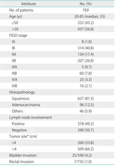
Seven hundred sixty nine are reviewed and their charac- teristics are given in Table 1. The median age of the patients
was 50 years (range, 20 to 85 years) and the median follow-up was 37 months (range, 1 to 162 months). Of 769 patients, 29 (3.8%), 50 (6.5%), and 25 patients (3.3%) had bladder invasion on CT, MRI scan, and cystoscopy, respectively. Rectal invasion was identified in 11 (1.4%), 14 (1.8%), and 7 patients (0.9%) on CT, MRI scan, and sigmoidoscopy, respectively. The results for each of the imaging modalities are given in Tables 2 and 3, with endoscopic findings considered as the gold standard. CT and MRI scans revealed bladder invasion in 15 and 12 patients, respectively, who had endoscopically confirmed bladder inva- sion (true-positive for bladder invasion) and CT and MRI both revealed rectal invasion in 6 patients, respectively, who had endoscopically confirmed rectal invasion (true-positive for rectal invasion). Thus, 14 patients demonstrated bladder inva- sion on CT scan only and 38 patients demonstrated bladder invasion on MRI scan only (false-positive for bladder invasion).
Five patients demonstrated rectal invasion on CT scan only and 8 patients demonstrated rectal invasion on MRI scan only (false-positive for rectal invasion). Seven and 3 patients who had cystoscopically confirmed invasion (false-negative blad- der invasion) did not show any invasion on CT and MRI scan, respectively. For each image modality, there was one patient who showed no invasion, but had sigmoidoscopically con- firmed invasion (false-negative rectal invasion). Finally, in 2 pa- tients showing no bladder involvement on CT and MRI scan, Table 1. Patient characteristics
Attribute No. (%)
No. of patients 769
Age (yr) 20-85 (median, 53)
≤50 332 (43.2)
>50 437 (56.8)
FIGO stage
IA 8 (1.0)
IB 314 (40.8)
IIA 134 (17.4)
IIB 207 (26.8)
IIIA 5 (0.7)
IIIB 60 (7.8)
IVA 25 (3.3)
IVB 16 (2.1)
Histopathology
Squamous 627 (81.5)
Adenocarcinoma 96 (12.5)
Others 46 (5.9)
Lymph node involvement
Positive 378 (49.2)
Negative 390 (50.7)
Tumor size* (cm)
≤4 260 (33.8)
>4 509 (66.2)
Bladder invasion 25/590 (4.2)
Rectal invasion 7/735 (1.0)
*Median, 4 cm; mean, 4.3±1.7 cm.
Table 3. Diagnostic ability of imaging modalities in rectal or bladder invasion
Sensitivity Specificity PPV NPV Accuracy
Bladder invasion
CT 68.2 96.4 51.7 98.2 94.9
MRI 88.0 93.1 35.6 99.4 92.8
CT & MRI 90.9 91.7 39.2 96.6 -
Rectal invasion
CT 85.7 98.9 54.5 99.8 98.7
MRI 85.7 98.9 42.9 99.7 98.6
CT & MRI 85.7 99.1 42.9 99.8 -
PPV, positive predictive value; NPV, negative predictive value.
Values are presented as percentage (%).
Table 2. Correlation of imaging findings with endoscopic findings for rectal and bladder invasion
Bladder invasion Rectal invasion
CT MRI CT MRI
True positive 15 12 6 6
False positive 14 38 5 8
True negative 375 509 466 701
False negative 7 3 1 1
bladder invasion was confirmed by cystoscopy. As shown in Table 3, CT scan showed a sensitivity of 68.2% and 85.7%, and a specificity of 96.4% and 98.9%, for detecting bladder and rectal invasion, respectively. CT scan had a low PPV (51.7% and 54.5%) and a high NPV (98.2% and 99.8%). MRI scan showed a sensitivity of 88.0% and 85.7%, and a specificity of 93.1% and 98.9% for detecting bladder and rectal invasion, respectively.
MRI scan had a low PPV (35.6% and 42.9%) and a high NPV (99.4% and 99.7%). The accuracies of the imaging modalities in detecting the bladder and rectal invasion are given in Table 3.
DISCUSSION
In FIGO staging, some studies concluded that the use of im- aging is not superior to physical examination [2,6]. In contrast, many studies have suggested that imaging is an important work-up tool and should be included in staging [3,7-11].
Cystoscopy and sigmoidoscopy, previously categorized as mandatory investigations, were reclassified as optional in- vestigations in a recent revision of FIGO staging [5]. Since the 2009 revision of FIGO staging, a few studies have explored the identification of patients who will need endoscopy [12-14].
The present study therefore had two objectives. The primary objective was to establish how to identify patients who re- quired cystoscopy or sigmoidoscopy according to the revised FIGO staging. The secondary objective was to demonstrate the accuracy of CT and MRI scans for pretreatment diagnosis of bladder and rectum invasion.
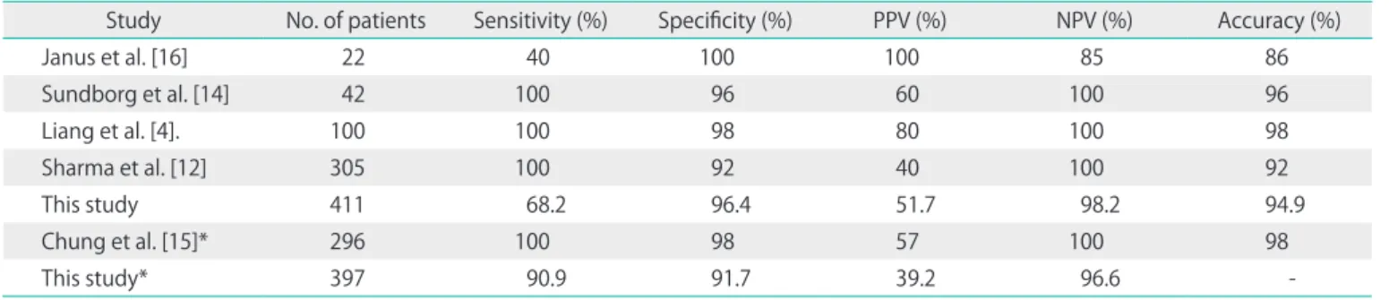
Based on previous studies, the sensitivities, specificities, PPVs, NPVs, and accuracies of CT and MRI scanning for bladder or rectal invasion were about 40-100%, 92-100%, 40-100%, 85- 100%, and 86-98%, respectively [4,12,14-17]. The results of this study correspond well with those of previous studies (Table 4), with the exception of a few differences in sensitivity. In this study,
the NPV was high enough so that additional invasive endos- copy was not necessary for patients who presented without invasion in imaging work-up (CT, MRI scan). Endoscopy should be considered as a tool for confirming invasion when patients were positive for invasion based on imaging work-up even though CT and MRI scans are not as effective for the purpose of diagnosis due to low sensitivity and PPV. These results sug- gest that there are advantages to using imaging modalities for the physician and patients, and that imaging offers additional cost benefits. There is no evidence that the low sensitivity of imaging can be increased by simultaneously using CT and MRI scans as shown in Table 3. MRI scans may be instrumental as a diagnostic tool to evaluate myometrial invasion or lymph node involvement, but it has no additional benefit as part of the confirmation process for bladder or rectal invasion.
Fig. 1 shows a false-negative image from an MRI scan and endoscopy. There is no evidence of bladder invasion on the CT and MRI scan, but cystoscopy revealed that the patient was positive for invasion. False negative in CT or MRI is a rare case in this study and in previous studies which conducted similar purpose with the current study. The false negative finding in this case may have been the result of poor image quality or focal invasion in between the CT slices, so additional invasive endoscopy is not recommended to check the invasion for pa- tients without invasion on imaging work-up.
The findings of this study are significant because they are based on a larger patient sample than those of previous stud- ies. In addition, we analyzed the accuracy and indications of two imaging modalities, CT and MRI, for the diagnosis of blad- der and rectal invasion.
There are some limitations to this single institution retro- spective study. First is that the number of patients in a specific group, such as stage IV, was too small for analysis and stage IIA was too large to bring about selection bias. Second is that this study does not include cervical cancer patients treated
Table 4. Summary of studies showing the capabilities of CT alone or CT and MRI in bladder invasion
Study No. of patients Sensitivity (%) Specificity (%) PPV (%) NPV (%) Accuracy (%)
Janus et al. [16] 22 40 100 100 85 86
Sundborg et al. [14] 42 100 96 60 100 96
Liang et al. [4]. 100 100 98 80 100 98
Sharma et al. [12] 305 100 92 40 100 92
This study 411 68.2 96.4 51.7 98.2 94.9
Chung et al. [15]* 296 100 98 57 100 98
This study* 397 90.9 91.7 39.2 96.6 -
PPV, positive predictive value; NPV, negative predictive value.
*Combined values for CT and MRI scans.
with surgery or chemotherapy only. Another limitation is that endoscopy was not performed in all patients, but was more likely to be used in patients who were suspected of having bladder or rectal invasion based on imaging work-up or physi- cal examination.
In conclusion, if there is no evidence of invasion on imaging work-up, endoscopy is not necessary as an invasive diagnos- tic modality. However, if there is any evidence of invasion on imaging work-up, endoscopy is necessary to obtain an accu- rate prognosis for appropriate treatment. Patients prefer non- invasive diagnostic methods, which have fewer side effects.
Therefore, future work should focus on the use of CT virtual endoscopy, which can be used in place of invasive endoscopy.
CONFLICT OF INTEREST
No potential conflict of interests relevant to this article was reported.
REFERENCES
1. Jemal A, Bray F, Center MM, Ferlay J, Ward E, Forman D.
Global cancer statistics. CA Cancer J Clin 2011;61:69-90.
2. Hricak H, Gatsonis C, Chi DS, Amendola MA, Brandt K, Schwartz LH, et al. Role of imaging in pretreatment evaluation of early invasive cervical cancer: results of the intergroup study American College of Radiology Imaging Network 6651-Gynecologic Oncology Group 183. J Clin Oncol 2005;23:9329-37.
3. Togashi K, Morikawa K, Kataoka ML, Konishi J. Cervical cancer. J Magn Reson Imaging 1998;8:391-7.
4. Liang CC, Tseng CJ, Soong YK. The usefulness of cysto- scopy in the staging of cervical cancer. Gynecol Oncol 2000;76:200-3.
5. Pecorelli S, Zigliani L, Odicino F. Revised FIGO staging for carcinoma of the cervix. Int J Gynaecol Obstet 2009;
105:107-8.
6. Hancke K, Heilmann V, Straka P, Kreienberg R, Kurzeder C. Pretreatment staging of cervical cancer: is imaging better than palpation? Role of CT and MRI in preoperative staging of cervical cancer: single institution results for 255 patients. Ann Surg Oncol 2008;15:2856-61.
7. Hawighorst H, Schoenberg SO, Knapstein PG, Knopp MV, Schaeffer U, Essig M, et al. Staging of invasive cervical carcinoma and of pelvic lymph nodes by high resolution MRI with a phased-array coil in comparison with pathological findings. J Comput Assist Tomogr 1998;22:75-81.
Fig. 1. A 37-year-old woman who was dia gnosed with squamous cell carcinoma of the uterine cervix. Magnetic resonance imaging (A, axial; B, sagittal) shows that the mass is focally in contact with the posterior wall of the bladder, and there is a fat plane and no definitive evidence of invasion. Cystoscopic finding (C) of an erythematous flat elevated lesion that is present on the bladder posterior wall.
8. Hricak H, Lacey CG, Sandles LG, Chang YC, Winkler ML, Stern JL. Invasive cervical carcinoma: comparison of MR imaging and surgical findings. Radiology 1988;166:623- 31.
9. Sheu MH, Chang CY, Wang JH, Yen MS. Cervical carci- noma: assessment of parametrial invasion and lymph node metastasis with magnetic resonance imaging.
Zhonghua Yi Xue Za Zhi (Taipei) 2000;63:634-40.
10. Subak LL, Hricak H, Powell CB, Azizi L, Stern JL. Cervical carcinoma: computed tomography and magnetic reso- nance imaging for preoperative staging. Obstet Gynecol 1995;86:43-50.
11. Hricak H, Yu KK, Powell CB, Subak LL, Stem J, Arenson RL.
Comparison of diagnostic studies in the pretreatment evaluation of stage Ib carcinoma of the cervix. Acad Radiol 1996;3 Suppl 1:S44-6.
12. Sharma DN, Thulkar S, Goyal S, Shukla NK, Kumar S, Rath GK, et al. Revisiting the role of computerized tomographic scan and cystoscopy for detecting bladder invasion in the revised FIGO staging system for carcinoma of the uterine cervix. Int J Gynecol Cancer 2010;20:368-72.
13. Nam H, Huh SJ, Park W, Bae DS, Kim BG, Lee JH, et al.
Prognostic significance of MRI-detected bladder muscle and/or serosal invasion in patients with cervical cancer treated with radiotherapy. Br J Radiol 2010;83:868-73.
14. Sundborg MJ, Taylor RR, Mark J, Elg SA. Cystoscopy after computed tomography scan to identify bladder invasion in cervical cancer. Obstet Gynecol 1998;92:364-6.
15. Chung H, Ahn HS, Kim YS, Lee EJ, Ryu HS, Chang KH, et al.
The value of cystoscopy and intravenous urography after magnetic resonance imaging or computed tomography in the staging of cervical carcinoma. Yonsei Med J 2001;42:527-31.
16. Janus CL, Mendelson DS, Moore S, Gendal ES, Dottino P, Brodman M. Staging of cervical carcinoma: accuracy of magnetic resonance imaging and computed tomo- graphy. Clin Imaging 1989;13:114-6.
17. Massad LS, Calvello C, Gilkey SH, Abu-Rustum NR. Assessing disease extent in women with bulky or clinically evident metastatic cervical cancer: yield of pretreatment studies.
Gynecol Oncol 2000;76:383-7.