Pandemic Novel Influenza A (H1N1) Virus in Korea:
The Experience from August to September 2009
Kyung-Ok Lee, Min-Young Park, Lyoung-Hyo Kim, Hye-Soon Seong, Bo-Hyun Park, and Su-Jin Jeong
Genome Research Center, Neodin Medical Institute, Seoul 133-847, Korea
Novel influenza A virus, subtype H1N1 of swine-lineage, has been transmitted rapidly to many regions of the world. Rapid detection of the virus is essential to instigate appropriate patient care and public health management and for disease surveillance. The aim of this study is to determine the prevalence of novel influenza A (H1N1) virus in Korea using reverse-transcription real time polymerase chain reaction (rRT-PCR). Novel H1N1 virus was detected in a total of 8,948 nasopharyngeal samples from patients with influenza-like illness throughout Korea from August to September 2009. RNA was extracted from 300 μl of sample using an RNA extraction kit (Zymo Research, CA, USA). In the present study, Genekam kit (Genekam, Duisburg, Germany) was used to detect novel H1N1 virus. Novel H1N1 virus was found in 1,130 samples from a total of 8,948 samples (12.6%). The highest frequency was found in 10- to 19-year-olds (M: 29.3% vs. F: 16.4%), followed by 20- to 29-year-olds (M: 17.9% vs. F: 15.4%), 40- to 49-year-olds (M: 6.5% vs. F: 8.1%), 50- to 59-year-olds (M: 6.0% vs. F: 5.5%), and 30- to 39-year-olds (M: 4.6% vs. F: 3.8%). The mean positive rate was higher in men than in women (M: 14.7% vs. F: 7.4%).
Novel H1N1 virus showed the lowest prevalence in patients over 60 years old. The positive rate increased daily and showed a significant high peak in mid-September 2009. In 19 provinces of Korea, Cheonan (41.1%), Busan (37.3%), Gangneung (33.3%), Jinju (32.1%), Ulsan (24.6%), Deajeon (23.7%) areas showed high frequencies and other provinces were found less than 10% of novel H1N1 virus. Since reverse-transcription real time PCR assay is rapid, accurate, and convenient, it may assist public health laboratories in detecting novel H1N1 virus. Moreover, these data could be useful for the management of patients with influenza-like illness.
Received 13 OCT 2009 / Returned for modification 11 DEC 2009 / Accepted 16 DEC 2009 Key Words : Novel H1N1 virus, Prevalence, Real-time PCR
I. INTRODUCTION
Influenza A viruses are medically important viral pathogens that cause significant mortality and morbidity throughout the world. Influenza A viruses, a genus of the
교신저자 : Kyung-Ok Lee. #2-3, Yongdap-Dong, Sungdong-gu, Seoul 133-847, Korea.
TEL : 02-2244-6500, 010-8728-0346 E-Mail : [email protected]
orthomyxoviridae family, are RNA viruses with a segmented genome comprising 8 negative-sense, single- stranded RNA segments that encode 11 proteins. Two surface glycoproteins, hemagglutinin (HA) and neura- minidase (NA), are the key antigens against which humoral immune responses are directed (Olsen, 2002).
Matrix protein 1 (M1) is the structural protein of the virus particle, while matrix protein 2 (M2) forms an envelope-spanning proton channel (Brockwell-Staats et al,
2009). Depending on the antigenicity of two envelope spikes, influenza A viruses are divided into 16 H (H1–
H16) and 9 N (N1–N9) groups, theoretically resulting in 16 × 9 serologic subtypes. Up to now, 105 influenza A virus subtypes have been discovered, all endemic in water birds. However, some subtypes have adapted to other birds (chickens) and mammals (pigs, horses, humans) in species-specific strains (Michaelis et al, 2009).
Recently, novel influenza A (H1N1) virus, designated
“novel H1N1 virus” was identified in humans (Brockwell- Staats et al, 2009). The subsequent phylogenetic chara- cterization of novel H1N1 virus showed a unique genome composition that had not been previously identified. Six genes (PB2, PB1, PA, HA, NP, and NS) were similar to those of viruses previously identified in pigs in North America. The hemagglutinin (HA) gene is similar to that of swine flu viruses present in pigs in the U.S. since 1999, while neuraminidase (NA) and matrix (M) genes resemble those of viruses present in pigs in Europe (Peiris et al, 2009). The transmissibility of novel H1N1 virus has been estimated to be higher than that of seasonal influenza viruses, since it is not transmitted from pigs to humans, but rather from person to person (Fraser et al, 2009). Clinical presentation and severity remains unclear, but most confirmed cases have been characterized by mild influenza-like illness, with symptoms including chills, fever, sore throat, muscle pains, severe headache, coughing, weakness, and general discomfort (Dawood et al, 2009). Since these symptoms are not specific to novel H1N1 viral infection, early in the pandemic, physicians were advised to consider novel H1N1 virus in the differential diagnosis of patients with acute febrile respiratory illness. However, a considerable proportion of patients reported vomiting or diarrhea, which is unusual in seasonal influenza (Dawood et al, 2009). To limit community or hospital transmission and to initiate antiviral therapy, rapid detection of the virus in suspected cases remains crucial. Therefore, the aim of this study is to determine the prevalence of novel H1N1 by using
reverse-transcription real time polymerase chain reaction (rRT-PCR) in Korean patients.
II. MATERIALS AND METHODS
1. RNA extraction from patient samples
The present study included 8,948 samples from patients with influenza-like illness throughout Korea. Patients ranged in age from 4 months to 83 years. Sample types tested included flocked nasopharyngeal (NP) swabs, NP aspirates and NP washes (Copan, Murrieta, CA, USA).
Samples were stored either refrigerated or frozen at −70
℃ until tested. All samples were collected according to a standard of care for routine diagnostic testing. Viral RNA was extracted from 300 µL of samples using a Viral RNA extraction kit (Zymo Research, CA, USA). Viral Gene- spin lysis buffer (250 µL) was added to sample and incubated at room temperature for 10 min before 350 µL of binding buffer was added. Lysate was loaded on a spin column and centrifuged at 13,000 rpm for 1 min. After the solution was discarded, 500 µL of washing buffer A was added to the column and centrifuged for 1 min at 13,000 rpm. After this solution was discarded, the spin column was placed in an RNase-free 1.5 mL micro- centrifuge tube, and elution buffer (30 µL) was added directly onto the membrane and incubated at room tem- perature for 1 min. The eluted solution (2∼5 µL) was used for the rRT-PCR assay.
2. Genome amplifications of novel H1N1 RNA This study used a Genekam kit (Genekam, Duisburg, Germany) to detect novel H1N1 virus. The H1N1 RNA received from the KCDC (Center of Disease Control and Prevention, Korea) was used for a positive control.
Amplification was performed in 20 μL of reaction mixture, and the PCR reaction consisted of one cycle of 60 min at 42℃ and 10 min at 70℃, followed by 50 cycles of 15 s at 95℃ and 60 s at 55℃. This assay was
Sex Age
Male Female Total
No. of samples
No. of Pos samples
Positive No. of samples
No. of pos samples
Positive Total samples
Positive samples
Positive
0∼3 464 15 3.2% 365 22 6.0% 829 37 4.6%
4∼9 628 42 6.7% 519 46 8.9% 1147 88 7.8%
10∼19 1,558 457 29.3% 1,319 216 16.4% 2,877 673 23.4%
20∼29 598 107 17.9% 654 101 15.4% 1,252 208 16.6%
30∼39 498 23 4.6% 524 20 3.8% 1,022 43 4.2%
40∼49 262 17 6.5% 309 25 8.1% 571 42 7.4%
50∼59 199 12 6.0% 255 14 5.5% 454 26 5.7%
60∼69 171 3 1.8% 175 2 1.1% 346 5 1.4%
>70 226 3 1.3% 224 5 2.2% 450 8 1.8%
Total 4,604 679 14.7% 4,344 451 10.4% 8,948 1,130 12.6%
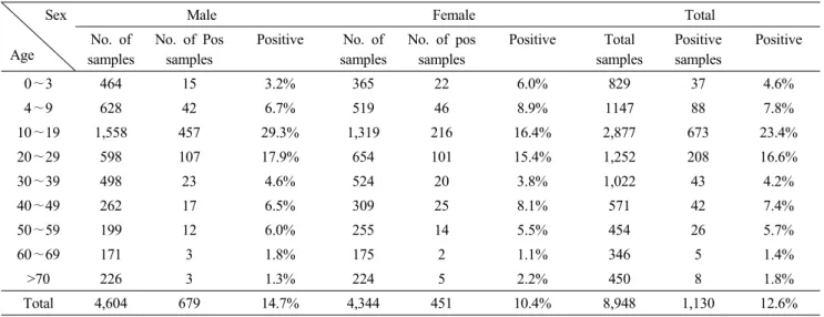
Table 1. Positive rates of novel influenza (H1N1) virus from August to September 2009 in groups according to age and sex (n=8,948).
determined by using a Roche LightCycler 480 Real-Time PCR System (Roche, Mannheim, Germany).
3. Statics
The usual Chi-square test was used to analyze the significance of the prevalence of H1N1 in group of age and sex. For all analyses, P<0.05 was considered statistically significant.
III. RESULTS
1. Prevalence of novel H1N1 virus in groups according to age and sex
The present study analyzed a total of 8,948 naso- pharyngeal samples obtained from patients showing influ- enza-like illness throughout Korea. The age distribution of samples ranged from 4 months to 83 years. The mean prevalence of novel H1N1 virus was 12.6% (1,130 out of 8,948) from August to September 2009. The positive rate of novel H1N1 virus was less than 10% in August, but increased daily to over 25% in mid-September 2009 (Fig.
2). The highest frequency was shown in 10- to
19-year-olds (M: 29.3% vs. F: 16.4%), followed by 20- to 29-year-olds (M: 17.9% vs. F: 15.4%), 40- to 49-year-olds (M: 6.5% vs. F: 8.1%), 50- to 59-year-olds (M: 6.0% vs.
F: 5.5%), and 30- to 39-year-olds (M: 4.6% vs. F: 3.8%).
The prevalence of novel H1N1 virus was less than 2% in patients over 60 years old (Table 1). Generally, the mean positive rate was higher in men than in women (M:
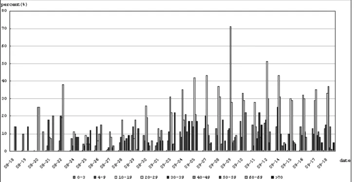
14.7% vs. F: 10.4%). The prevalence of novel H1N1 virus in 4 to 9-year olds were higher than 0- to 3-year olds (7.8% vs 4.5%). The highest frequency (71.0%) was found in 10 to 19-year olds on September 9, 2009 (Fig.
3). Generally the positive rate was higher in Men, however, it was high in four age groups of women, including 0- to 3-year-olds, 4- to 9-year-olds, 40- to 49-year-olds and over 70 years old. The prevalence of novel H1N1 virus with age and sex was statistically significant (P<0.0001) (Fig. 1)
2. Prevalence of novel H1N1 virus in 19 provinces of Korea
The 8,948 patient samples analyzed in this study were collected from 19 provinces of Korea. Fig. 4 shows the prevalence of novel H1N1 virus in each province of
P<.0001; for sex Fig 1. Prevalence of novel influenza (H1N1) virus from August to September 2009 according to age group and sex (n=8,948).
P<.0001; for age Fig 2. Prevalence of novel influenza (H1N1) virus according to the date (n=8,948).
Korea. Generally, Cheonan (41.1%), Busan (37.3%), Gangneung (33.3%), Jinju (32.1%), Ulsan (24.6%), Dea- jeon(23.7%) areas showed high frequencies and other provinces were found less than 10% of novel H1N1 virus in this analysis. During in August, Jinju showed the highest prevalence. In early to mid-September, the positive rate was high in Pusan. Kwangju showed the highest prevalence during September 12 to 18, 2009. The
prevalence of novel H1N1 virus with periods and provinces was statistically significant (P<0.0001) (Fig. 4)
IV. DISCUSSION
The rapid detection and differentiation of novel H1N1 virus from seasonal influenza is essential to instigate
Fig 3. Prevalence of novel influenza (H1N1) virus from August to September, 2009 by age groups and date (n=8,948).
appropriate patient care and public health management as well as for disease surveillance. The recent emergence of novel H1N1 virus poses a serious health threat (Mahony et al, 2009), prompting an urgent necessity for molecular tests to rapidly detect the virus. At the onset of the H1N1 influenza pandemic, no specific or well-validated diagnostic test was available. In the clinical diagnosis of influenza, nucleic acid testing by RT-PCR has widely replaced traditional virus culture due to shorter turn around times and increased sensitivity (Petric et al, 2006).
Although broadly reactive RT-PCR assays are indeed capable of detecting novel H1N1 virus (Leo et al, 2009), they may lack sensitivity (Carr et al, 2009). The multiplex PCR method (Choi et al 2002; He et al, 2009; Mahony et al, 2009) and real-time PCR assay (rRT-PCR) (Carr et al, 2009; Panning et al, 2009; Whiley et al, 2009) have been used for detecting novel H1N1 virus. Although the multiplex PCR assay has an advantage in its ability to simultaneously detect several amplified products, it may also lack sensitivity (Ginocchio et al, 2009). The rRT-PCR
assay is a rapid and convenient method, and its sensitivity is higher than previous amplification methods due to the use of fluorescence-tagged probes in the amplification reaction (Panning et al, 2009).
A suspected case of novel H1N1 was initially defined as a patient with acute respiratory symptoms and a history of travel to any other affected country within seven days before the onset of symptoms, or a history of close contact with a confirmed or probable case. However, this definition has been updated owing to the rapid spread of infection and the presence of laboratory-confirmed cases in patients who had not traveled outside the country. The current definition of a suspected case includes a history of travel to any affected country or acute respiratory illness requiring hospitalization (Castro-Jiménez et al, 2009).
In Korea, a 51-year-old man died with H1N1 infection, on August 15, 2009, after traveling to Thailand (http://www.CDC.go.kr). After that, human-to-human transmission was also reported in people who had not traveled to other countries, and referrals for tests for novel
P<.0001; for periods, P<.0001; for provinces Fig 4. Incidence of novel influenza (H1N1) virus in 19 provinces of Korea.
H1N1 virus has rapidly increased. In this study, novel H1N1 virus was found most frequently in 10- to 19-year-olds in Korea (Table 1). This high prevalence could be due to the active community lives characteristic of youth in this age group, including school attendance.
The next highest frequency was seen in 20- to 29-year-olds, 40- to 49-year-olds, followed by 50- to 59-year-olds. When these data were compared with those from the KCDC, the prevalence rates for different age groups were similar. The frequency of novel H1N1 infection was highest for 10- to 19-year-olds, while the old age group showed a lower frequency. Moreover, the prevalence of novel H1N1 virus was higher in men than in women (http://www.CDC.go.kr). The reason for this higher prevalence in men is not clear, but infection seems to occur more frequently when patients participate actively in the social community. In Japan, the age group with the highest frequency of infection was the same as in our study (10- to 19-year-olds) (Nishiura et al, 2009).
However, even though the frequency of novel H1N1 viral infection was low in the old age group, the death rate for this group was higher than that for the younger group.
Older people are more at risk for novel H1N1 viral infection due to the presence of adult diseases including cardiovascular disease, diabetes, severe respiratory illness, and so forth. Until October 13, 2009, 15 patients had died with novel H1N1 viral infections in Korea. Among these patients, ten cases were over 65 years old with chronic diseases, diabetes, cancer or renal failure. The most common cause of death was pneumonia, which is a major complication of novel H1N1 viral infection (http://
www.CDC.go.kr).
The frequencies of novel H1N1 virus in 19 provinces of Korea were determined. Generally, the prevalence of novel H1N1 virus was high in the Cheonan, Jinju, and Pusan areas. Kangneung also showed a high frequency, but the sample number was too low to have clinical significance (Fig. 3). In New Zealand, the pandemic accelerated markedly in June, reached a peak within four to six weeks, and has been declining since mid-July (Baker et al, 2009). By contrast, the prevalence of the virus in Korea peaked in mid-September (Fig. 2). In Korea, because of its location in the northern hemisphere, novel H1N1 viral infection could continue from autumn to
the winter of 2009. Based on these results, we believe this molecular test can play an important role as a sentinel test to detect novel H1N1 virus in patients presenting with influenza-like illness. Moreover, these results act as an early warning system for the detection of future pandemic influenza threats to the Korean population.
REFERENCES
1. Baker MG, Wilson N, Huang QS, Paine S, Lopez L, Bandaranayake D, Tobias M, Mason K, Mackereth GF, Jacobs M, Thornley C, Roberts S, McArthur C.
Pandemic influenza A (H1N1) in New Zealand: the experience from April to August 2009. Euro- surveillance 14(34):1-3, 2009.
2. Brockwell-Staats C, Webster RG, Richard J. Webby RJ. Diversity of influenza viruses in swine and the emergence of a novel human pandemic influenza A (H1N1). Influenza Other Respi Viruses 1;3(5):207- 213, 2009.
3. Carr MJ, Gunson R, Maclean A, Coughlan S, Fitzgerald M, Scully M, O’Herlihy B, Ryan J, O’Flanagan D, Connell J, Carman WF, Hall WW.
Development of a real-time RT-PCR for the detection of Swine-lineage Influenza A (H1N1) virus infections.
J Clin Virol 45:196-199, 2009.
4. Castro-Jiménez MA, Castillo-Pabón JO, Rey-Benito GJ, Pulido-Domínguez PO, Barbosa-Ramírez J, Velandia- Rodriguez DA, Angulo-Martínez ES. Epidemiologic analysis of the laboratory-confirmed case of influenza A(H1N1) virus in Colombia. Eurosurveillance 14(30):
1-4, 2009.
5. Choi YK , Goyal SM, Kang SW, Farnham MW, Joo HS. Detection and subtyping of swine influenza H1N1, H1N2 and H3N2 viruses in clinical samples using two multiplex RT-PCR assays. J Virol Methods 102:53-59, 2002.
6. Dawood FS, Jain S, Finelli L, Shaw MW, Lindstrom
S, Garten RJ. Emergence of a novel swine-origin influenza A (H1N1) virus in humans. N Engl J Med 360(25):2605-2615, 2009.
7. Fraser C, Donnelly CA, Cauchemez S, Hanage WP, Van Kerkhove MD, Hollingsworth TD. Pandemic potential of a strain of influenza A (H1N1). Science 324(5934):1557-1561, 2009.
8. Ginocchio CC. Zhang F, Manji R, Arora S, Born- freund M, Fork L, Lotlikar M. Evaluation of multiple test methods for the detection of the novel 2009 influenza A (H1N1) during the New York City out break. J Clin Virol 45(3):191-195, 2009.
9. He J, Bose ME, Beck ET, Fan J, Tiwari S, Metallo J, Jurgens LA, Kehl SC, Ledeboer N, Kumar S, Weisburg W, Henrickson KJ. Rapid multiplex reverse transcription-PCR typing of influenza A and B virus, and subtyping of Influenza A Virus into H1, 2, 3, 5, 7, 9, N1 (Human), N1 (Animal), N2, and N7, including typing of novel swine origin influenza A (H1N1) virus, during the 2009 outbreak in Milwaukee, Wisconsin. J Clin Microvirol 47(9):2772- 2778, 2009.
10. Leo L, Poon M, Chan KH, Smith GJ, Leung SWC, Guan Y, Yuen KY, Peiris1 JSM. Molecular detection of a novel Human Influenza (H1N1) of pandemic potential by conventional and real-time quantitative RT-PCR assays. Clin Chem 8:1555-1558, 2009.
11. Mahony JB, Hatchette T, Ojkic D, Drews SJ, Gubbay J, Low DE, Petric M, Tang P, Chong S, Luinstra K, Petrich A, Smieja M. Multiplex PCR tests sentinel the appearance of pandemic influenza viruses including H1N1 swine influenza. J Clin Virol 45:200-202, 2009.
12. Michaelis M, Doerr HW, Cinatl J. Novel swine-origin infuenza A virus in humans: another pandemic knock- ing at the door. Med Microbiol Immunol 198:175-183, 2009.
13. Nishiura H, Castillo-Chavez C, Safan M, Chowell G.
Transmission potential of the new influenza A (H1N1) virus and its specificity in Japan. Euro- surveillance 14(22):1-4, 2009.
14. Olsen W. The emergence of novel swine influenza viruses in North America. Virus Research 85:199-210, 2002.
15. Panning M, Eickmann M, Landt O, Monazahian M, Olschlager S, Baumgarte S, Reischl U, Wenzel JJ, Niller HH, Gunther S, Hollmann1 B, Huzly1 D, Drexler JF, Helmer A, Becker S, Matz B, Eis- Hubinger AM, Drosten C. Detection of influenza (H1N1) virus by real-time PCR. Eurosurveillance 14(36):7-10, 2009.
16. Peiris JSM, Poona LLM, Guan Y. Emergence of a novel swine-origin influenza A virus (S-OIV) H1N1 virus in humans. J Clin Virol 45:169-173, 2009.
17. Petric M, Comanor L, Petti CA. Role of the labo- ratory in diagnosis of influenza during seasonal epide- mics and potential pandemics. J Infect Dis 194(2):
98-110, 2006.
18. Whiley DM, Bialasiewicz S, Bletchly C, Faux CE, Harrower B, Goulde AR, Lambert SB, Nimmo GR, Nissen MD, Sloots TP. Detection of novel influenza A(H1N1) virus by real-time RT-PCR. J Clin Virol 45:203-204, 2009.