DOI : 10.3341/jkos.2009.50.2.308
= 증례보고 =
마이토마이신을 이용한 라섹 수술 후 지연성으로 발생한 비대 각막 반흔 2예
김동윤⋅김명준⋅윤삼영⋅신철진⋅김경훈⋅차흥원 울산대학교 의과대학 서울아산병원 안과학교실
목적: 마이토마이신을 이용한 라섹 수술 후 지연성으로 발생한 비대 각막 반흔 2예를 경험하였기에 보고하고자 한다.
증례요약: 내원 15개월 전 mitomycin C를 이용한 라섹 수술을 받은 34세 환자가 우안의 각막혼탁으로 의뢰되었다. 수술 후 별다른 문제 없이 지내오던 환자는 수술 후 11개월째 우안의 시력 저하가 발생하였다. 본원 내원 시 시력은 0.03이었고, 중심각막에서 비대 반흔이 관찰되었으며, 중심각막두께는 828 m였다. 두번째 증례로 Mitomycin C를 이용한 라섹 수술을 받은 23세 여자 환자가 술 후 12개월째에 발생한 각막 혼탁으로 내원하였다. 본원 내원 시 시력은 0.2였으며, 각막 중심부에 반흔이 관찰되었고, 중심각막두께는 794 m였다.
두 증례 모두에서 각막반흔절제술을 시행하였다. 증례1에서 수술 후 시력은 0.63, 증례2에서 원시성 난시를 교정한 교정시력은 0.63으로 모두 호전되었다.
결론: 저자들은 mitomycin C를 이용한 라섹 수술 후 시력에 심각한 영향을 줄 수 있는 지연성 비대 각막 반흔의 증례를 경험하고, 드물지만 중요한 합병증이라 생각하여 보고하는 바이다.
<대한안과학회지 2009;50(2):308-312>
■ 접 수 일: 2008년 5월 28일 ■ 심사통과일: 2008년 8월 29일
■ 통 신 저 자: 차 흥 원
서울시 송파구 풍납동 388-1 울산대학교 서울아산병원 안과 Tel: 02-3010-3674, Fax: 02-470-6440 E-mail: [email protected]
* 본 논문의 요지는 2008년 대한안과학회 제98회 추계학술대회에서 포스터로 발표되었음.
라섹은 라식과 PRK의 장점을 섞어놓은 형태의 수술로서, 라식에서 나타날 수 있는 각막절편 유리, 각막절삭기의 불 완전한 통과, 각막절편의 주름, 각막상피안내 증식, 미만성 층판각막염 등의 합병증과 PRK 후에 나타날 수 있는 통증, 유루, 시력회복 지연 등을 줄일 수 있는 장점이 있다. 또한 얇은 각막 두께를 갖는 사람이나 운동선수, 군인처럼 외상 의 가능성이 많은 사람들에게 적합한 수술방법으로 알려져있 다.1-3하지만 일부 환자에서 라섹 수술 후, 시력저하를 유발하 는 각막 혼탁이 발생하는 경우가 있다. 이를 방지하기 위해 vitamin A나 E, collagenase inhibitor, amino acids, amniotic membrane, ubiquinone Q10 등이 사용되고, 최근에는 스테로 이드 점안제와 mitomycin C가 사용된다. Mitomycin C는 상 피 하 각막세포의 증식을 억제함으로써 각막혼탁의 발생을 줄 인다고 알려져 있다. 실제 mitomycin C를 이용한 라섹 수술에 서 각막혼탁의 발생이 줄었다는 다수의 보고가 있다.4-13저자 는 mitomycin C를 이용한 라섹 수술 후, 약 1년이 경과한 후에 발생한 비대 각막 반흔 2예를 경험하였기에 보고하고자 한다.
증례보고
증례 1
내원 15개월 전 mitomycin C 를 이용한 양안 라섹 수술을 받은 34세 남자 환자가 우안의 시력저하를 주소로 내원하였 다. 라섹 수술 후 환자의 나안시력은 우안 1.0, 좌안 1.0이었 다. 수술 후 11개월째 우안의 시력저하가 발생하였고, 이와 함께 우안의 각막 혼탁이 심해져서 본원으로 전원 되었다.
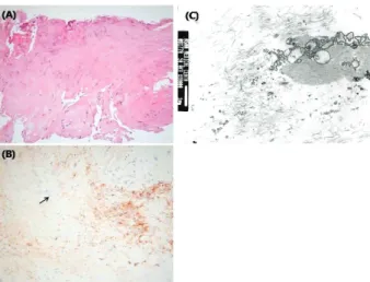
본원 내원 당시 환자의 나안시력은 우안 0.03, 좌안 1.0이었 으며, 공기 안압계로 측정한 안압은 우안 21 mmHg, 좌안 10 mmHg였다. 세극등 검사상 좌안은 정상 소견을 보였으나,우 안은 시축을 가리는 각막 상피하의 백색의 비대성 반흔 소견 이 보였으며, 병변은 각막 상피 아래쪽과 각막 실질의 앞쪽 에 위치하고 있었다(Fig. 1A). 비대성 반흔을 제거하기 위해 반흔 절제술을 시행하였다. 수술 시 비대 각막 반흔은 주변 정상 각막과 비교적 쉽게 분리 되었다. 광학 현미경 소견상 각막 실질부의 경화 소견을 보였으며(Fig. 2A), 면역 형광 염 색 검사상 smooth muscle actin 양성 소견을 보였다(Fig.
2B). 전자 현미경 검사 소견에서는 20 nm 크기의 콜라겐 섬 유가 관찰 되었다(Fig. 2C).수술 1개월 후 환자의 나안 시력 은 0.63으로 호전되었으며, 현성 굴절 검사상 정시 소견을 보였다. 세극등 검사상 일부 각막 상피 하 혼탁이 남아 있었 으나 시축을 가리는 비대 각막 반흔은 사라졌다(Fig. 1B).
Figure 3. (A) Anterior segment photographs at the first visit. Anterior segment photograph of the her left eye shows dense whitish subepithelial corenal opacity.
(B) Ultrasoud biomicroscope shows anterior stromal opacity. (White arrow) (C) After manual debridement, there was decreased corneal opacity which had covered visual axis.
Figure 2. (A) Light microscope shows stromal sclerosis. (H & E stain) (B) Smooth muscle actin positive. (Black arrow) (C) Electomicroscope shows 20 nm diameter collagen fibers. (Black arrow)
Figure 1. (A) Anterior segment photographs at the first visit. Anterior segment photograph of the right eye shows whitish subepithelial corenal opacity. (B) After manual debridement, there was decreased corneal opacity.
증례 2
내원 14개월 전 mitomycin C를 이용한 양안 라섹 수술 을 받은 23세 여자 환자가 좌안의 시력저하를 주소로 내원 하였다. 라섹 수술 후 환자의 나안 시력은 우안 1.0, 좌안 1.0이었다.
수술 12개월째 좌안의 각막 혼탁과 함께 시력저하가 진 행되어 본원으로 전원되었다. 내원 시 환자의 나안 시력은 우안 0.8, 좌안 0.2였으며, 공기 안압계로 측정한 안압은 우 안 15 mmHg, 좌안 17 mmHg였다. 세극등 검사상 우안은 정상 소견을 보였으나 좌안에서는 시축을 가리는 각막상피 하의 백색의 비대성 반흔 소견이 관찰되었다(Fig. 3A). 초 음파생체현미경(ultrasound biomicroscope)으로 각막 실질
의 앞쪽에 위치한 병변을 확인할 수 있었다(Fig. 3B). 비대 각막 반흔 제거술을 시행하였으며. 수술 시 일부의 비대 각 막 반흔이 남았으나 대부분 제거되었다. 광학 현미경 소견 상 퇴행성 콜라겐 섬유가 관찰되었고(Fig. 4A), 면역 형광 염색 검사상 Smooth muscle actin 양성 소견을 보였다 (Fig. 4B). 전자 현미경 검사에서는 콜라겐 섬유와 퇴행성 섬유아세포(Fibroblast) 소견이 관찰되었다(Fig. 4C). 수술 1개월 후 환자의 나안 시력은 0.16이었으며, 교정시력은 0.63으로 호전되었다. 현성 굴절 검사결과는 -0.25 D sph 였으며, 세극등 검사상 각막 상피하의 혼탁이 일부 남았으 나 시축을 가리는 비대 각막 반흔은 사라졌다(Fig. 3C).
Figure 4. (A) Light microscope shows degenerative fibrous tissue sclerosis. (H & E stain) (B) Smooth muscle actin positive. (Black arrow) (C) Electo- microscope shows collagen fibrils and degenerated fibroblast-like cells, consistent with corneal stroma.
고 찰
라섹 이후 시력에 영향을 주는 각막 혼탁의 발생은 8-10% 정도로 보고되고 있다.14-16 시력에영향을 미치는 각막 혼탁은, 각막 상피의 파괴로 분비되는 interleukin-1, transforming growth factor (TGF)-β, interleukin 6, epithelial growth factor 등의 사이토카인에 의한 각막 상 피 재생과 각막기질세포의 세포자멸사(apoptosis)에 기인
한다.17-21이러한 사이토카인들은 남아있는 각막 세포의 증
식과 활성화를 촉진시켜 새로운 각막 세포들을 증식시키고, 증식된 각막 세포들은 섬유아세포단계를 거쳐 근육섬유아 세포로 변형되는데, 이로 인해 각막기질에 콜라겐 같은 세 포 외 물질이 축적되고 그 배열에 변화가 생기므로 각막기 질의 혼탁도와 두께가 증가 한다고 알려져 있다.22,23 라섹 수술 후 발생하는 각막 혼탁을 예방하기 위해서는 수술 시 각막 상피의 손상을 최소화해야 하는데, 이를 위해서 최저 농도의 알코올을 최소 시간 동안 노출 시켜야 하며, 각막상 피절삭기(epikeratome)를 사용하여 각막 상피 및 보우만 층의 정상 구조를 유지 시키는 방법을 시도하고 있다.24수 술 후 발생하는 각막 혼탁을 예방하기 위해서 수술 중 mitomycin C를 사용하는데, mitomycin C는 각막 상피 하 각막기질세포의 분화를 억제하여 수술 후 각막 혼탁의 발 생을 예방한다고 알려져 있다.10-13
앞의 두 증례는 mitomycin C를 이용한 라섹 수술 1년 후 에 발생한 비대 각막 반흔이라는 특징을 보이고 있었으며, 굴절 교정 수술 이후 각막혼탁의 원인이 될 수 있는 Large treatment zone, 아토피 피부염, 자가 면역 질환, 수술 후
과도한 자외선의 노출 등이 없었으며 켈로이드(Keloid) 체 질 또한 없었다.25,26라섹 수술 후에 사이토카인에 대한 각 막기질세포의 반응으로 발생하는 각막 혼탁은 수술 1~3개 월 후에 최고 반응을 보이는데 mitomycin C는 이러한 각막 기질 세포 반응을 억제함으로써 수술 후 발생하는 각막 혼 탁의 발생을 예방한다. 두 증례 모두 각막 혼탁의 발생을 예방하기 위해 mitomycin C를 사용하였으나 수술 후 1년 이 경과 한 시점에서 과도한 비대성 반흔이 발생하였다. 이 는 mitomycin C가 세포자멸사에 의해 발생되는 사이토카 인의 분비 시점을 지연시켜, 수술 1년 후 지연성으로 과도 한 각막 기질 세포 반응이 발생하여 비대성 각막 반흔이 발 생한 것이라 생각된다.27
본 두 증례를 통해 라섹 수술 시 각막 혼탁의 발생을 예 방하기 위해 사용하는 mitomycin C가 시력에 심각한 영향 을 줄 수 있는 비대 반흔을 발생 시킬 수 있음을 확인할 수 있었다. Qazi et al은 Mitomycin C를 이용한 양안 라섹 수 술 이후 17개월째 발생한 단안의 비대 각막 반흔을 보고하 였으며, 각막 반흔 절제술 및 레이져각막절제술(Photo- therapeutic keratectomy)을 통하여 성공적으로 치료하였 다.27Qazi et al이 보고한 증례에서도 본 증례와 같이 수술 이후 각막혼탁의 원인이 될 수 있는 Large treatment zone, 아토피 피부염, 자가 면역 질환, 수술 후 과도한 자외선의 노출 등의 과거력은 없었다.
Mitomycin C를 이용한 라섹 수술 후 비대 각막 반흔이 발생한 경우는 전세계적으로 1례가 보고된 바 있으며 국내 에서는 최초로 보고되는 것이다.27저자들은 라섹 수술 후 발생한 지연성 비대 각막 반흔의 증례를 경험하고, 이는 드 물게 발생하지만 시력에 심각한 영향을 줄 수 있는 중요한 합병증이라 생각되어 이를 보고하는 바이다.
참고문헌
1) Taneri S, Zieske JD, Azar DT. Evolution, techniques, clinical outcomes, and pathophysiology of LASEK: Review of the literature. Surv Ophthalmol 2004;49:576-602.
2) Camellin M. Laser epithelial keratomileusis for myopia. J Refract Surg 2003;19:666-70.
3) Claringbold TV II. Laser-assisted subepithelial keratectomy for the correction of myopia. J Cataract Refract Surg 2002;28:18-22.
4) Brancato R, Schiavone N, Siano S, et al. Prevention of corneal keratocyte apoptosis after argon fluoride excimer laser irradia- tion with the free radical scavenger ubiquinone Q10. Eur J Ophthalmol 2000;10:32-8.
5) Wang MX, Gray TB, Park WC, et al. Reduction in corneal haze and apoptosis by amniotic membrane matrix in excimer laser photoablation in rabbits. J Cataract Refract Surg 2001;27: 310-9.
6) Vinciguerra P, Camesasca FI, Ponzin D. Use of amino acids in refractive surgery. J Refract Surg 2002;18:374-7.
7) Corbett MC, O'Brart DP, Patmore AL, Marshall J. Effect of collagenase inhibitors on corneal haze after PRK. Exp Eye Res 2001;72:253-9.
8) Vetrugno M, Maino A, Cardia G, et al. A randomised, double masked, clinical trial of high dose vitamin A and vitamin E supplementation after photorefractive keratectomy. Br J Ophthalmol 2001;85:537-9.
9) Brancato R, Fiore T, Papucci L, et al. Concomitant effect of topical ubiquinone Q10 and vitamin E to prevent keratocyte apoptosis after excimer laser photoablation in rabbits. J Refract Surg 2002;18:135-9.
10) Yee RW, Yee SB. Update on laser subepithelial keratectomy (LASEK). Curr Opin Ophthalmol 2004;15:333-41.
11) Kim ES, Jin KH. Evaluation of the prophylactic use of mitomycin to inhibit haze formation after LASEK. J Korean Ophthalmol Soc 200748:623-9.
12) Verweij J, Pinedo HM. Mitomycin C: mechanism of action, usefulness and limitations. Anticancer Drugs 1990;1:5-13.
13) Xu H, Liu S, Xia X, et al. Mitomycin C reduces haze formation in rabbits after excimer laser photorefractive keratectomy. J Refract Surg 2001;17:342-9.
14) Rouweyha RM, Chuang AZ, Mitra S, et al. Laser epithelial keratomileusis for myopia with the autonomous laser. J Refract Surg 2002;18:217-24.
15) Chalita MR, Tekwani NH, Krueger RR. Laser epithelial keratomileusis:
outcome of initial cases performed by an experienced surgeon. J Refract Surg 2003;19:412-5.
16) Kim JK, Kim SS, Lee HK, et al. Laser in situ keratomileusis versus laser assisted subepithelial keratectomy for the correction of high myopia. J Cataract Refract Surg 2004;30:1405-11.
17) Baldwin HC, Marshall J. Growth factors in corneal wound healing following refractive surgery: a review. Acta Ophthalmol Scand 2002;80:238-47.
18) Nakamura K, Kurosaka D, Bissen-Miyajima H, Tsubota K.
Intact corneal epithelium is essential for the prevention of stromal haze after laser assisted in situ keratomileusis. Br J
Ophthalmol 2001;85:209-13.
19) Wilson SE, Mohan RR, Hong JW, et al. The wound healing response after laser in situ keratomileusis and photorefractive keratectomy; elusive control of biological variability and effect of custom laser vision correction. Arch Ophthalmol 2001;
119:889-96.
20) Kaji Y, Soya K, Amano S, et al. Relation between corneal haze and transforming growth factor-beta1 after photorefractive keratectomy and laser in situ keratomileusis. J Cataract Refract Surg 2001;27:1840-6.
21) Kuo IC, Seitz B, LaBree L, McDonnell PJ. Can zinc prevent apoptosis of anterior keratocytes after superficial keratectomy.
Cornea 1997;16:550-5.
22) Corbett MC, Prydal JI, Verma S, et al. An in vivo investi- gation of the structures responsible for corneal haze after photorefractive keratectomy and their effect on visual function.
Ophthalmology 1996;103:1366-80.
23) Moller-Pedersen T, Cavanagh HD, Petroll WM, Jester JV. Stromal wound healing explains refractive instability and haze development after photorefractive keratectomy; a 1-year confocal microscopic study. Ophthalmology 2000;107:1235-45.
24) Pallikaris IG, Naoumidi II, Kalyvianaki MI, Katsanevaki VJ.
Epi-LASIK: comparative histological evaluation of mechanical and alcohol assistedepithelial separation. J Cataract Refract Surg 2003;29:1496-501.
25) Lin N, Yee SB, Mitra S, et al. Prediction of corneal haze using an ablation depth/corneal thickness ratio after laser epithelial keratomileusis. J Cataract Refract Surg 2004;20:797-802.
26) Cua IY, Pepose JS. Late corneal scarring after photorefractive keratectomy concurrent with the development of systemic lupus erythematosus. J Cataract Refract Surg 2002;18:750-2.
27) Qazi MA, Johnson TW, Pepose JS. Development of late-onset subepithelial corneal haze after laser-assisted subepithelial keratectomy with prophylactic intraoperative mitomycin-C. J Cataract Refract Surg 2006;32:1573-8.
=ABSTRACT=
Late-onset Hypertrophic Corneal Scars After Laser-assisted Subepithelial Keratectomy With Mitomycin C
Dong Yoon Kim, MD, Myoung Joon Kim, MD, PhD, Sam Young Yoon, MD, Chul Jin Shin, MD, Kyoung Hoon Kim, MD, Hungwon Tchah, MD, PhD
Department of Ophthalmology, University of Ulsan College of Medicine, Asan Medical Center, Seoul, Korea
Purpose: To report late-onset hypertrophic corneal scars after laser epithelial keratomileusis (LASEK) with mitomycin C.
Case summary: Case 1. A 34-year-old man who had undergone LASEK with mitomycin C 15 months prior was referred to our clinic because of corneal opacity of his right eye. After LASEK, there have been no abnormalities in either of his eyes. However, 11 monthsafter LASEK, he experienced decreased visual acuity in his right eye. The visual acuity was 0.03 in his right eye and 1.0 in his left eye. On slit lamp examination there was a whitish, hypertrophic scarin his right cornea. The lesion was located in the corneal center and the subepithelial space. Central corneal thickness was 828 µm. Case 2. A 23-year-old woman who had undergone LASEK with mitomycin C 14 months before was referred our clinic because of corneal opacity of her left eye.
After LASEK, there had been no abnormalities in either of her eyes. However, 12 months after LASEK she experienced decreased visual acuity in her left eye. The visual acuity was 1.0 in her right eye and 0.2 in her left eye. On slit lamp examination there was a whitish, hypertrophic scar in her left cornea. Central corneal thickness was 794 µm.
Conclusions: Manual debridement was performed to remove the hypertrophic scar in both cases. Case 1. After manual debridement, visual acuity of the right eye improved to 0.63. Case 2. After manual debridement, best-corrected visual acuity of the left eye was 0.63.
J Korean Ophthalmol Soc 2009;50(2):308-312 Key Words: Corneal opacity, LASEK, Mitomycin
Address reprint requests to Hungwon Tchah, MD, PhD
Department of Ophthalmology, University of Ulsan College of Medicine
#388-1 Pungnap-2dong, Songpa-gu, Seoul 138-736, Korea
Tel: 82-2-3010-3674, Fax: 82-2-470-6440, E-mail: [email protected]