ISSN 0378-6471 (Print)⋅ISSN 2092-9374 (Online)
https://doi.org/10.3341/jkos.2017.58.11.1234
Original Article
안면화염혈관종 환자에서의 녹내장 발생양상 및 치료
Clinical Presentation and the Treatment of Glaucoma in Patients with a Facial Port-wine Stain
김미진1,2⋅이원준1,2⋅박기호1,2⋅김태우1,3⋅이은지1,3⋅유영석1,2⋅정진욱1,2
Mi Jin Kim, MD1,2, Won June Lee, MD1,2, Ki Ho Park, MD, PhD1,2, Tae-Woo Kim, MD, PhD1,3, Eun Ji Lee, MD, PhD1,3, Young Suk Yu, MD, PhD1,2, Jin Wook Jeoung, MD, PhD1,2
서울대학교 의과대학 안과학교실1, 서울대학교병원 안과2, 분당서울대학교병원 안과3 Department of Ophthalmology, Seoul National University College of Medicine1, Seoul, Korea
Department of Ophthalmology, Seoul National University Hospital2, Seoul, Korea Department of Ophthalmology, Seoul National University Bundang Hospital3, Seongnam, Korea
Purpose: To characterize the development of glaucoma, age of glaucoma onset, and treatments for patients with a facial port-wine stain (PWS).
Methods: We performed a retrospective analysis of the medical records of 58 patients (116 eyes) with facial PWS between January 2000 and August 2016. We noted patients’ age at the initial examination, cup-to-disc ratio, corneal diameter, occurrence of ocular hypertension, development of glaucoma, age of glaucoma onset, and treatments. We compared the clinical features of eyes that developed glaucoma with those that did not develop glaucoma. Among those eyes with glaucoma, we investigated the differences between eyes that underwent surgery and those that did not undergo surgery.
Results: Among the 58 patients with a facial PWS (116 eyes), glaucoma was diagnosed in 38 patients (46 eyes; 39.66%). Of these, 26 patients (27 eyes; 58.69%) underwent glaucoma surgery. PWS-associated glaucoma usually developed by the age of 2 years (85.61%). In all patients, glaucoma developed on the same side of the face as the PWS. Of the 58 patients, 19 (32.76%) showed neurological symptoms, including seizures, developmental delays, intellectual disabilities, or hemiplegia, and 32 (55.17%) were di- agnosed with Sturge-Weber syndrome. The mean number of glaucoma surgeries was 1.55 ± 0.93. The initial surgery included tra- beculectomy (7 eyes), trabeculotomy (5 eyes), combined trabeculotomy/trabeculectomy (13 eyes), and aqueous drainage device insertion (2 eyes). The mean age at the first surgery was 35.14 ± 50.91 months. In 18 of 27 eyes (66.67%), the postoperative intra- ocular pressure (IOP) was controlled to below 21 mmHg, but 9 eyes (33.33%) showed elevated IOP and required a reoperation.
Conclusions: PWS can be accompanied by ocular hypertension or glaucoma, so patients require regular ophthalmic examinations.
When glaucoma occurs, it often does not respond to medication, making it difficult in some cases to control the IOP, so appro- priate glaucoma surgery is necessary.
J Korean Ophthalmol Soc 2017;58(11):1234-1241
Keywords: Facial angioma, Facial port-wine stain, Glaucoma, Nevus flammeus, Sturge-Weber syndrome
■Received: 2017. 7. 6. ■ Revised: 2017. 9. 18.
■Accepted: 2017. 10. 20.
■Address reprint requests to Jin Wook Jeoung, MD, PhD Department of Ophthalmology, Seoul National University Hospital, #101 Daehak-ro, Jongno-gu, Seoul 03080, Korea Tel: 82-2-2072-2438, Fax: 82-2-741-3187
E-mail: neuroprotect@gmail.com
*Conflicts of Interest: The authors have no conflicts to disclose.
ⓒ2017 The Korean Ophthalmological Society
This is an Open Access article distributed under the terms of the Creative Commons Attribution Non-Commercial License (http://creativecommons.org/licenses/by-nc/3.0/) which permits unrestricted non-commercial use, distribution, and reproduction in any medium, provided the original work is properly cited.
안면화염혈관종(Port wine stain, Nevus flammeus)은 선 천적 혈관성 병변으로 신생아의 약 0.3-0.5%에서 발생한 다.1 주로 삼차 신경의 제1분지와 제2분지가 분포하는 부 위에서 편측으로 나타나지만 제3분지 및 상부 경신경이 분포하는 부위까지 침범할 수 있고 10-20%는 양측성으로 발생한다.2
대부분 산발적으로 발생해서 피부 병변 단독으로 있지 만 몇몇은 눈 또는/그리고 동측 연막의 혈관성 기형이 같 이 발생하여 결막이나 상공막 혈관 확장, 맥락막 혈관종, 홍채이색증, 포도막 흑색종, 연질뇌막 다발성혈관종 등이 동반되기도 한다.3,4 또한 안면화염혈관종, 안구의 혈관종, 두개강내혈관종 등의 특징적인 징후를 보이는 스터지-웨 버 증후군(Sturge-Weber syndrome)이나 클리펠-트레노우 네이-웨버 증후군(Klippel-Trenaunay-Weber syndrome)에 서 나타나는 소견이기도 하다.5,6
특히 안압이 상승하거나 녹내장을 동반할 수 있고 약물 이나 수술로 치료하기 어렵다는 연구들이 많아 소아에서 안면화염혈관종이 확인되는 경우 안과적 검진을 시행하 고, 녹내장이 진단되는 경우에는 적절한 치료를 시행하는 것이 중요하다.7-9 그러나 현재까지 안면화염혈관종 환자 에서의 녹내장 발생에 대한 국내 보고는 많지 않다. 안기 형을 동반한 스터지-웨버 증후군 환자 증례보고10-14, 스터 지-웨버 증후군에 동반된 녹내장 환자에서 섬유주절제술 을 시행한 증례보고가 있었으나15-18 각각 1명의 환자를 대 상으로 했다. 이후 녹내장이 발생한 스터지-웨버 증후군 환자에 대한 임상양상과 치료방법에 대한 보고가 있었으 나 섬유주절제술을 시행 받은 환자만을 대상으로 시행한 연구였다.19 본 연구에서는 안면화염혈관종 환자에서의 안 과적 임상양상, 녹내장 발생여부와 시기, 치료방법 등을 조사하여 보고하는 바이다.
대상과 방법
2000년 1월부터 2016년 8월까지 서울대학교병원과 분 당서울대학교병원에서 안면화염혈관종으로 안과검진이 의뢰된 77명, 154안 중 관찰 기간이 6개월 미만 또는 외래 관찰 횟수가 2번 미만인 경우 대상에서 제외하고 58명, 116안의 의무기록을 후향적으로 검토하였다. 이들을 대상 으로 초진 시 연령, 시신경유두함몰 비율, 각막 직경 및 안 압 상승여부, 녹내장의 발생여부와 시기, 수술을 시행한 환아에서 첫 수술 시 연령, 술 전 및 술 후 안압, 수술 후 안압약의 사용 유무 등을 조사하였다. 이 연구는 본원 의 학연구윤리 심의위원회의 승인하에 진행되었다(IRB No, H-1703-152-840, B-1708-417-403). 안압은 8세 이상의 협 조가 되는 환아에서는 골드만압평안압계(Goldman’s ap- planation tonometer)로 측정하였고, 8세 미만 또는 골드만 압평안압계로 협조가 안 되는 경우 TonoPen® 안압계 (Reichert Inc., Depew, NY, USA), 2012년 이후부터는 리 바운드 안압계(I-Care tonometer, Icare®, Helsinki, Finland) 로 측정하였다.
녹내장 진단은 1) 안압이 21 mmHg 이상, 2) 시신경유 두의 녹내장성 변화(시신경유두함몰비가 0.4 이상이거나 양안 차이가 비대칭적으로 0.2 이상인 경우, 각막 혼탁으 로 안저검진이 불가능한 경우 제외), 3) 수평각막 직경이 1세 미만의 경우 10.5 mm 초과, 1세 이상의 경우 12.0 mm 초과로, 1)을 충족하면서 2) 또는 3)을 동반하는 경우 로 진단하였다.20
외래 경과관찰 중 점안제재의 최대 허용 약물요법(베타 차단제, 프로스타글란딘, 탄산탈수효소억제제)으로 안압 이 목표 안압(21 mmHg) 범위 내로 조절되지 않거나 연속 된 시신경유두 검진에서 수직유두함몰비의 증가, 시신경 유두테의 얇아짐 등의 시신경 손상 진행이 관찰될 때, 녹 내장 전문의의 판단하에 녹내장 수술 여부를 결정하였다. 1차 수술 후 안압이 조절되는 경우에는 안압하강제를 모 두 중단하고 경과관찰 하였으나, 수술 후에도 안압이 21 mmHg 미만으로 조절되지 않거나 시신경 손상의 진행, 시 야 소실 같은 합병증이 발생하는 경우 안압하강제를 지속 적으로 점안하면서 정기적인 추적관찰을 시행하거나 추 가적인 녹내장 수술을 시행하였다.
Statistical package for social science version 16.0 pro- gram (IBM Corp., Armonk, NY, USA)을 이용하여 통계 분석을 시행하였다. 수술 전후 안압 비교는 Paired t-test를 이용하였고, 그 외 연속형 변수는 independent t-test, 범주형 변수는 Pearson’s chi-squared test로 분석하였다. Analysis of variance (ANOVA) 분석을 통해 안면화염혈관종 침범이 없는 눈, 안면화염혈관종 침범과 녹내장이 있는 눈, 안면 화염혈관종 침범이 있지만 녹내장이 없는 눈의 첫 외래 시 각막 직경, 시신경유두함몰 비율, 안압, 마지막 외래 시 안압의 평균차이를 비교하고, 사후분석(post hoc)을 실시 하여 유의수준을 적절히 보정한 상태에서 전체 유의수준 5%를 지키기 위해 Tukey 방법을 사용해 세 군을 비교했 다. 통계적 유의성은 p<0.05로 정의하였다.
결 과
대상환자 58명 116안이었으며, 남자는 27명, 여자는 31 명이었다. 안면화염혈관종이 편측에서 발생한 경우가 46 명(79.31%), 양측에서 발생한 경우가 12명(20.68%)이었으 며 모든 환자에서 삼차신경의 제1분지를 침범하였다(Fig.
1). 대상환자 58명 중 19명(32.76%)에서 경련, 발달지연, 정신지체, 편마비 등의 신경학적 증상을 동반하고, 32명 (55.17%)에서 스터지-웨버증후군을 진단 받았다.
단안 또는 양안에 안면화염혈관종이 있는 환자 58명의 116안 중에서 한 눈이라도 녹내장을 진단 받은 환자는 38
A B
C
Figure 1. Sturge-W eber syndrome patient who underwent trabeculectomy (patient 6). (A) Distribution of left-sided facial port-wine
stain that does not cross the midline, and left-sided glaucoma. (B) One-week postoperative appearance. Mildly engorged con- junctival and episcleral vessels with hypovascular bleb. (C) Optic disc photography revealing deep cupping with a cup/disc ratio of 0.9 in the left eye compared to right eye.Table 1. Clinical characteristics of patients with a port-wine stain
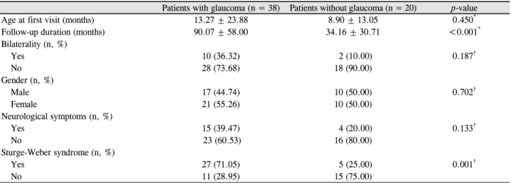
Patients with glaucoma (n = 38) Patients without glaucoma (n = 20) p-value
Age at first visit (months) 13.27 ± 23.88 8.90 ± 13.05 0.450*
Follow-up duration (months) 90.07 ± 58.00 34.16 ± 30.71 <0.001*
Bilaterality (n, %)
Yes 10 (36.32) 2 (10.00) 0.187†
No 28 (73.68) 18 (90.00)
Gender (n, %)
Male 17 (44.74) 10 (50.00) 0.702†
Female 21 (55.26) 10 (50.00)
Neurological symptoms (n, %)
Yes 15 (39.47) 4 (20.00) 0.133†
No 23 (60.53) 16 (80.00)
Sturge-Weber syndrome (n, %)
Yes 27 (71.05) 5 (25.00) 0.001†
No 11 (28.95) 15 (75.00)
Values are presented as the mean ± SD or n (%) unless otherwise indicated.
*A t-test was used for continuous variables; †A chi-square test was used for categorical variables.
명(65.52%), 녹내장을 진단 받지 않은 환자는 20명 (34.48%)이었다. 녹내장을 진단 받은 환자와 녹내장을 진 단 받지 않은 환자의 비교는 Table 1과 같다. 녹내장이 발 생한 군에서 그렇지 않은 군에 비해 유의하게 경과관찰 기간이 길었으며(p<0.001) 스터지-웨버 증후군이 더 많이
동반되었다(p<0.001).
눈을 기준으로 정리해 보았을 때 116안 중 46안(39.66%) 에서 녹내장이 발생하고 70안(60.34%)에서 녹내장이 발 생하지 않았다. 안면화염혈관종이 있는 눈과 없는 눈, 안 면화염혈관종이 있는 눈 중에서도 녹내장이 발생한 눈과
Table 2. Clinicopathological characteristics summarized by eyes of patients with PW S (n = 116)
Eyes withoutPWS involvement
(n = 46)1
Eyes with PWS involvement (n = 70)
P1 P2
Total
Eyes with glaucoma (n = 46)2
Eyes without glaucoma (n = 24)3
Cornea diameter at first visit (mm) 10.59 ± 0.86 11.42 ± 1.99 12.03 ± 0.92 10.45 ± 1.01 <0.001 0.005*, 0.014†, <0.001‡ C/D ratio at first visit 0.30 ± 0.10 0.53 ± 0.19 0.61 ± 0.16 0.31 ± 0.07 <0.001 <0.001*, 0.955†, <0.001‡ IOP baseline (mmHg) 16.87 ± 8.56 21.11 ± 10.41 24.24 ± 9.72 15.09 ± 9.12 <0.001 <0.001*, 0.725†, <0.001‡ IOP last follow-up (mmHg) 13.52 ± 2.91 15.66 ± 3.80 16.22 ± 4.15 14.55 ± 2.72 0.001 <0.001*, 0.477†, 0.142‡ Values are presented as mean ± SD unless otherwise indicated. ‘P1’ means ‘comparison between 1 & 2 & 3 (analysis of variance [ANOVA])’, and ‘P2’ means ‘*Comparison between 1 and 2 by post hoc test; †Comparison between 1 and 3 by post hoc test; ‡Comparison between 2 and 3 by post hoc test’.
PWS = port wine stain; C/D = cup/disc; IOP = intraocular pressure.
Table 3. Treatment of glaucoma eyes with port-wine stain
Eyes with glaucoma (46 eyes) %
No surgical intervention 19 17.39
Surgical intervention 27 58.69
TLO 7 25.93
TLE 5 18.52
TLO + TLE 13 48.15
Valve surgery only 2 7.41
Operation ≥2 9 33.33
No. of medications following surgery
No therapy 6 22.22
Monodrug therapy 8 29.63
Multidrug therapy 13 48.15
TLO = trabeculotomy; TLE = trabeculectomy.
발생하지 않은 눈의 첫 검진 시 차이는 Table 2와 같다.
안면화염혈관종 침범이 없는 눈, 안면화염혈관종 침범이 있고 녹내장이 있는 눈, 안면화염혈관종 침범이 있지만 녹 내장이 없는 눈의 첫 외래 시 각막 직경, 시신경유두함몰 비율, 평균안압, 마지막 외래 시 평균안압을 비교했을 때 4가지 변수에서 모두 통계적으로 유의한 차이가 있었고 (Table 2, P1) 사후 분석 결과, 안면화염혈관종이 있으면 서, 녹내장이 있는 눈과 없는 눈 사이에 첫 외래 시의 각 막 직경, 시신경유두함몰 비율, 안압에서 유의한 차이가 있었다(Table 2, P2).
녹내장을 진단 받은 46안 중 38안(82.61%)은 2세 이전 에 녹내장 진단을 받았고 8안(17.39%)은 그 이후에 진단 받았다. 녹내장 수술까지 시행한 경우는 27안(58.69%)으 로 첫 수술나이는 평균 35.14 ± 50.91개월, 평균 수술횟수 는 1.55 ± 0.93회였다. 양측에 안면화염혈관종이 관찰된 12명의 환자 중 10명에서 녹내장이 생겼고, 8명은 양안에, 2명은 단안에 녹내장이 발생했다. 녹내장은 안면화염혈관 종이 존재하는 동측에서 발생했고, 단안에서만 녹내장이 있었던 환자에서 경과관찰 기간 동안 반대편 눈에서 녹내 장이 발생하지는 않았다.
첫 수술로 섬유주절개술 7안(25.93%), 섬유주절제술 5안 (18.52%), 섬유주절개술 및 섬유주절제술 병합 13안(48.15%), 방수유출장치삽입술 2안(7.41%) 시행하였다(Table 3). 수술 받은 눈의 수술 전 안압하강제의 사용개수는 평균 1.72 ± 0.84개(1-3개)였으며, 수술 후에는 평균 1.29 ± 0.99개(0-3 개)였다. 18안(66.67%)은 수술 후 안압하강제를 사용하더라 도 안압이 21 mmHg 미만으로 조절되었으나 9안(33.33%) 은 안압하강제를 사용하더라도 안압이 21 mmHg 미만으 로 조절되지 않거나 시신경 손상의 진행, 시야 소실 같은 합병증이 발생하여 추가적인 녹내장 수술을 하였다.
녹내장으로 진단 받은 총 46안 중, 수술을 받은 27안의 수술 직전 외래에서 측정한 안압은 평균 27.66 mmHg, 마 지막 외래에서 측정한 안압은 평균 16.22 mmHg, 약물치 료만 받은 19안의 약물치료 전 안압은 평균 21.75 mmHg, 마지막 외래에서 측정한 안압은 평균 16.64 mmHg로 각각 유의하게 감소했다(p<0.001, p=0.010, paired t-test). 녹내장 수술을 받은 눈과 약물치료로 조절된 눈(Supplementary Table 1)의 녹내장 진단시기, 첫 외래 방문 시 각막 직경, 시 신경유두함몰 비율, 첫 외래 평균안압, 마지막 외래 평균안압 (p=0.363, p=0.265, p=0.341, p=0.147, p=0.573, Independent
t-test) 및 마지막 외래 방문 시 점안 안약개수(p=0.145, Fischer’s exact test)의 차이는 없었고, 첫 외래 방문 시 점 안 안약개수만 유의하게 차이가 있었다(p=0.039, Fischer's exact test). 적어도 10년 이상 추적관찰 중인 환자 13명 중 약물치료만 받고 있는 환자는 2명, 녹내장 수술을 시행한 환자는 11명(Supplementary Table 2)으로, 수술 후 안압은 21 mmHg 미만으로 조절되고 있다.
고 찰
안면화염혈관종은 삼차신경이 지배하는 부위에서 주로 나타나는 양성의 피부혈관 기형으로 대개 출생 시부터 저 명하게 관찰되고 매끄러운 표면의 분홍색을 띠다가 나이 가 들수록 색깔이 짙어지고 비후되며 울퉁불퉁한 결절성 병변으로 진행한다.1 신생아 1,000명당 3명의 빈도로 발생 하고 비교적 흔한 질환이나, 이 환자 중 녹내장의 동반 여 부에 대한 정확한 조사 및 임상적 특징에 대해 연구한 적 은 없었다. 이에 안면화염혈관종 환자의 임상적 특징과 녹 내장 발생, 치료 및 예후 예측 등에 대해 도움을 주고자 본 연구를 시행하였다.
연구에 따라 다양한 빈도로 보고되고 있지만 안면화염 혈관종 환자에서 약 30%의 빈도로 녹내장이 발생하는 것 으로 알려져 있다.1 본 연구에서는 안면화염혈관종이 있는 환자를 대상으로 녹내장 발생에 대해 조사하였고 이는 문 헌 고찰상 국내에서는 유일한 보고이다. 안면화염혈관종 이 있는 58명 116안 중 38명 46안(39.66%)에서 녹내장이 발생하였고 모두 녹내장이 발생한 눈과 같은 방향에서 안 면화염혈관종을 보였다. 이 결과는 안면화염혈관종 환자 가 내원할 시 혈관종이 분포하는 눈을 더욱 주의 깊게 검 진할 필요가 있음을 의미한다. 이는 안면화염혈관종 환자 에서 녹내장 발생이 유의한 관련이 있다는 연구와도 일치 한다.1 본 연구에서도 안면화염혈관종 침범이 있으면서 녹 내장이 있는 눈과 없는 눈의 비교에서 뿐만 아니라, 안면 화염혈관종 침범이 없는 눈과 녹내장은 없지만 안면화염 혈관종 침범이 있는 눈의 비교에서 각막 직경의 유의한 차이가 있었으므로(Table 2, p=0.014, post hoc test) 각막 직경 측정을 포함한 면밀한 검진을 해야겠다. 뿐만 아니라 녹내장 진행으로 인한 시기능 손상을 피하고 삼출성 박리 등의 망막 합병증을 빨리 발견하기 위해 추적관찰에서도 완전한 안과검진을 하는 것이 중요하겠다.
본 연구에서는 화염상모반으로 내원한 환자들의 녹내 장의 발생시기를 알아보고자 하였고, 상당수(85.61%)가 2 세 이전에 녹내장 진단을 받았다. 어느 시기에도 발생할 수 있지만 약 60%는 선천적으로 또는 만 2세 이전에, 약
40%는 그 이후에 발생한다는 이전의 보고와도 일치하였
다.1,7,20,21 또한 당장의 녹내장이 없더라도 추적 관찰 기간
이 길어질수록 녹내장 발생 위험이 높아진다는 연구22를 바탕으로 정기적인 경과관찰 및 평가가 필요하다는 것을 알 수 있다.
안면화염혈관종이 있는 환아가 신경학적 증상을 보이 거나 뇌자기공명영상에서 연수막 혈관이상을 동반할 때 스터지-웨버 증후군을 의심한다.9,23 신경학적 증상으로는 경련발작이 흔하고 두통, 반신마비, 발달지연, 지적장애 등이 발생하기도 한다.3 특히 윗눈꺼풀을 침범하면 눈과 중추신경계 이상소견을 가질 위험성이 증가한다.5,24,25 본 연 구에서는 58명 중 19명(32.76%)에서 경련, 발달지연, 정신 지체, 편마비 등의 신경학적 증상을 동반하고, 32명(55.17%)에 서 스터지-웨버 증후군을 진단 받았다. 문헌26,27에서 안면 화염혈관종 환자의 약 10-20%에서 스터지-웨버 증후군이 확인된다는 것과 비교하여 그 빈도가 높았는데 이는 소아 과, 신경과, 피부과 등과 협진을 통한 다각적인 접근을 했 기 때문으로 판단된다. 안 증상이 동반될 경우, 신경학적 증상 및 스터지-웨버 증후군의 동반 여부에 대한 추가적 인 검사가 필요할 수 있음을 시사한다.
안면화염혈관종 환자에서 발생한 녹내장은 안압을 조 절하기 어렵고7-9 약물치료에 잘 반응하지 않아서 최대약 물요법을 유지하며 지켜보거나 수술을 하는 경우가 빈번 하다. 본 연구에서도 녹내장을 진단 받은 환자 중 녹내장 수술까지 시행한 경우는 58.69%, 평균 수술 횟수도 1.55 회였다. 특히 스터지웨버증후군을 동반한 환자에서의 녹 내장 수술은 얕은전방, 전방출혈, 맥락막박리, 맥락막삼출, 망막박리 등의 합병증이 발생할 수 있어 각 환자의 특성 에 따라 전방각절개술, 섬유주절개술, 섬유주절제술, 섬모 체파괴술, 방수유출장치 삽입술 등 적합한 치료 방향을 설 정하는 것이 필요하겠다.19,20
Enjolras et al27은 삼차 신경의 제1분지가 분포하는 부 위에 혈관종이 없는 환자에서는 눈의 이상이나 뇌의 연수 막 혈관이상이 동반되지 않았음을 보고하였다. 본 연구에 서는 모든 증례에서 삼차신경의 제1분지를 침범하고 있어 얼굴뿐만 아니라 목, 몸체, 사지 등의 분포에 따른 녹내장 의 발생 위험도 차이는 확인하지 못했다. 또한 안면화염혈 관종 환자에서 발생한 녹내장에서 수술 성적의 보고는 적 은 대상 환자 수로 해석에 제한점이 있을 수 있고, 약물치 료만 시행한 환자의 경우 경과관찰 기간이 동일하지 않았 으며, 약물치료 후 10년 이상 경과관찰한 환자의 수가 제 한되어 이를 해석하는 데 제한이 있을 수 있다. 소아 환자 를 대상으로 하다 보니 환아의 협조도나 깨어있는 상태 또는 마취 상태에서 측정한 경우가 안압 수치에 영향을
끼칠 수 있으므로 안압 측정오차를 보정한 충분한 대상자 의 결과를 분석해 보는 전향적인 연구가 필요하겠다.
본 연구는 국내 최초로 세심한 병력청취와 안과적 검사 를 바탕으로 안면화염혈관종 환자에서의 녹내장 발생에 대해 보고하였다. 안면화염혈관종 환자의 약 40%에서 녹 내장이 발생하였고, 이와 동반된 녹내장은 주로 2세 미만 의 어린 나이에 발생하며 수술적 치료가 필요한 경우가 많았다. 그러므로 안면화염혈관종이 확인되는 경우 정기 적인 안과적 검진을 시행하고 적절한 치료를 늦지 않게 시작하는 것이 중요할 것으로 사료된다.
REFERENCES
1) Khaier A, Nischal KK, Espinosa M, Manoj B. Periocular port wine stain: the great ormond street hospital experience. Ophthalmology 2011;118:2274-8.e1.
2) Dutkiewicz AS, Ezzedine K, Mazereeuw-Hautier J, et al. A pro- spective study of risk for Sturge-Weber syndrome in children with upper facial port-wine stain. J Am Acad Dermatol 2015;72:473-80.
3) Ch'ng S, Tan ST. Facial port-wine stains - clinical stratification and risks of neuro-ocular involvement. J Plast Reconstr Aesthet Surg 2008;61:889-93.
4) Sullivan TJ, Clarke MP, Morin JD. The ocular manifestations of the Sturge-Weber syndrome. J Pediatr Ophthalmol Strabismus 1992;29:349-56.
5) Mazereeuw-Hautier J, Syed S, Harper JI. Bilateral facial capillary malformation associated with eye and brain abnormalities. Arch Dermatol 2006;142:994-8.
6) Hofeldt AJ, Zaret CR, Jakobiec FA, et al. Orbitofacial angiomatosis.
Arch Ophthalmol 1979;97:484-8.
7) Iwach AG, Hoskins HD Jr, Hetherington J Jr, Shaffer RN. Analysis of surgical and medical management of glaucoma in Sturge-Weber syndrome. Ophthalmology 1990;97:904-9.
8) Hennedige AA, Quaba AA, Al-Nakib K. Sturge-Weber syndrome and dermatomal facial port-wine stains: incidence, association with glaucoma, and pulsed tunable dye laser treatment effectiveness.
Plast Reconstr Surg 2008;121:1173-80.
9) Ong T, Chia A, Nischal KK. Latanoprost in port wine stain related paediatric glaucoma. Br J Ophthalmol 2003;87:1091-3.
10) Kim SU, Kim YZ, Hong YJ, Kim HB. A case of Sturge-Weber syndrome. J Korean Ophthalmol Soc 1981;22:273-6.
11) Chung I, Jang SG, Lew HM. A case of Sturge-Weber syndrome. J Korean Ophthalmol Soc 1986;27:723-8.
12) Choi JS, Yi KP, Hong KY. A case of Sturge-Weber syndrome. J Korean Ophthalmol Soc 1989;30:459-64.
13) Chung IH, Kim MH. Sturge-Weber syndrome with congenital ocu- lar anomaly. J Korean Ophthalmol Soc 1995;36:2266-70.
14) Lee H, Choi SS, Kim SS, Hong YJ. A case of glaucoma associated with Sturge-Weber syndrome and Nevus of Ota. Korean J Ophthalmol 2001;15:48-53.
15) Kim JW, Park CH, Lee CH. Clinical experience of treatment in a case of Sturge-Weber syndrome with bilateral glaucoma. J Korean Ophthalmol Soc 1996;37:908-12.
16) Bae JH, Cho HD, Woo SC. Serous retinal detachment following trabeculectomy in a case of Sturge-Weber syndrome with glaucoma.
J Korean Ophthalmol Soc 1996;37:2150-3.
17) Lee SH, Jung DY. Choroidal effesuion after trabeculectomy in glaucoma with Sturge-Weber syndrome. J Korean Ophthalmol Soc 1999;40:1164-8.
18) Yuen NS, Wong IY. Congenital glaucoma from Sturge-Weber syn- drome: a modified surgical approach. Korean J Ophthalmol 2012;
26:481-4.
19) Park JH, Lim SH, Cha SC. Clinical features and surgical outcomes of Sturge-Weber syndrome with glaucoma. J Korean Ophthalmol Soc 2013;54:1737-47.
20) Sharan S, Swamy B, Taranath DA, et al. Port-wine vascular mal- formations and glaucoma risk in Sturge-Weber syndrome. J AAPOS 2009;13:374-8.
21) Parsa CF. Sturge-weber syndrome: a unified pathophysiologic mechanism. Curr Treat Options Neurol 2008;10:47-54.
22) Miller SJ. Symposium: the Sturge-Weber syndrome. Proc R Soc Med 1963;56:419-21.
23) Mantelli F, Bruscolini A, La Cava M, et al. Oculr manifestations of Sturge-Weber syndrome: pathogenesis, diagnosis, and management.
Clin Ophthalmol 2016;10:871-8.
24) Dorairaj S, Ritch R. Encephalotrigeminal angiomatosis (Sturge- Weber syndrome, Klippel-Trenaunay-Weber Syndrome): a review.
Asia Pac J Ophthalmol (Phila) 2012;1:226-34.
25) Tallman B, Tan OT, Morelli JG, et al. Location of port-wine stains and the likelihood of ophthalmic and/or central nervous system complications. Pediatrics 1991;87:323-7.
26) Patient perspectives: what is a port-wine stain (also known as a port-wine birthmark)? Pediatr Dermatol 2016;33:447-8.
27) Enjolras O, Riche MC, Merland JJ. Facial port-wine stains and Sturge-Weber syndrome. Pediatrics 1985;76:48-51.
Supplementary Table 1. Clinicopathlological characteristics in glaucoma treatment group (n = 46)
Surgery group (n = 27) Topical medication group (n = 19) p-value
Glaucoma diagnosis age (months) 12.38 ± 23.84 19.68 ± 29.94 0.363*
Cornea diameter at first visit (mm) 11.93 ± 1.02 12.24 ± 0.70 0.341*
C/D ratio at first visit 0.64 ± 0.16 0.58 ± 0.16 0.265*
IOP (mmHg)
Initial visit 26.00 ± 10.96 21.75 ± 7.16 0.147*
Last follow-up 15.92 ± 4.33 16.64 ± 3.96 0.573*
Number of medication
Initial visit 1.34 ± 0.70 0.94 ± 0.80 0.039†
Last follow-up 1.29 ± 0.99 1.93 ± 0.99 0.145†
Values are presented as mean ± SD unless otherwise indicated.
C/D = cup to disc; IOP = intraocular pressure.
*An Independent t-test was used for continuous variables; †Fischer’s exact test was used for categorical variables.
Supplementary Table 2. Details of 11 patients who have undergone glaucoma surgery and at least 10 years of follow-up
PatientAge (years)/
Sex Side
Age of first attack (months)
Age of first surgery (months)
Initial IOP (mmHg)
Preop IOP (mmHg)
Final IOP (mmHg)
Glaucoma surgery
Preop C/D ratio
Final C/D ratio
Final BCVA (decimal)
Associated
findings Syndrome
Patient 1 15/F Left I (18.2) J (48.7) 11 36 14 TLE 0.5 0.5 0.5 Seizure
Hemiplegia Brain lesion
SWS
Patient 2 15/M Left I (5.0) I (10.6) 10 32 11 TLO 0.7 0.7 0.01 None SWS
Patient 3 10/F Right I (0.1) I (0.5) 32 32 14 TLO 0.4 0.7 Light
perception
Seizure Brain lesion
SWS
Patient 4 10/F Left I (0.1) I (0.5) 32 28 17 TLO 0.3 0.7 0.4 Seizure
Brain lesion SWS
Patient 5 17/F Left J (81.2) J (91.5) 28 23 10 TLE +
TLO
0.8 0.8 0.9 Brain lesion SWS
Patient 6 13/F Left I (10.4) J (132.9) 22 33 7 TLE 0.8 0.9 0.3 Seizure
Brain lesion SWS
Patient 7 22/F Right J (85.4) J (211.1) 26 31 17 Ahmed 0.7 0.8 0.6 Epilepsy
Hemiplegia Brain lesion
SWS
Patient 8 17/F Right I (0.3) J (57.9) 43 40 13 TLE
TLE Ahmed
0.9 0.9 0.2 Brain lesion SWS
35 31
Patient 9 12/M Right I (1.0) I (1.9) 32 17 15 TLE +
TLO
0.8 0.9 0.04 Seizure
Brain lesion SWS Patient 10 12/F Right I (0.1) I (5.8 ) 22 23 18 TLE +
TLO
0.5 0.6 0.01 Brain lesion SWS
Patient 11 11/M Left I (0.1) I (6.6) 29 31 17 TLE +
TLO
0.5 0.6 0.06 Brain lesion SWS
‘Age’ means ‘age in 2016; Patients 8 underwent glaucoma surgery three times’.
IOP = intraocular pressure; Preop = preoperative; C/D = cup to disc ratio; BCVA = best corrected visual acuity; F = female; M = male;
I = infantile onset (<2 years of age); J = juvenile onset (>2 years of age); TLE = trabeculectomy; SWS = sturge-weber syndrome; TLO
= trabeculotomy.
= 국문초록 =
안면화염혈관종 환자에서의 녹내장 발생양상 및 치료
목적: 안면화염혈관종 환자의 녹내장 발생여부 및 시기, 치료방법에 대해 알아보고자 하였다.
대상과 방법: 2000년 1월부터 2016년 8월까지 안면화염혈관종 환자 58명 116안을 대상으로 후향적으로 의무기록을 분석하였다. 환자 들의 초진 시 연령, 시신경유두함몰 비율, 각막 직경 및 안압 상승여부, 녹내장의 발생여부와 시기, 치료 방법 등을 조사하였다. 녹내장 이 발생한 눈과 녹내장이 발생하지 않은 눈의 임상적 특징을 비교하였으며, 추가적으로 녹내장이 발생한 눈 중에서 수술을 받은 눈과 그렇지 않은 눈으로 나누어 그 차이를 비교하였다.
결과: 안면화염혈관종 환자 58명 116안 중에서 녹내장을 진단 받은 것은 38명 46안(39.66%)이었고, 이 중에서 녹내장 수술을 시행한 경우는 26명 27안(58.69%)이었다. 대상환자 58명 중 19명(32.76%)에서 경련, 발달지연, 정신지체, 편마비 등의 신경학적 증상을 동반 하고, 32명(55.17%)에서 스터지-웨버증후군을 진단 받았다. 안면화염혈관종과 연관된 녹내장은 2세 이전에 대부분 발병하였고 (85.61%), 모든 환자에서 녹내장이 발생한 눈과 같은 방향에서 안면화염혈관종을 보였다. 녹내장 수술의 평균 횟수는 1.55 ± 0.93회 로 첫 수술로 섬유주절개술 7안(25.93%), 섬유주절제술 5안(18.52%), 섬유주절개술 및 섬유주절제술의 병합 13안(48.15%), 방수유출 장치삽입술 2안(7.41%)을 시행하였다. 첫 수술나이는 평균 35.14 ± 50.91개월이었고, 27안 중에서 18안(66.67%)은 수술 후 안압이 21 mmHg 미만으로 조절되었으나 9안(33.33%)은 안압 상승으로 추가적인 녹내장 수술을 시행하였다.
결론: 안면화염혈관종 환자에서는 안압이 상승하거나 녹내장이 동반될 수 있으므로 정기적인 안과검진이 필요하다. 또한 발생하는 녹내장은 약물치료에도 안압을 조절하기 어려운 경우가 있으며, 이 경우 적절한 녹내장 수술 시행이 필요하다.
<대한안과학회지 2017;58(11):1234-1241>