https://doi.org/10.47955/jkbs.20.0001
CASE REPORT
화상을 동반한 당뇨발 환자에게 Razor Blade를 이용한 부분층피부조각 이식술의 효용성
박철흠ㆍ최만기ㆍ강찬수ㆍ김태곤
영남대학교 의과대학 성형외과학교실Fragmented Split-Thickness Skin Graft Using a Razor Blade in Burn Induced Diabetic Foot
Cheol-Heum Park, M.D., Manki Choi, M.D., Chan-Su Kang, M.D. and Tae-Gon Kim, M.D., Ph.D.
Department of Plastic and Reconstructive Surgery, College of Medicine, Yeungnam University, Daegu, Korea
Diabetic patients have an increased risk of burn injuries on foot. Because of their diabetic neuropathy, they could contact with hot water or warming device without being aware of it. Split-thickness skin graft (STSG) is successful in treatment of various wound types; however, donor site wounds are sometimes problematic, and complications such as pain and impaired healing often occur. Although, donor site wounds in healthy young individuals can rapidly heal without complications, the wound-heal- ing capacity of elderly patients or those with a comorbidity has been reported to be low. The dermatome is the most commonly used tool because it can harvest a large skin graft in one attempt. However, it is difficult to harvest tissues if the area is not flat.
Furthermore, because the harvested skin is usually rectangular, additional skin usually remains after skin grafting. Therefore, use of razor blade and fragmented STSG on a large defect area is advantageous for harvesting a graft with a desired size, shape, and thickness. From January 2018 to July 2018, fragmented STSG was used in 9 patients who suffered from burn in- duced open wound on foot with diabetic neuropathy. With this approach, healing process was relatively rapid. The mean age of patients was 70 (57∼86 years) and all of 9 patients had diabetes mellitus type 2. In all patients, the skin graft on the defect site healed well and did not result in complications such as hematoma or seroma. (J Korean Burn Soc 2020;23:20-24)
Key Words: Diabetic foot, Burns, Skin transplantation, Lower extremity, Wound healing
Received: 2020. 3. 4, Revised: 2020. 3. 30, Accepted: 2020. 4. 23 Corresponding author: Tae-Gon Kim, Department of Plastic
and Reconstructive Surgery, College of Medicine, Yeungnam University, 170 Hyeonchung-ro, Nam-gu, Daegu 42415, Korea
Tel: 82-53-620-3483, Fax: 82-53-626-0705 E-mail: [email protected]
This study was funded by the 2017 Yeungnam University research grants.
INTRODUCTION
Diabetic patients have an increased risk of burn injuries on foot. Because of their diabetic neuropathy, they could contact with hot water or warming device without being aware of it
1,2). A split-thickness skin graft (STSG) is com- monly used for reconstructing a lower extremity skin de- fect induced by trauma or burns
3,4). Although the STSG
successfully treats various wound types, a donor site wound is sometimes problematic, and complications such as pain, impaired healing, infection, itching, hypo- pigmentation, and hyperpigmentation often occur
5).
Donor site wounds in healthy, young individuals can rapidly heal without problems; however, the wound-heal- ing capacity of elderly patients or those with a co- morbidity has been reported to be reduced
6). Various ap- proaches to facilitating the healing of a donor site wound have been introduced in several studies
4,7-9), including an approach involving re-grafting extra skin after grafting onto the donor site wound, which may induce rapid re-epithelialization and decrease the likelihood of hyper- trophic scarring
6).
The authors believe that although the aforementioned
approach has produced favorable outcomes by re-graft-
ing, starting out with making multiple small donor site
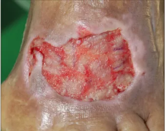
Fig. 1. Preoperative skin defect. A 4×2 cm
2 open wound on the dorsum of the left foot.Fig. 3. Immediate postoperative donor site wound. After a site
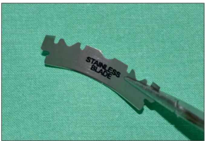
the same size as the defect (4×2 cm2) was marked, approximately 50% of the skin graft was harvested.Fig. 2. Razor blade method. After the razor blade was cut into
two pieces, the halved blade was fixated with a mosquito holder and curved so that it would have adequate contact with the skin.wounds and harvesting fragmented skin rather than mak- ing one large would induce less post-operative pain and faster wound healing. An electrical dermatome, air der- matome, scalpel, razor blade, Weck knife, or other tools may be used to harvest the STSG
10,11). The dermatome is the most commonly used because it can harvest a large skin graft in one attempt. However, it is difficult to har- vest tissues if the area around the donor site interferes with the dermatome or is not flat; in addition, it is diffi- cult to harvest small grafts. Because the shape of the har- vested skin is usually rectangular, extra skin usually re- mains after skin grafting. Additionally, the set-up and clean-up before and after surgery are time-consuming, and the using dermatome is expensive.
The authors report the patients with a comorbidity and various skin defects of the lower extremity who under- went treatment with a fragmented skin graft harvested with a sterilized razor blade. There was no large wound at the donor site, and healing at the defect sites and do- nor sites was favorable.
CASE REPORT
A 64-year-old woman with previous diabetes mellitus type 2 presented to Yeungnam University Medical Center with a chief complaint of a 2-degree contact burn on her left foot dorsum sized 4×2 cm
2(Fig. 1). After a donor site (include the thigh, area around the lower limb defect, skin surface of a fasciocutaneous flap, sole of the foot,
and scalp) of the same size as the skin defect was marked,
a mix of 1% lidocaine with 1:100,000 epinephrine was in-
jected in the subdermal layer. A razor blade was cut into
two pieces, and the halved blade was fixated using a
Mosquito forceps (or needle holder). The blade was bent
into a curved shape so that it would have adequate con-
tact with the skin (Fig. 2). Skin grafts approximately 1×1
cm
2were harvested by moving the blade back and forth
(Fig. 3). The size and thickness of the skin grafts were
controlled by adjusting the degree of curvature of the
blade and the downward pressure on the donor site. The
Table 1. Patient Data
No. Age (yr) Sex Wound
location
Wound size (cm2)
The time to complete re-epithelialization on
recipient site (day)
The time to complete re-epithelialization on
donor site (day)
1 57 M Rt. lateral malleolar area 4×1.5 4 8
2 58 F Lt. calf 4×4 6 8
3 61 M Rt. knee 4×3 6 8
4 62 M Lt. foot 8×3 4 10
5 64 F Lt. foot dorsum 4×2 6 10
6 73 F Lt. calf 4×3 6 10
7 82 F Rt. foot dorsum 5×3 8 8
8 84 M Lt. pretibial area 3×3, 4×1 8 8
9 86 F Lt. foot dorsum 5×4 8 10
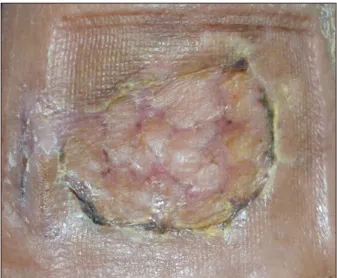
Fig. 5. A recipient site on day 4 after surgery. The gap between
the grafted skin narrowed as epithelialization progressed.Fig. 4. Immediate postoperative recipient site wound. After the
fragmented STSGs were placed at an interval of 3∼4 mm, the wound was compressed with wet gauze and fixated without suturing.harvested skin grafts were carefully unfolded and placed over the defect site with an adequate space of approx- imately 3∼4 mm (Fig. 4). Fixation is possible using com- pression without a fixation suture. The entire wound area including the skin graft is compressed with wet gauze, so discharges or fluid remaining in the wound can be removed. The use of fibrin glues with spray mode pro- vides better fixation. For our patient, a compressive dress- ing was applied using foam or transparent film at both the recipient and donor sites and the wound dressing was changed every other day.
From January 2018 to July 2018, fragmented STSG were performed on 9 patients (Table 1). The mean age of pa- tients was 70 years (range, 57∼86 years) and the mean defect size was 14.0 cm
2(range, 6∼20 cm
2). All of 9 pa-
tients had diabetes mellitus type 2. In all patients, the skin graft on the defect site healed well and did not result in problems such as hematoma or seroma. 4 days after the operation, the surgical site was opened for the first time and dressing was performed every 2 days thereafter. On day 4 after surgery, the gap between the grafted skin nar- rowed as epithelialization progressed (Fig. 5). On mean day 8.9 after surgery (range, 8∼10 days), healing was complete, and an acceptable scar remained (Fig. 6, 7).
DISCUSSION
A razor blade is easy to use and does not require addi-
tional equipment. Although it is difficult to control the
depth of the donor skin, it costs less than a dermatome,
and does not require set-up and clean-up time. Because
of these advantages, the use of a razor blade has been re-
Fig. 7. Complete healing on day 10 after surgery (donor site).
Fig. 6. Complete healing on day 6 after surgery (recipient site).
ported for skin grafting for small facial wounds
12,13). The authors extended the use of a razor blade to lower ex- tremity skin grafting and used a fragmented STSG on a larger defect area. This approach is advantageous for har- vesting a graft with a desired size, shape, and thickness, and it involves adjusting the tilting angle, downward pressure, and degree of curvature of the razor blade such that there is no need for trimming or defatting
14). Additionally, this procedure was able to be conducted under local anesthesia for patients who had poor general condition and comorbidities.
For a skin graft to successfully take, hematoma, sero- ma, and infection should not occur. With larger skin grafts, the rate of successful graft-taking may be de- creased because hematoma and seroma are not easy to drain. To counter this difficulty, fenestration may be per- formed with a slit incision, or mesh skin grafting may be performed. Because the fragmented skin grafts used for the patient in this study were small (approximately 1×1 cm
2), they were fixated well, without the need for a slit incision or mesh, and graft failure due to a hematoma or seroma did not occur. Even without the use of a large skin graft, the gaps between small fragmented skin grafts were confirmed to be decreased as epithelialization pro- ceeded to the surrounding area. Although there is no need for suture fixation, fibrin glue may be used to en- sure fixation.
The healing mechanism of a donor site wound involves activation, migration, and proliferation of keratinocytes.
Keratinocytes generate healing as they move along the wound surface from the basal layer of the epidermis and adnexal structures, which are in the same dermal layer as the sebaceous glands and hair follicles
15,16).
With the approach taken by the authors, healing oc- curred relatively quickly. The donor site wound had a cluster of several individual wounds approximately 1 cm
2, and a 3- to 4-mm gap was exposed in the recipient site wound. A fragmented STSG induces spontaneous re-epithelialization and decreases the likelihood of hyper- trophic scarring during healing of the recipient site be- cause an individual STSG has independent re-epitheliali- zation potential. Wound healing in our patient was rapid because re-epithelialization in the recipient and donor site wounds was influenced by the stem cells in the adnexal structure that originated from a healthy skin margin, and because the areas of the dermis layer exposed in the frag- mented STSG were small.
In the present study, a razor blade was used to re-
construct a relatively large skin defect in the lower ex-
tremities using a fragmented STSG. The defect site and
donor site wounds healed well. There was less pain dur-
ing wound dressing and no complications such as im-
paired healing, hematoma, seroma, and graft loss. We fur-
ther used this method for a total of 9 patients, and all
patients healed well without complications. Possible do-
nor sites include the thigh, area around the lower limb
defect, skin surface of a fasciocutaneous flap, sole of the
foot, and scalp. The donor site can be determined based on the state of the defect site and the patient’s medical condition
17).
Altogether, using a fragmented STSG harvested with a razor blade can be an easy, simple, and safe surgical method for resurfacing the skin of the lower extremity.
DECLARATIONS
No potential conflict of interest relevant to this article was reported. The study was performed in accordance with the principles of the Declaration of Helsinki. Written informed consents were obtained. The patient provided written informed consent for the publication and the use of his images.
CONFLICTS OF INTEREST
The authors have no financial interests in any of the products, devices, or drugs mentioned in this article.
REFERENCES
1) Nerone VS, Springer KD, Atway SA. Diabetic foot burns: a case series. J Foot Ankle Surg 2014;53:453-454.
2) Barsun A, Sen S, Palmieri TL, Greenhalgh DG. A ten-year review of lower extremity burns in diabetics: small burns that lead to major problems. J Burn Care Res 2013;34:255-260.
3) Lin Rakel BA, Bermel MA, Abbott LI, et al. Split-thickness skin graft donor site care: a quantitative synthesis of the research. Appl Nurs Res 1998;11:174-82.
4) Fowler A, Dempsey A. Split-thickness skin graft donor sites.
J Wound Care 1998;7:399-402.
5) Brölmann FE, Eskes AM, Goslings JC, et al. Randomized clinical trial of donor-site wound dressings after split-skin grafting. Br J Surg 2013;100:619-27.
6) Bradow BP, Hallock GG, Wilcock SP. Immediate regrafting of the split thickness skin graft donor site assists healing.
Plast Reconstr Surg Glob Open 2017;5:e1339.
7) Blight A, Fatah MF, Datubo-Brown DD, et al. The treatment of donor sites with cultured epithelial grafts. Br J Plast Surg 1991;44:12-4.
8) Toru M, Yasuo H, Akihiko S. Minced skin grafting for promoting epithelialization of the donor site after split- thickness skin grafting. Burns 2017;43:819-23.
9) Hur SW, Kim TG. Management of the accidental STSG donor site injury-Case reports. J Kor Burn Society 2015;18:31-4.
10) Snow SN, Stiff M, Lambert D, et al. Freehand technique to harvest partial-thickness skin to repair superficial facial defects. Dermatol Surg 1995;21:153-7.
11) Johnson T, Zide MF. Freehand full-thickness grafting for facial defects: a review of methods. J Oral Maxillofac Surg 1997;55:1050-6.
12) Grabski WJ, Salasche SJ, Mulvaney MJ. Razor-blade surgery.
J Dermatol Surg Oncol 1990;16:1121-6.
13) Goldberg LH, Segal RJ. Surgical pearl: a flexible scalpel for shave excision of skin lesions. J Am Acad Dermatol 1996;35:
452-3.
14) Andrew PK, Zheng Q, Nadia SU, et al. The use of a flexible razor blade in skin graft harvesting. Dermatol Surg 2009;
35:120-3.
15) Ito M, Liu Y, Yang Z, et al. Stem cells in the hair follicle bulge contribute to wound repair but not to homeostasis of the epidermis. Nat Med 2005;11:1351-4.
16) Levy V, Lindon C, Zheng Y, et al. Epidermal stem cells arise from the hair follicle after wounding. FASEB J 2007;21:1358-66.
17) Sinha S, Schreiner AJ, Biernaskie J, et al. Treating pain on skin graft donor sites: Review and clinical recommendations. J Trauma Acute Care Surg 2017;83:954-64.