Comparative Effectivenesses of Pulsed Radiofrequency and Transforaminal Steroid Injection for Radicular Pain due to Disc Herniation: A Prospective Randomized Trial
Transforaminal Epidural steroid injections (TFESI) have been widely adopted to alleviate and control radicular pain in accord with current guidelines. However, sometimes repeated steroid injections have adverse effects, and thus, this prospective randomized trial was undertaken to compare the effectivenesses of pulsed radiofrequency (PRF) administered to a targeted dorsal root ganglion (DRG) and TFESI for the treatment of radicular pain due to disc herniation. Subjects were recruited when first proved unsuccessful (defined as a score of > 4 on a visual analogue scale (VAS; 0-10 mm) and of > 30% according to the Oswestry Disability Index (ODI) or the Neck Disability Index (NDI)). Forty-four patients that met the inclusion criteria were enrolled. The 38 subjects were randomly assigned to receive either PRF (PRF group; n = 19) or additional TFESI (TFESI group; n = 19) and were then followed for 2, 4, 8, and 12 weeks. To evaluate pain intensity were assessed by VAS. ODI and NDI were applied to evaluate functional disability. Mean VAS scores for cervical and lumbar radicular pain were significantly lower 12 weeks after treatment in both study groups. NDI and ODI scores also declined after treatment. However, no statistically significant difference was observed between the PRF and TFESI groups in terms of VAS, ODI, or NDI scores at any time during follow-up. PRF administered to a DRG might be as effective as TFESI in terms of attenuating radicular pain caused by disc herniation, and its use would avoid the adverse effects of steroid.
Keywords: Pulsed Radiofrequency Treatment; Transforaminal Epidural Steroid Injection;
Radicular Pain Dong Gyu Lee,1 Sang-Ho Ahn,2
and Jungwon Lee2
1Department of Physical Medicine & Rehabilitation, Dongsan Medical Center, Keimyung University School of Medicine, Daegu, Korea; 2Department of Physical Medicine & Rehabilitation, Yeungnam University School of Medicine, Daegu, Korea Received: 21 April 2016
Accepted: 23 June 2016 Address for Correspondence:
Sang-Ho Ahn, MD
Department of Rehabilitation Medicine and Spine Center, Yeungnam University, College of Medicine, 170 Hyunchung-ro, Nam-gu, Daegu 42415, Korea
E-mail: [email protected]
Funding: This work was supported by the 2015 Yeungnam University Research Grant.
http://dx.doi.org/10.3346/jkms.2016.31.8.1324 • J Korean Med Sci 2016; 31: 1324-1330
INTRODUCTION
Steroids are powerful anti-inflammatory agents, and effectively reduce nerve root inflammation produced by disc herniation or a disc pathology near the epidural space. An initial observa- tional study reported transforaminal epidural steroid injection (TFESI) offered a treatment option for radicular pain due to lumbar disc herniation and that it provided > 50% pain reduc- tion in 75% of treated patients (1,2). A randomized prospective study also showed that TFESI had a success rate of 84% after a follow-up of 1.4 years (3).
Epidural steroid injections are widely and conventionally used to alleviate and control radicular pain effectively. However, some- times, single and/or repeat steroid injections cause adverse ef- fects, such as, spinal cord infarct, epidural fat hypertrophy, men- strual changes, and adrenal suppression (4,5). In particular, care should be taken to control blood sugar levels in patients with diabetes mellitus after an epidural steroid injection (6).
Radiofrequency (RF) treatment involves continuous stimula- tion and ablates nerves and tissues by increasing temperature
around the RF needle tip (7), and thus, RF treatment involves nerve ablation. However, pulsed radiofrequency (PRF) uses a brief stimulation period followed by a long resting phase, which exposes target nerves and tissues to an electric field without producing sufficient heat to cause structural damage (8,9). Sev- eral studies have reported PRF stimulation modulates suscepti- bility to radicular pain without causing tissue damage, and ob- servational studies on PRF application to dorsal root ganglia (DRGs) also concluded the technique appears both effective and relatively safe for the treatment of cervical and lumbar ra- dicular pain (10). However, few systemic studies have been con- ducted to compare the effectiveness of PRF and TFESI for ra- dicular pain. Because PRF procedure does not need require the injection of any material, it is certain to be free of the adverse effects associated with TFESI, and thus, offers the possibility of providing a method of treating radicular pain in a safer manner.
For these reasons the present randomized controlled study was aimed at determining the effectiveness of PRF for treating radicular pain due to disc herniation and comparing its outcomes with those of TFESI.
Rehabilitation & Sports Medicine
MATERIALS AND METHODS Materials
During the period from March 2013 to February 2015, 193 pa- tients were underwent TFESI for the treatment of spinal radicu- lar pain. Patients received TFESI initially, which was conducted using 2 mL of 0.125% bupivacaine mixed with 5 mg dexameth- asone. If, after first TFESI, patients still presented with a Visual Analogue Scale (VAS; 0-10 mm) of > 4 and an Oswestry Disabil- ity Index (ODI) or Neck Disability Index (NDI) of > 30%, then PRF and additional TFESI after initial TFESI were randomly al- located and conducted within from 2 to 6 weeks later after 1st TFESI (10,11). Each procedure was conducted by the physician who had experience spine interventions over more than 25 years.
Subjects with cervical and lumbar radicular pain was proved by physical examination and imaging studies corresponding with clinical manifestations. Exclusion criteria of instability is defined as >10 degrees sagittal-plane angulation a >3 mm sagittal-plane displacement on flexion-extension radiograph (12).
Inclusion criteria
• Age between 20 and 70 years
• Presentation with symptomatic cervical or lumbar radicular pain
• Imaging findings of a cervical or lumbar intervertebral disc pathology compatible with pain symptoms
• Severe cervical or lumbar radicular pain than cervical or lum- bar axial pain
• Presentation with a VAS of > 4 and an ODI or Neck Disability Index NDI of > 30% after first TFESI
Exclusion criteria
• Severe allergy to injectants
• History of spine surgery
• Spinal instability
• Spinal stenosis or degenerative spondylolisthesis
• Infection on the spine
• Tumor or tumor metastasis in the involved spinal area
• Pregnancy TFESI procedures
Strict aseptic technique was adopted for TFESI procedure (13).
Patients were supine for cervical procedure and prone position for lumbar under C-arm fluoroscopy (Siemens, Erlangen, Ger- many). To focus the target, C-arm was rotated toward the re- gion and controlled the cranial-caudal angle for focusing the intervertebral foramen. A 26-gauge with 90 mm spinal needle with a bend at the tip was inserted into the skin and advanced to the anterior half of superior articular process at cervical spine and to the 6 o’clock position below pedicle at lumbar spine. Then, the depth of needle tip checked by anterior posterior view and
lateral view of C-arm. Test dose of contrast medium (0.2-0.3 mL) was injected to figure out whether needle tip was placed at proper position. Then, the further injection of contrast medium was performed under real-time fluoroscopic monitoring. Fi- nally, patients received 2 mL of 0.125% bupivacaine mixed with 5 mg dexamethasone as 1st TFESI was conducted.
PRF procedures
Aseptic techniques were adopted for PRF therapy. For cervical procedures, the patient was laid in a supine position for C-arm fluoroscopy (Siemens), and a 22-gauge curved-tip cannula (SMK Pole needle 54 mm with a 4 mm active tip, Cotop International BV, Amsterdam, the Netherlands) was placed around the DRG (10). For lumbar procedures, the patient was laid in a prone po- sition for C-arm fluoroscopy (Siemens), and an 18-gauge curved- tip cannula (SMK Pole needle 100 mm with a 10 mm active tip, Cotop International BV) was placed around the DRG.
The catheter needle (active tip electrode) was inserted and a sensory stimulation test was carried out using an RF generator (Cosman G4, Burlington, MA, USA). The catheter needle was then advanced toward the DRG until the patient reported a tin- gling sensation and/or dysesthesia at less than 0.3V. PRF treat- ment was administered at 5 Hz and a 5 ms pulsed width for 240 seconds at 45V under the constraint that the electrode tip tem- perature not exceed 42°C (14).
Outcome measurements
Pain intensities were assessed by VAS for arm and leg radiating pain, before treatment, and 2, 4, 8, and 12 after treatment. ODI and NDI were obtained to evaluate functional disabilities asso- ciated with lumbar and cervical radicular pain, respectively, at the same times.
Adverse events
Adverse effects were carefully evaluated at each visit to detect pain flare-up and newly developed neurologic deficits after the procedures.
Statistical analysis
Statistical analysis was conducted using SPSS ver. 23 for win- dow and clinical course was analyzed using two-way factor re- peated measures analysis of variance. Statistical significance was accepted for P values < 0.05.
Ethics statement
This prospective randomized study was conducted at a spine specialist clinic in a university hospital after obtaining institu- tional review board approved for the study protocol (YUMC 2010-01-023), which also complied with the tenets of the Hel- sinki declaration. All study subjects provided written informed consent before study commencement.
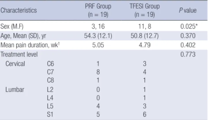
Table 1. Demographic characteristics Characteristics PRF Group
(n = 19) TFESI Group
(n = 19) P value
Sex (M.F) 3, 16 11, 8 0.025*
Age, Mean (SD), yr 54.3 (12.1) 50.8 (12.7) 0.370
Mean pain duration, wk† 5.05 4.79 0.402
Treatment level 0.773
Cervical C6 C7 C8
1 8 1
3 4 1
Lumbar L2
L4 L5 S1
0 0 4 5
1 1 3 6
PRF, pulsed radiofrequency; TFESI, transforaminal epidural steroid injection.
*Statistically significant (P < 0.05); †Pain duration before 1st TFESI injection.
Table 2. Statistical results of cervical procedures by two-factor repeated measures analysis
Variable Group Time, Mean (SD) F (P value)
Pre-treatment 2 wk 4 wk 8 wk 3 mon Time Group Time*Group
VAS PRF (n = 10) TFESI (n = 8)
5.3 (1.2) 4.9 (0.8)
4.2 (1.3) 3.6 (1.2)
3.3 (1.1) 2.8 (1.3)
2.4 (0.9) 2.5 (2.1)
2.0 (0.8) 2.4 (2.3)
20.472 (0.000)*, Pre-treatment
> 2,4,8 wk and 3 mon†
0.200 (0.661) 0.743 (0.566) NDI PRF (n = 10)
TFESI (n = 8) 38.7 (8.3)
39.1 (11.6) 28.3 (14.7)
28.6 (9.7) 22.2 (11.6)
19.4 (11.2) 17.6 (6.8)
18.8 (15.7) 14.0 (7.0)
17.0 (14.3) 19.90 (0.000)*, Pre-treatment
> 2,4,8 wk and 3 mon† 0.013 (0.912) 0.238 (0.916) PRF, pulsed radiofrequency; TFESI, transforaminal epidural steroid injection; VAS, visual analogue scale; NDI, neck disability index.
*Statistically significant (P < 0.05); †Multiple comparison result by contrast.
Fig. 1. Patient flow schematic.
PRF, pulsed radiofrequency; TFESI, transforaminal steroid injection.
193 Patients underwent single TFESI d/t disc herniation
44 Patients enrolled and randomized
Lost on follow up (n = 6) drop-out (n = 5) flare up pain (n = 1)
19 Patients
underwent PRF 19 Patients
underwent TFESI Excluded (n = 149) Back surgery (n = 19)
Relieved pain after single TFESI (n = 59) Did not visit after single TFESI (n = 69) Declinded to participate (n = 2)
RESULTS
Among 193 patients underwent TFESI for the treatment of spi- nal radicular pain, Forty-four patients (mean age: 52.4 ± 12.3, range 23-70) that met the study inclusion criteria and were en- rolled (Fig. 1). However, five patients were lost to follow up and one patient in the PRF group experienced a pain flare up and dropped out. Accordingly, 38 of the 44 screened patients were followed up for 3 months after PRF or TFESI (PRF group, n = 19;
TFESI group n = 19). The demographic characteristics of the subjects were shown in Table 1. Although subjects of our study were randomly allocated, PRF and TFESI group had statistical differences in sex ratios.
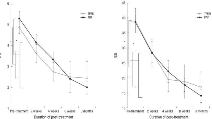
Changes in VAS and NDI in cervical radicular patients Mean VAS and NDI decreased over the 3-month follow up pe- riod after TFESI and PRF. Mean VAS and NDI for cervical radic- ular pain were significantly improved at 2, 4, 8, and 12 weeks af- ter TFESI and PRF (P < 0.001) (Table 2, Fig. 2). However, no sig- nificant intergroup difference was observed, although mean VAS and NDI showed marginally greater improvement after PRF at 3 months after treatment.
Changes in VAS and ODI in lumbar radicular patients Mean VAS and ODI for lumbar radicular pain also declined af-
ter TFESI and PRF, and were significantly improved at 2, 4, 8, and 12 weeks after TFESI and PRF (P < 0.001) (Table 3, Fig. 3).
However, no significant intergroup difference was observed at any time point.
Adverse events
One patient complained of aggravated radicular pain at 4 weeks post-PRF, and exited the study.
DISCUSSION
In this study, we evaluated the comparative effectivenesses of TFESI and PRF after 1st TFESI for the treatment of radicular pain due to disc herniation. Mean VAS, NDI, and ODI scores continuously declined after both procedures. However, the ef- fectivenesses of the two modalities were similar over the first 3 post-procedural months.
Herniated nucleus pulposus have been reported to cause in- flammation and ectopic firing in affected DRGs and spinal nerves, such as, glial activity in the spinal cord and the release of pain- modulating substances by activated glia (15,16), which are in- volved in the development and maintenance of chronic neuro- pathic pain associated with central sensitization (17,18). Ac- cordingly, activated glia are considered modulators of nocicep- tion and neuropathic pain.
The rationale behind epidural steroid injection for disc her- niation stems from the observation that inflammatory and no-
Table 3. Statistical results of lumbar procedures by two-factor repeated measures analysis
Variable Group Time, Mean (SD) F (P value)
Pre-treatment 2 wk 4 wk 8 wk 3 mon Time Group Time*Group
VAS PRF (n = 9)
TFESI (n = 11) 4.8 (0.8)
5.0 (1.0) 3.9 (0.8)
3.7 (1.0) 3.3 (0.7)
3.0 (1.3) 2.9 (1.0)
2.6 (1.8) 2.8 (1.2)
2.5 (1.5) 17.234 (0.000)*, Pre-treatment
> 2,4,8 wk and 3 mon† 0.242 (0.629) 0.177 (0.949) ODI PRF (n = 9)
TFESI (n = 11) 46.6 (8.4)
43.8 (10.0) 36.2 (10.4)
33.4 (12.2) 29.1 (10.1)
26.0 (11.8) 25.4 (8.9)
20.7 (14.7) 21.8 (12.9)
21.1 (14.3) 21.78 (0.000)*, Pre-treatment
> 2,4,8 wk and 3 mon† 0.587 (0.454) 0.114 (0.977) PRF, pulsed radiofrequency; TFESI, transforaminal epidural steroid injection; VAS, visual analogue scale; ODI, Oswestry disability index.
*Statistically significant (P < 0.05); †Multiple comparison result by contrast.
Fig. 2. Visual Analogue Scale (VAS) and Neck Disability Index (NDI) scores of cervical radicular pain patients.
TFESI, transforaminal steroid injection; PRF, pulsed radiofrequency.
*Statistically significant (P < 0.05).
VAS
Duration of post-treatment
Pre-treatment 2 weeks 4 weeks 8 weeks 3 months 6
5
4
3
2
1
* *
TFESI PRF
NDI
Duration of post-treatment
Pre-treatment 2 weeks 4 weeks 8 weeks 3 months 45
40
35
30
25
20
15
10
* *
TFESI PRF
ciceptive mediators concentrate around herniated discs in the epidural space (19-21). Steroid treatment is well known to have excellent anti-inflammatory effects that ca decrease inflamma- tion in DRGs, spinal nerves, and in the epidural space. Accord- ingly, steroid treatment probably inhibits neuroglial activation in spinal cords with acute disc herniation and/or attenuate glial activation, and for these reasons, TFESI is widely used as a con- ventional means of controlling and alleviating radicular pain (22). One study showed steroid injection effectively reduces the gadolinium enhancement (an indicator of nerve inflammation) of spinal nerves associated with herniated discs (23). Accord- ingly, TFESI was chosen in the present study as an initial treat- ment option for severe radicular pain after disc herniation.
The transforaminal approach, unlike the interlaminar and caudal approaches, enables steroid to be administered to target sites (21,24-26). In a systemic review of the effectiveness of TFE- SI, it was reported the effectiveness of lumbar TFESI achieved
the II-1 level of evidence in the short term and level II-2 in the long term (22), and in a systemic review of the effectiveness of cervical TFESI, it was found approximately 50% of patients achi- eved 50% pain relief for at least 4 weeks (27). In another study, fluoroscopically guided TFESI was found to be effective at alle- viating radicular pain and reducing need for surgery (28-30).
We are advocates of TFESI because we believe it is an effec- tive modality for the management of spinal radicular pain. How- ever, despite its effectiveness, the adverse effects of TFESI raise safety issues. The majority of these adverse effects concern the administration of steroid and contrast media (5,27). The side effects of steroid administration include facial flushing, high blood sugar, and transient headaches, and the major complica- tions of repeated steroid injection include suppression of pitu- itary adrenal axis, hypocorticism, Cushing’s syndrome, osteo- porosis, steroid myopathy, and epidural lipomatosis.
Catastrophic adverse events have also been reported, even
when TFESI is conducted by well trained physicians, the injec- tion of particulate steroid into an artery around the spinal canal can occlude capillaries and arterioles and cause spinal cord and cerebellar infarction resulting in permanent motor and sensory deficits (31). Recently, the non-particulate steroid, dexametha- sone, was used to minimize or eliminate embolic events after TFESI (32,33). Nevertheless, meticulous studies are required to confirm the safety of dexamethasone for TFESI.
On the other hand, PRF does not require the injection of ste- roid, contrast material, or local anesthetic, and thus, is not liable to the catastrophic adverse effects associated with vascular oc- clusion. Instead, PRF uses pulses of high voltage that produces an electric field around a needle tip and then allows heat to dis- sipate, and thus, stimulates the targeted dorsal root ganglion and the dorsal horn. Resultantly, PRF causes changes in C and Aδ fibers that transmit nociceptive and neuropathic pain (34).
It has been shown application of PRF at a DRG, but not at the sciatic nerve, caused the up- regulation of activating transcrip- tion factor 3 (an indicator of cellular stress) in DRG neurons (35). In another study, pulsed RF stimulation caused neuronal changes at targeted dorsal root ganglia and in neurons of the superficial dorsal horn, which could be associated with pain processing (36).
In a rat model of lumbar disc herniation, DRG stimulation using PRF attenuated microglial activation in the ipsilateral dorsal horn and reduced pain-related behavior as evidence by
reduced mechanical withdrawal thresholds. Therefore, it ap- pears DRG stimulation by PRF influences neural systems in- volving in pain processing by modulating glial activities closely associated with progression and maintenance of central sensi- tization, and thus, DRGs are frequently chosen as targets to mod- ulate electrophysiological change and modulate central sensiti- zation after disc herniation. Although observed histochemical changes and increases in neural markers after DRG stimulation by PRF do not constitute a mechanism for PRF, these observa- tions do show that PRT-induced electric fields induce neural system and gene expressional changes in DRGs and the dorsal horn (37).
Several studies that addressed chronic radicular pain have reported DRG stimulation by PRF appears to offer an effective and safe intervention for cervical and lumbar radicular pain.
Choi et al. (10) reported that 71% patients with chronic cervical radicular pain refractory to repeated TFESI were satisfied with the effectiveness DRG targeted PRF. Similarly, Bozem et al. (38) reported that 55% patients with chronic intractable lumbosa- cral radicular showed substantial pain improvements at 6 months after PRF, and Koh et al. (39) reported that the combined appli- cation of PRF and TFESI achieved higher treatment efficacies than TFESI alone in patients with chronic refractory radicular pain. These encouraging outcomes for the treatment of chronic radicular pain might suggest central sensitization can be mod- ulated by suppressing glia activity in the dorsal horn. However, Fig. 3. Visual Analogue Scale (VAS) and Oswestry disability index (ODI) scores of lumbar radicular pain patients.
TFESI, transforaminal steroid injection; PRF, pulsed radiofrequency procedure.
*Statistically significant (P < 0.05).
VAS
Duration of post-treatment
Pre-treatment 2 weeks 4 weeks 8 weeks 3 months 6
5
4
3
2
1
* * **
TFESI PRF
NDI
Duration of post-treatment
Pre-treatment 2 weeks 4 weeks 8 weeks 3 months 55
50 45 40 35 30 25 20 15
* * *
TFESI PRF
the above-mentioned studies did not include comparable con- trols or validate the benefits of DRG stimulation by PRF in sub- acute radicular limb pain.
Our study shows the clinical outcomes of patients treated with PRF for radicular pain was not inferior to those treated by TFESI at 3 months after treatment, and that TFESI and PRF both have significant treatment effects. Patients included in the pres- ent study had sustained radicular pain of > 4 by VAS and of
> 30% by ODI or NDI, despite receiving TFESI for severe radic- ular pain. Thus, our study subjects might have exhibited incom- plete suppression of inflammation around DRGs and spinal nerves after 1st TFESI, which we believe may have produced similar outcomes in the two groups. Disc herniation increases potential for generating ectopic discharges at dorsal root gan- glion, which produces central sensitization. As 1st TFESI par- tially suppressed inflammation around the nerve and epidural space, central sensitization at DRG and dorsal horn of spinal cord was processing, which could explain no inferior effective- ness of PRF at DRG comparing to additional TFESI.
There were randomized double-blind comparative studies for effectiveness of PRF at occipital neuralgia and adjuvant PRF with TFESI at chronic lumbosacral radicular pain (39,40). How- ever, this is the first randomized, controlled study directly to compare the effectivenesses of DRG stimulation by PRF and TFESI and validate the alternatives for the treatment of subacute radicular pain.
The majority of studies conducted on the effectiveness of PRF for the treatment of radicular pain have reported no seri- ous adverse events, but several authors have reported flare up of pain or temporary pain aggravation (10,39,40). In the present study, one patient experienced temporary aggravated radicular pain, which subsided after several days. Although PRF is a less destructive procedure than conventional radiofrequency treat- ment and does not cause neurologic deficits, PRF can cause microscopic neuronal damage, endoneural edema, and patho- logic changes in myelin, possibly due to the heat generated at the electrode tip. We presume that these effects could explain the temporary aggravation of radicular pain experienced on oc- casion.
In conclusion, this study showed that effectiveness of PRF at DRG stimulation with respect to the treatment of recalcitrant radicular pain after first TFESI was not inferior to TFESI but was not enough to alleviate pain. Although PRF is also minimally invasive procedure that has a possibility to destruct structure, we suggest if repeated TFESI is needed or a patient has a medi- cal condition requiring consideration with respect to the admin- istration of steroid, such as, uncontrolled diabetic mellitus, PRF be considered a useful clinical option for the control of subacute radicular pain that helps reduce or avoid the possible catastroph- ic adverse effects of TFESI.
DISCLOSURE
All of the authors have no potential conflicts of interest to dis- close.
AUTHOR CONTRIBUTION
Study concept and design: Lee DG, Ahn SH. Data acquisition:
Lee DG, Ahn SH, Lee J. Data analysis: Lee DG, Ahn SH. Writing:
Lee DG, Ahn SH. Revision: Lee DG, Ahn SH, Lee J. Final approv- al: all authors.
ORCID
Dong Gyu Lee http://orcid.org/0000-0002-4787-4448 Sang-Ho Ahn http://orcid.org/0000-0003-2921-7820 Jungwon Lee http://orcid.org/0000-0001-9230-3583
REFERENCES
1. Weiner BK, Fraser RD. Foraminal injection for lateral lumbar disc hernia- tion. J Bone Joint Surg Br 1997; 79: 804-7.
2. Lutz GE, Vad VB, Wisneski RJ. Fluoroscopic transforaminal lumbar epi- dural steroids: an outcome study. Arch Phys Med Rehabil 1998; 79: 1362- 6.
3. Vad VB, Bhat AL, Lutz GE, Cammisa F. Transforaminal epidural steroid injections in lumbosacral radiculopathy: a prospective randomized study.
Spine 2002; 27: 11-6.
4. Botwin KP, Gruber RD, Bouchlas CG, Torres-Ramos FM, Freeman TL, Slaten WK. Complications of fluoroscopically guided transforaminal lum- bar epidural injections. Arch Phys Med Rehabil 2000; 81: 1045-50.
5. Baker R, Dreyfuss P, Mercer S, Bogduk N. Cervical transforaminal injec- tion of corticosteroids into a radicular artery: a possible mechanism for spinal cord injury. Pain 2003; 103: 211-5.
6. Kim WH, Sim WS, Shin BS, Lee CJ, Jin HS, Lee JY, Roe HJ, Kim CS, Lee SM.
Effects of two different doses of epidural steroid on blood glucose levels and pain control in patients with diabetes mellitus. Pain Physician 2013;
16: 557-68.
7. van Kleef M, Spaans F, Dingemans W, Barendse GA, Floor E, Sluijter ME.
Effects and side effects of a percutaneous thermal lesion of the dorsal root ganglion in patients with cervical pain syndrome. Pain 1993; 52: 49- 53.
8. Vatansever D, Tekin I, Tuglu I, Erbuyun K, Ok G. A comparison of the neu- roablative effects of conventional and pulsed radiofrequency techniques.
Clin J Pain 2008; 24: 717-24.
9. Cosman ER Jr, Cosman ER Sr. Electric and thermal field effects in tissue around radiofrequency electrodes. Pain Med 2005; 6: 405-24.
10. Choi GS, Ahn SH, Cho YW, Lee DG. Long-term effect of pulsed radiofre- quency on chronic cervical radicular pain refractory to repeated transfo- raminal epidural steroid injections. Pain Med 2012; 13: 368-75.
11. Spiegel MA, Lafage R, Lafage V, Ryan D, Marascalchi B, Trimba Y, Ames C, Harris B, Tanzi E, Oren J, et al. Developing the total disability index based on an analysis of the interrelationships and limitations of oswestry and
neck disability index. Spine (Phila Pa 1976) 2016; 41: 74-81.
12. Kanemura A, Doita M, Kasahara K, Sumi M, Kurosaka M, Iguchi T. The influence of sagittal instability factors on clinical lumbar spinal symptoms.
J Spinal Disord Tech 2009; 22: 479-85.
13. Bogduk N; International Spine Intervention Society. Practice Guidelines for Spinal Diagnostic and Treatment Procedures. 2nd ed. San Francisco, CA: International Spine Intervention Society, 2013.
14. Rohof OJ. Caudal epidural of pulsed radiofrequency in post herpetic neu- ralgia (PHN); report of three cases. Anesth Pain Med 2014; 4: e16369.
15. Park HW, Ahn SH, Kim SJ, Seo JM, Cho YW, Jang SH, Hwang SJ, Kwak SY.
Changes in spinal cord expression of fractalkine and its receptor in a rat model of disc herniation by autologous nucleus pulposus. Spine 2011;
36: E753-60.
16. Ito T, Ohtori S, Inoue G, Koshi T, Doya H, Ozawa T, Saito T, Moriya H, Taka- hashi K. Glial phosphorylated p38 MAP kinase mediates pain in a rat model of lumbar disc herniation and induces motor dysfunction in a rat model of lumbar spinal canal stenosis. Spine 2007; 32: 159-67.
17. Watkins LR, Maier SF. Glia: a novel drug discovery target for clinical pain.
Nat Rev Drug Discov 2003; 2: 973-85.
18. Watkins LR, Maier SF. Beyond neurons: evidence that immune and glial cells contribute to pathological pain states. Physiol Rev 2002; 82: 981-1011.
19. Byröd G, Rydevik B, Nordborg C, Olmarker K. Early effects of nucleus pulp- osus application on spinal nerve root morphology and function. Eur Spine J 1998; 7: 445-9.
20. Brisby H, Olmarker K, Larsson K, Nutu M, Rydevik B. Proinflammatory cytokines in cerebrospinal fluid and serum in patients with disc hernia- tion and sciatica. Eur Spine J 2002; 11: 62-6.
21. Ploumis A, Christodoulou P, Wood KB, Varvarousis D, Sarni JL, Beris A.
Caudal vs transforaminal epidural steroid injections as short-term (6 mon- ths) pain relief in lumbar spinal stenosis patients with sciatica. Pain Med 2014; 15: 379-85.
22. Cohen SP, Bicket MC, Jamison D, Wilkinson I, Rathmell JP. Epidural ste- roids: a comprehensive, evidence-based review. Reg Anesth Pain Med 2013; 38: 175-200.
23. Tak HJ, Jones R, Cho YW, Kim EH, Ahn SH. Clinical evaluation of transfo- raminal epidural steroid injection in patients with gadolinium enhancing spinal nerves associated with disc herniation. Pain Physician 2015; 18:
E177-85.
24. Lee JH, Moon J, Lee SH. Comparison of effectiveness according to differ- ent approaches of epidural steroid injection in lumbosacral herniated disk and spinal stenosis. J Back Musculoskeletal Rehabil 2009; 22: 83-9.
25. Lee JH, An JH, Lee SH. Comparison of the effectiveness of interlaminar and bilateral transforaminal epidural steroid injections in treatment of patients with lumbosacral disc herniation and spinal stenosis. Clin J Pain 2009; 25: 206-10.
26. Van Zundert J, Huntoon M, Patijn J, Lataster A, Mekhail N, van Kleef M;
Pain Practice. 4. Cervical radicular pain. Pain Pract 2010; 10: 1-17.
27. Engel A, King W, MacVicar J; Standards Division of the International Spine Intervention Society. The effectiveness and risks of fluoroscopically guid- ed cervical transforaminal injections of steroids: a systematic review with comprehensive analysis of the published data. Pain Med 2014; 15: 386- 402.
28. Kumar N, Gowda V. Cervical foraminal selective nerve root block: a ‘two- needle technique’ with results. Eur Spine J 2008; 17: 576-84.
29. Kolstad F, Leivseth G, Nygaard OP. Transforaminal steroid injections in the treatment of cervical radiculopathy. A prospective outcome study.
Acta Neurochir (Wien) 2005; 147: 1065-70.
30. Razzaq AA, O’Brien D, Mathew B, Bartlett R, Taylor D. Efficacy and dura- bility of fluoroscopically guided cervical nerve root block. Br J Neurosurg 2007; 21: 365-9.
31. Derby R, Lee SH, Kim BJ, Chen Y, Seo KS. Complications following cervi- cal epidural steroid injections by expert interventionalists in 2003. Pain Physician 2004; 7: 445-9.
32. Park CH, Lee SH, Kim BI. Comparison of the effectiveness of lumbar trans- foraminal epidural injection with particulate and nonparticulate cortico- steroids in lumbar radiating pain. Pain Med 2010; 11: 1654-8.
33. El Abd O, Amadera J, Pimentel DC, Gomba L. Immediate and acute ad- verse effects following transforaminal epidural steroid injections with dexamethasone. Pain Physician 2015; 18: 277-86.
34. Erdine S, Bilir A, Cosman ER, Cosman ER Jr. Ultrastructural changes in axons following exposure to pulsed radiofrequency fields. Pain Pract 2009; 9: 407-17.
35. Hamann W, Abou-Sherif S, Thompson S, Hall S. Pulsed radiofrequency applied to dorsal root ganglia causes a selective increase in ATF3 in small neurons. Eur J Pain 2006; 10: 171-6.
36. Higuchi Y, Nashold BS Jr, Sluijter M, Cosman E, Pearlstein RD. Exposure of the dorsal root ganglion in rats to pulsed radiofrequency currents acti- vates dorsal horn lamina I and II neurons. Neurosurgery 2002; 50: 850-5.
37. Cho HK, Cho YW, Kim EH, Sluijter ME, Hwang SJ, Ahn SH. Changes in pain behavior and glial activation in the spinal dorsal horn after pulsed radiofrequency current administration to the dorsal root ganglion in a rat model of lumbar disc herniation: laboratory investigation. J Neurosurg Spine 2013; 19: 256-63.
38. Van Boxem K, de Meij N, Kessels A, Van Kleef M, Van Zundert J. Pulsed radiofrequency for chronic intractable lumbosacral radicular pain: a six- month cohort study. Pain Med 2015; 16: 1155-62.
39. Koh W, Choi SS, Karm MH, Suh JH, Leem JG, Lee JD, Kim YK, Shin J. Treat- ment of chronic lumbosacral radicular pain using adjuvant pulsed radio- frequency: a randomized controlled study. Pain Med 2015; 16: 432-41.
40. Cohen SP, Peterlin BL, Fulton L, Neely ET, Kurihara C, Gupta A, Mali J, Fu DC, Jacobs MB, Plunkett AR, et al. Randomized, double-blind, compara- tive-effectiveness study comparing pulsed radiofrequency to steroid in- jections for occipital neuralgia or migraine with occipital nerve tender- ness. Pain 2015; 156: 2585-94.