교신저자: 황 금
220-701, 강원도 원주시 일산동 162번지 원주기독병원 신경외과
Tel: 82-33-741-0592, Fax: 82-33-746-2287 E-mail: [email protected]
*2006년 대한신경외과 춘계학술대회에서 구연 발표한 바 있음.
두부 외상 후 지속적 두개강내 출혈에 대한 위험인자 및 예후
연세대학교 원주의과대학 신경외과학교실
강지훈ㆍ한용표ㆍ홍순기ㆍ허 철ㆍ변진수ㆍ황 금
Risk Factors and Outcomes of Progressive Intracranial Hemorrhage after Traumatic Head Injury
Ji Hoon Kang, M.D., Yong Pyo Han, M.D., Soon Ki Hong, M.D., Chul Hu, M.D., Jhin Soo Pyen, M.D., and Kum Whang, M.D.
Department of Neurosurgery, Wonju College of Medicine, Yonsei University, Wonju, Korea
Objectives: The purpose of this study is to find a better solution to treatment of patients with head trauma by analyzing the clinical progress and factors related to progressive intracranial hemorrhage (PIH) through comparisons between initial and follow up brain CT.
Methods: Of all acute head trauma patients admitted from November, 2003 to November 2005, 122 patients with EDH, SDH, ICH, and SAH seen on initial brain CT were selected. We retrospectively assessed brain CT and clinical data of all the patients, and sex, age, initial GCS, interval from initial injury to initial brain CT, interval from initial CT to follow CT, presence of skull fractures, GOS scores, and CT image findings were analyzed. We divided these patients into the two groups; patients with increased hemorrhage on follow up brain CT, and those without any interval change. Possible initiating factors and prognosis were analyzed in both groups.
Results: Of the 122 patients, 48 patients showed increased hemorrhage on follow up CT (39.3%), and 74 patients did not (60.7%).
The mean age of patients with increased hemorrhage was 49 yrs. 31 patients were male, and 17 patients were female in the increased hemorrhage group. The increased hemorrhage group had an older mean age and a higher ratio of male patients (p<0.05, p<0.05).
In the increased hemorrhage group, the type of hemorrhage were as follows: 16 patients with an ICH, 12 patients with a SDH, 11 patients with an EDH, and 9 patients with a traumatic SAH. The average interval between initial trauma and initial brain CT was 3.8±0.62 hrs in the increased hemorrhage group, and 5.3±1.24 hrs in the no interval change group. Average interval between initial and follow up brain CT was 13.4±2.41 hrs in the increased hemorrhage group, and 23.48±0.95 hrs in the no interval change group. Patients with increased hemorrhage showed a shorter interval between all studies, but only the interval between the initial and follow up brain CT showed a statistical significant correlation (p>0.05, p<0.05). Initial GCS, presence of skull fractures, and hemorrhage lesion did not show a significant difference between the two groups.
Conclusion: The patients age, sex, increased PT (INR) were risk factors of PIH, and the interval between initial and follow up brain CT were shorter in these cases. Therefore early brain CT follow up can help identify and adequately treat patients with PIH.
Key Words: Brain CT․Clinical outcome․Head trauma․Predictor․Progressive intracranial hemorrhage
서 론
전세계적으로 연간 1,000만 건의 외상성 뇌 손상으로 인해 주로 15~35세 가량의 젊은 연령층이 사망하거나 불구가 된 다26). 이는 한 개인의 불행한 문제이기도 하지만, 국가 전체
Table 1. PIH vs. Clinical Factors
Clinical Factors PIH (No. of pts)
p value
(+) (-)
Age(yr) Sex(M:F) Initial GCS
Mean Range Mean
48.63±14.20 12-70 31:17 8.64±2.55
41.52±10.90 20-74 44:30 9.10±3.20
p<0.05 p<0.05 p>0.05 Cause of injury Traffic accident
Slip Fall down Assault
36 5 5 2
47 18 6 3
p>0.05
Total 48 74
Interval of Brain CT (hrs)
Trauma to 1st 1st to 2nd
3.8±0.62 13.4±2.41
5.3±1.24 23.48±0.95
p>0.05 p<0.05 PIH: Progressive intracranial hemorrhage
의 문제이기도 하다. 이러한 두부 외상은 사회적 생산 활동 이 왕성한 젊은 연령층에서 대부분이 발생하여 이에 대한 경 제적인 비용이 높기 때문이다5,13).
두부 외상에서 치료의 주된 흐름은 1차 손상보다는 2차 손상을 줄이는데 많은 노력을 기울이고 있다. 이는 2차 손상 의 정도와 기간에 따른 환자의 예후가 많은 차이를 보이기 때문이다. Kobayashi 등10)과 Cooper 등3)은 좋은 예후와 새로 운 병변의 부재간의 상관관계와 나쁜 예후와 새로운 병변의 발달간의 상관관계가 있는 것으로 보고하고 있으며, 이러한 2차 손상은 외상성 뇌 손상 후 병원 내 사망의 주요 원인이
다8,27). 따라서 두부 외상 치료의 주요 관점은 2차 손상을 최
소화하는데 있다. 두부외상 후 시행한 초기 뇌전산화단층촬 영(CT)으로부터 환자가 비가역적으로 신경학적 손상이 발생 하기 이전에 추적 뇌 CT 검사를 시행하여 정확한 진단으로 환자에 대한 치료 방법을 결정하는 것이 필요하다.
본 연구에서는 Progressive intracranial hemorrhage (PIH)를 사고 후 24시간 이내에 시행한 추적 뇌 CT 검사상 두개강내 출혈부위(intracranial hemorrhagic lesion)의 크기가 25% 이상 증가한 것으로 정의하고15). 이러한 PIH 환자들 중에서 그 출 혈량이 많은 경우(>30 cc)에는 수술적 치료를 시행하였다.
본 연구에서는 내원 초기 치료에도 불구하고, 의식상태가 악화되거나, 기대되는 것보다 의식상태가 나아지지 않았던 환자를 대상으로 하여 추적 뇌 CT를 시행하여 PIH가 관찰된 환자들을 대상으로 PIH에 관련된 위험인자를 알아보고, PIH 와 환자의 예후와의 상관관계를 분석하여 치료에 있어서 좀 더 나은 개선점을 찾고자 하였다.
대상 및 방법
2003년 11월부터 2005년 11월까지 본원에서 두부손상으 로 입원하여 응급수술을 하지 않은 122명의 환자에 대한 초 기 및 추적 뇌 CT와 경과기록을 후향적으로 분석하였다.
본원에서는 추적 뇌 CT의 적응증으로 첫 번째 Glascow coma scale (GCS)가 악화되었을 때, 두 번째 초기 뇌 CT로부 터 기대되는 것보다 환자의 의식상태가 나아지지 않을 때, 세 번째 정기적 경과 뇌 CT로 정하였다.
환자에 대한 초기 평가는 GCS로 하였고, 환자의 예후는 Glascow outcome scale (GOS)로 good recovery와 moderate disability 를 좋은 예후로 severe disablility, persistent vegetative status와 death를 나쁜 예후로 나누어 평가하였다.
통계 분석
모든 요소에 대한 통계분석은 Chi squire test와 T-test로 하 였고, P value가 0.05 미만 일 때만 통계적으로 유의한 것으 로 하였다. 모든 통계 자료는 SPSS 10.0 version for windows 를 이용하여 분석하였다.
결 과
1. PIH와 임상적 요소의 상관관계
조사 기간 중 두부 손상으로 입원한 122명의 환자에 대한
Table 2. PIH vs. Laboratory Factors
Laborary Factors PIH (No. of pts) p value
(+) (-)
PT (INR)
normal prolonged mean
40 8 1.08±0.12
73 1 0.98±0.41
p<0.05
PTT normal prolonged mean (sec)
47 1 13.45±1.32
73 1
12.93±2.01 p>0.05 Platelet
count
mean (x 103/mm3)
231±42.41 223±23.31 p>0.05
PT: Prothrombin time, PTT: Partial thromboplastin time
Table 3. PIH vs. brain CT
Radiologic Factors PIH (No. of pts) p value
(+) (-)
Location of hemorrhage
F-T-P Frontal Temporal Occipital Cerebellar
23 13 5 5 2
30 23 12 5 4
p>0.05
Total 48 74
Type of main hemorrhage
ICH SDH EDH SAH
16 12 11 9
30 6 28
10 p>0.05
Total 48 74
Coincident skull Fx. 24 25 p>0.05 F-T-P: frontotemporoparietal, ICH: intracerebral hemorrhage, EDH: epidural hemorrhage, SAH: subarachnoid hemorrhage, SDH: subdural hemorrhage
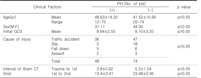
임상적 요소와 PIH의 상관관계는 다음과 같았다(Table 1). PIH (+)군의 평균연령은 48.63 (12~70)세이었고, PIH(-)군의 평균 연령은 41.52 (20~70)세였다(p<0.05). 남녀 성비는 PIH(+) 군에 서 31:17이었고, PIH(-)군에서는 44:30이었다(p<0.05). 초기 GCS는 PIH(+)군에서 평균 8.64±2.55이었으며, PIH(-)군에서 는 9.10±3.20이었다(p>0.05).
두부 손상의 원인으로는 중 교통사고가 각각의 군에서 36 명(29.5%)과 47명(38.6%)으로 가장 많았으며, 다음으로 미끄 러짐 사고와 추락사고 가해자에 의한 폭력 순이었다(p>0.05).
두부 외상 후 초기 뇌 CT를 시행 할 때까지의 시간과 초 기 뇌 CT 검사 이후 추적 뇌 CT 검사까지의 시간을 비교 분 석한 바, 두부 외상 이후 초기 뇌 CT 검사까지의 시간은 PIH (+)군에서 3.8±0.62 hrs이고, PIH(-)군에서는 5.3±1.24 hrs 이 였으며(p>0.05), 초기 뇌 CT로부터 추적 뇌 CT 검사까지 걸 린 시간은 PIH(+)군에서 13.4±2.41 hrs 이었고, PIH(-)군에서 는 23.48 ±0.95 hrs 이었다(p<0.05).
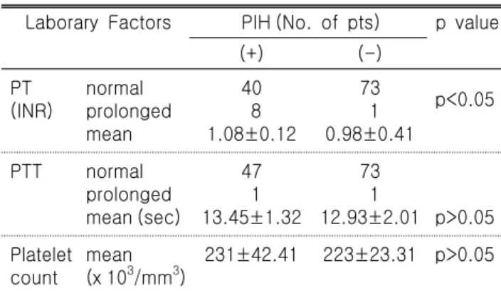
2. PIH와 혈액학적 검사 결과와의 상관관계 Prothrombin time (PT: international normalized ratio)은 PIH(+)/
PIH(-)군에서 정상인 환자는 40명/73명이었으며, 증가된 경우 는 8명/1명이었다(Table 2, p<0.05). 평균 PT (INR)는 1.08±0.12/
0.96±0.41이었다(p>0.05). Partial thromboplastin time (PTT)은 PIH(+)/PIH(-)군에서 정상인 환자는 47명/73명이었고, 증가한 환자는 1명/1명 이었다(p>0.05). 평균 PTT는 13.45±1.31 sec/
12.93±23.31 sec이었다(p>0.05). 평균 혈소판 수치는 PIH(+)/PIH(-) 군에서 231±42.41(×103 per mm3)/223±23.31(×103 per mm3)이었 다(p>0.05).
3. PIH와 뇌 CT와의 상관관계
출혈부위는 전두-측두-두정엽에서 23명(18.8%)/30명(24.6%) 으로 가장 많았으며, 전두엽, 측두엽, 후두엽, 소뇌 순이었다 (Table 3, p>0.05). 또한 동반된 두개골 골절은 24명(19.6%)/
25명(20.5%)으로 전체 122명 환자 중 49명으로 40.1%를 차지 하였다(p>0.05).
입원 당시의 출혈부위와 PIH(+/-)군 간의 상관관계는 뇌실 질내 출혈이 PIH(+/-)군 각각에서 16명/30명으로 가장 많았으 며, 그 다음이 경막외출혈, 경막하출혈, 지주막하출혈 순이었 다(p>0.05).
4. PIH군과 GOS간의 상관관계
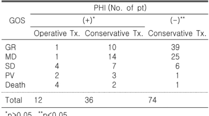
PIH(-)군 74명의 평균 GOS는 4.39±0.81이었고 PIH(+)군 48
명의 평균 GOS는 3.42±1.30이었다(Table 4)(p<0.05). 하지만 PIH로 수술을 받은 12명의 평균 GOS는 2.42±1.31이었고, PIH이지만 수술을 받지 않고 대증적 치료를 받은 36명의 평 균 GOS는 3.75±1.13이었다(p>0.05).
고 찰
1891년 Otto Bollinger2)가 두부 외상 후 2차 두개강내 출혈 (secondary intracranial hematoma)에 대해서 처음 기술한 이후, 1938년 Doughty6)가 “Delayed intracranial hemorrhage"라는 용 어를 도입하였고, 이는 1970년대 CT 기술의 발달로 좀 더 보
Table 4. PIH vs. GOS
GOS
PHI (No. of pt)
(+)* (-)**
Operative Tx. Conservative Tx. Conservative Tx.
GR MD SD PV Death
1 1 4 2 4
10 14 7 3 2
39 25 6 1 1
Total 12 36 74
*p>0.05, **p<0.05
GD: good recovery, MD: moderate disability, SD: severe disability, PV: persistent vegetative status, Tx: treatment
편화되었다. Fowein 등7)이 외상 이후 초기 뇌 CT의 시간을 기준으로 외상 후 2~3시간 이후가 출혈부위를 확인하는데 매우 중요한 것으로 결론지었다. 최근 PIH에 대한 많은 연구 에서 외상 후 24~72시간 이내에 두 번의 연속된 CT를 시행 한 경우에 환자의 23~47.5%에서 두개강내 출혈이 지속적으 로 커지는 소견이 관찰되었다16,25,29).
본 연구에서는 두부 외상으로 본원에 입원한 122명의 환 자들 대부분은 사고, 이후 대략 5시간 이내에 초기 뇌 CT검 사를 시행하였고, 이들 중 48명(39.3%)의 환자에게서 추적 뇌 CT를 시행하였을 때 PIH가 관찰되었다. PIH의 위험인자 로는 환자가 고령인 경우와 남성인 경우, 초기 뇌 CT로부터 추적 뇌 CT까지의 시간이 짧은 경우 그리고 PT(INR)이 연장 된 경우였다. 이외에 환자의 나이나 PTT나 혈소판 수치 및 두부외상의 원인이나 출혈의 위치나 형태, 두개골 골절의 동 반유무는 PIH와 상관관계가 없는 것으로 나타났다.
고령에 따른 PIH와 연관성은 혈관의 경직성과 취약성의 증 가와 관련이 있다. 고혈압, 당뇨, 유전분증 등은 혈관의 취약 성과 연관이 있으며, 고령의 환자에게서 흔하고 출혈성 뇌졸 중의 중요한 원인이 된다1,17,22). 하지만 이러한 요소들이 두부 외상 후 PIH를 유발하는 것인지에 대해서는 명확하지 않다.
환자가 남성인 경우 PIH와 상관관계가 있는 것으로 관찰 되었는데, 이는 여성의 estrogen이나 progesterone의 신경보호 효과일 가능성이 높다. estrogen은 뇌혈류를 증가시키거나 lipid peroxidation과 세포막 파괴를 감소시키고, 혈소판 응집 과 계획된 세포사멸을 줄이는 효과가 있다21). progesterone의 신경보호 효과는 주로 membrane stabilization, glutamate receptor inhibition 그리고 γ-aminobutyric acid receptor potentiation로 인
한 것이다20).
본 연구에서는 혈액응고장애를 확인하기 위해 PT (INR), PTT, 혈소판 등의 parameter를 이용하였다. PT (INR)가 증가 한 경우 PIH와 상관관계가 있는 것으로 관찰되었지만, 이에 대해서는 여러 가지 다른 견해가 있다. Stein 등29,30)은 PT, PTT 가 증가되고 혈소판이 감소한 경우 PIH의 예견인자라는 견 해를 냈지만, 다른 연구에서는 이러한 것들이 PIH와 상관관 계가 없는 것으로 관찰되었다4,23,32). 또 다른 연구에서는 diffuse intravascular coagulation (DIC)이 PIH와 상관관계가 있는 것으 로 관찰되었다9,11,33). 따라서 이 부분에 대해서는 좀 더 많은 연 구가 있어야 될 것으로 생각된다.
대부분의 저자들은 초기 뇌 CT를 2시간 이내에 시행한 경 우 추적 뇌 CT를 24~36시간 이내에 시행할 것을 권고하고
있다14,19,28,31). 본 연구에서는 환자의 두부손상 이후 초기 뇌
CT까지 걸린 시간은 PIH의 유무와 상관이 없었지만, 추적 뇌 CT까지 걸린 시간은 PIH(+)군에서 13시간 가량으로 PIH (-)군의 23시간에 비해 짧은 간격을 나타내어 환자의 상태가 빠른 시간 내에 나빠짐을 알 수 있다. 이는 PIH(+)군의 환자 가 재출혈로 인한 급격한 의식소실로 PIH(-)군의 환자보다 빨리 뇌 CT 검사를 시행함에 따라 시간의 차이가 생긴 것으 로 생각된다.
PIH 유무에 따른 환자들의 GOS를 비교해 볼 때 PIH가 생긴 군에서는 보전적 치료와 수술적 치료에 따른 예후의 차이가 없었고, PIH 유무에 따라서 환자의 예후에 차이가 나타났다.
중증 뇌손상 환자에서 두개골 골절의 발생 빈도는 3~65
%까지 매우 다양하며24), 두개골 골절과 뇌 손상 정도와의 관 계에 대해서는 논란이 많다. Mendelow 등18)은 일반적으로 골 절은 상당한 충격을 의미하며, 두개강내 혈종발생의 중요한 예견인자로서 중증 뇌손상 및 두개강내 감염의 위험성이 높 다고 했다. Levi 등12)은 소아 두부 손상군에서 두개강내 병변 의 발생빈도는 두개골 골절이 있을 때나 없을 때나 서로 비 슷하여, 골절은 두개강내 병변을 의미하는 소견이 아니라고 하였다. 본 연구에서는 49명(40.16%)의 환자에게 두개골 골 절이 있었으며, PIH(+)/(-)군은 24명과 25명으로 두개골 골절과 PIH 와는 상관관계가 없는 것으로 관찰되었다.
따라서 이러한 고위험군에 속한 환자들에게는 추적 뇌 CT 검사를 저위험군 보다 조기에 시행하여 환자의 상태를 확인하는 것이 환자의 치료 및 예후에 도움이 될 것으로 생 각한다.
결 론
본 연구에서는 환자의 상태가 고령, 남성, PT (INR)이 연 장된 경우에 PIH가 잘 발생하며, 이러한 경우에 추적 뇌 CT 검사까지의 시간이 짧은 것으로 나타났다. 또한 PIH가 발생 한 경우 수술적 혹은 대증적 치료에도 불구하고, 환자의 예 후는 PIH가 발생하지 않은 군에 비해 좋지 않았다. 따라서 이러한 고험군에 속하는 환자들은 두부 외상 후 추적 뇌 CT 검사를 약 13시간 이내에 시행하여 PIH 유무를 확인하여 집 중적인 치료를 하는 것이 환자의 예후에 도움이 될 것으로 사료된다.
참 고 문 헌
1. Arboix A, Garcia-Eroles L, Massons J, et al: Hemorrhagic lacunar stroke. Cerebrovasc Dis 10:229-234, 2000
2. Bollinger O: Ueber traumatische Spät-Apoplexie: Ein Beitragyur Lehre von der Hirnerschlütterung, in Internationale Beiträge zur wissenshcaftlichen Medizin; Festschrift, Rudolf Virchow gewidmet zur vollendung seines 70. Lebensjahres. Berlin:
Hirschwald, Vol 2:457-470, 1891
3. Cooper PR, Maravilla K, Moody S, Clark WK: Serial compu- terized tomographic scanning and the prognosis of severe head injury. Neurosurgery 5(5):566-569, 1979
4. Crone KR, Lee KS, Kelly DL Jr: Correlation of admission fibrin degradation products with outcome and respiratory failure in patients with severe head injury. Neurosurgery 21:
532-536, 1987
5. Diamond PT: Brain injury in the commonwealth of Virgi- nia: An analysis of central registry data, 1988-1993. Brain Inj 10:413-419, 1996
6. Doughty RG: Posttraumatic delayed intracerebral hemorrhage.
South Med J 31:254-256, 1938
7. Frowein RA, Schiltz F, Stammler U: Early post-traumatic intracranial hemetoma. Neurosurg Rev 12 (Suppl 1):184-187, 1989
8. Ghajar J: Traumatic brain injury (seminar). Lancet 356:923- 929, 2000
9. Kaufman HH, Moake JL, Olson JD, et al: Delayed and recurrent intracranial hematomas related to disseminated intravascular
clotting and fibrinolysis in head injury. Neurosurgery 7:445- 449, 1980
10. Kobayashi S, Nakazawa S, Otsuka T: Clinical value of serial computed tomography with severe head injury. Surg Neurol 20(1):25-29, 1983
11. Kumura E, Sato M, Fukuda A, et al: Coagulation disorders following acute head injury. Acta Neurochir 85:23-28, 1987 12. Levi L, Guilburd JN, Lemberger A, et al: Diffuse axonal
injury: Analysis of 100 patients with radiological signs. Neu- rosurgery 27:429-432, 1990
13. Luerssen TG, Klauber MR, Marshall LF: Outcome from head injury related to patient's age: A longitudinal prospec- tive study of adult and prediatric head injury. J Neurosurg 68:409-416, 1988
14. Mandavia DP, Villagomez J: The importance of serial neu- rologic examination and repeat cranial tomography in acute evolving epidural heamtoma. Pediatr Emerg Care 17(3):193- 195, 2001
15. Matthias O, Daniel FK, David M, W. John B, Thomas CG, Jae Hong Lee, et al: Progressive hemorrhage after head trau- ma: Predictors and consequences of the evolving injury. J Neurosurg 96:109-116, 2002
16. McBride DQ, Patel AB, Caron M: Early repeat CT scan:
Importance in detection surgical lesions after closed head injury. J Neurotrauma 10 (Suppl 1):S227, 1993 (Abstract) 17. McCarron MO, Nicoll JA, Ironside JW, et al: Cerebral amyloid
angiopathy-related hemorrhage. Interaction of APOE epsilon2 with putative clinical risk factors. Stroke 30:1643-1646, 1999 18. Mendelow AD, Teasdale G, Jennett B, et al: Risks of intra-
cranial hematoma in head injured adults. Br Med J 287:
1173-1176, 1983
19. Oertel M, Kelly DF, McArthur D, et al: Progressive hemorr- hage after head trauma: Predictors and consequences of the evolving injury. J Neurosurg 96(1):109-116, 2002
20. Roof RL, Duvdevani R, Stein DG: Gender influences out- come of brain injury: Progesterone plays a protective role.
Brain Res 607:333-336, 1993
21. Roof RL, Hall ED: Gender differences in acute CNS trauma and stroke: Neuroprotective effects of estrogen and proges- terone. J Neurotrauma 17:367-388, 2000
22. Roses AD, Saunders A: Head injury, amyloid beta and
Alzheimer's disease. Nat Med 1:603-604, 1995 (Letter) 23. Sawada Y, Sadamitsu D, SAkamoto T, et al: Lack of correla-
tion between delayed traumatic intracerebral haematoma and disseminated intravascular coagualtion. J Neurol Neurosurg Psychiatry 46:1125-1127, 1984
24. Servadei F, Ciucci G, Morichetti A, et al: Skull fracture as a factro of increased risk in minor head injuries. Surg Ne- urol 30:364-369, 1988
25. Servadei F, Nasi MT, GiulianiG, et al: CT prognostic factors in acute subdural haematomas: The value of the `worst` CT scan. Br J Neurosurg 14:110-116, 2000
26. Signorini DF, Andrews PJ, Jones PA, Miller JD. Predicting survival using simple clinical variables: A case study in trau- matic brain injury. J Neurol Neurosurg Psychiatry 66:20- 25, 1999
27. Signorini DF, Andrews PJ, Jones PA, Miller JD. Adding insult to injury: The prognostic value of early secondary insults for survival after traumatic brain injury. J Neurol Neu- rosurg Psychiatry 66:26-31, 1999
28. Smith HK, Miller JD: The danger of an ultra-early com- puted tomographic scan in a patient with an evolving acute epidural hematoma. Neurosurgery 29(2):258-260, 1991 29. Stein SC, Spettell C, Young G, et al: Delayed and progres-
sive brain injury in closed-head trauma: radiological demons- tration. Neurosurgery 32:25-31, 1993
30. Stein SC, Young GS, Talucci RC, et al: Delayed brain injury after head trauma: Significance of coagulopathy. Neurosur- gery 30:160-165, 1992
31. Sullivan TP, Jarvik JG, Cohen WA. Follow-up of conserva- tively managed epidural hematomas: Implications for timing of repeat CT. Am J Neuroradio 20(1):107-113, 1999 32. Touho H, Hirakawa K, Hino A, et al: Relationship between
abnormalities of coagulation and fibrinolusis and postopera- tive intracranial hemorrhage in head injury. Neurosurgery 19:523-531, 1986
33. van der Sande JJ, Emeis JJ, Lindeman J: Intravascular coa- gulation: A common phenomenon in minor experimental head injury. J Neurosurg 54:21-25, 1981