Peripheral lymph nodes, including the cubital, axil- lary, inguinal and popliteal type, have been reported to be largest in neonates and to become gradually smaller
throughout life, showing minor differences in size change according to location. The replacement of lym- phatic parenchyma by fatty tissue (fatty change) is more characteristic of peripheral lymph nodes, which usually receive little antigenic stimulation (1).
In clinical work-up involving MR imaging of the knee, we have frequently noticed popliteal lymph nodes in pa- tients with internal derangement, or rheumatoid or de- generative arthritis. Several studies have reported the
─ 665 ─
Magnetic Resonance Appearance of Normal Popliteal Lymph Nodes: Location and Relationship of Number,
Fatty change, and Size of the Lymph nodes with Aging
1Hee Jung Moon, M.D., Jin-Suck Suh, M.D., Sang Hoon Lee, M.D.
Purpose: To investigate the location of popliteal lymph nodes and the relationship be- tween patient age and their number, size, and the occurrence of fatty chang.
Materials and Methods: We retrospectively evaluated the magnetic resonance (MR) images of 222 patients 〔age range, 8-79 (mean, 47.1) years〕who had undergone MRI of the knee after its internal derangement. Images were obtained in the axial, coronal, and sagittal planes. A lymph node was defined as ‘observed’if it was visible in at least two planes, such as axial and sagittal or axial and coronal. With regard to location, nodes were classified as anteromedial, anterolateral, posteromedial, or posterolateral, depending upon their relationship with the popliteal vein. To determine their size, the smallest diameter was measured.
Results: Popliteal lymph nodes were present in 116 of 222 examinations (52.3%), and their total number was 158 (mean, 1.36). Patients’age correlated negatively with their presence (R square=0.826), and positively with the occurrence of fatty change (R square=0.840). Sixty- five of 158 lymph nodes (41.1%) were located anteromedially, 58 (36.7%) posterolaterally, 27 (17.1%) anterolaterally, and eight (5.1%) posteromedially.
Their distance from the most distal femoral articular surface was 4.6±1.4 cm (mean ± SD), and their mean diameter was 4.96±2.4 mm (mean ±SD; range, 4-8 mm).
Conclusion: The number of popliteal lymph nodes decreased with age, while the inci- dence of fatty change increased. Nodes were most frequently anteromedial or posteri- lateral to the popliteal vein.
Index words : Lymph nodes
Lymphatic system, MR Extremities, MR
1Department of Diagnostic Radiology, Yonsei University College of Medicine
Received August 16, 2002 ; Accepted November 6, 2002
Address reprint requests to : Jin-Suck Suh, M.D., Department of Diagnostic Radiology, Yonsei University College of Medicine, 134 Shinchon-dong Seodaemun-gu, Seoul 120-752, Korea.
Tel. 82-2-361 5840 Fax. 82-2-393-3035 E-mail: [email protected]
imaging findings of peripheral lymph nodes at axillary and inguinal sites but not, to our knowledge, those of popliteal lymph nodes. In this reports we describe popliteal lymph nodes in terms of their prevalence, loca- tion, and size, and the occurene of fatty change.
Materials and Methods
Between January 2000 and February 2001, 249 MR imagings procedures were performed for the evaluation of arthralgia of the knee. The findings of 27 cases were excluded due to presence of metal artifact (n=19) or rheumatoid arthritis (n=8); those remaining related to 115 females and 107 males〔mean age, 47.1 (range, 8- 79) years〕in their first (n=4), second (n=23), third (n=42), fourth (n=34), fifth (n=40), sixth (n=40), sev- enth (n=28), or eighth (n=11) decade, and are described in Table 1. The most common MR diagnosis was menis- cal tear or degeneration, revealed by two or more radio-
logic findings in one-third of the patients. Other findings included ganglion cyst, fibrous cortical defect, bipartite patella, hemarthrosis, Baker’s cyst, bone infarction, os- teochondral lesion, and medial patellar plicae syn- drome.
For all examinations, a 1.5-T MR imager (Signa;
General Electric Medical Systems, Milwaukee, Wis., U.S.A.) was used, together with a dedicated knee coil.
The patients were placed in the magnet with the knee in
Table 1. MR Imaging Interpretation Results of the 222 Patients Meniscal tear or degeneration 141
Ligament tear 43
Contusion 19
Chondromalacia 42
Discoid meniscus 24
Normal 21
Others 20
ligament; anterior and posterior crucate, medial and lateral collat- eral ligaments
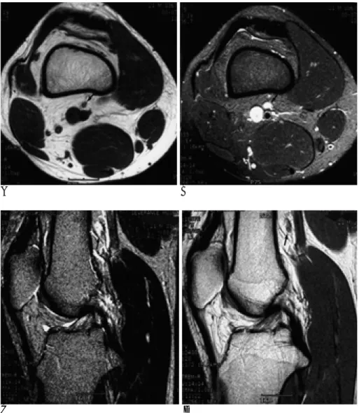
A B
Fig. 1. A 21 year-old-man with bipartite patella.
The popliteal lymph node shows inter- mediate signal intensity on T1 weight- ed images (A), high signal intensity on T2 weighted axial and sagittal images (B, C), and low signal intensity on pro- ton weighted sagittal images (D).
C D
the neutral position, and the following sequences were obtained: axial T1-weighted (TR/TE/number of excita- tions: 600/14/2; field of view, 14×14 cm; matrix, 256×
192; slice thickness, 4mm; interslice gap, 0 mm); axial fat-suppressed T2-weighted (TR/TE/number of excita- tions: 4000/50/2; field of view, 14×14 cm; matrix, 256×
192; slice thickness, 4mm; interslice gap, 0 mm); sagittal T2-weighted (TR/TE/number of excitations: 2000/80/1;
field of view, 14×14 cm; matrix, 256×256; slice thick- ness, 3 mm; interslice gap, 1 mm); sagittal proton densi- ty-weighted (TR/TE/number of excitations: 2000/20/1;
field of view, 14×14 cm; matrix, 256×192; slice thick- ness, 3 mm; interslice gap, 1 mm); and coronal fat-sup- pressed fast spin-echo T2-weighted (TR/TE/number of excitations: 4000/50/2; field of view, 14×14 cm; matrix, 256×256; slice thickness, 3 mm; interslice gap, 1 mm).
All images were reviewed for the presence of popliteal lymph nodes by two of the authors (H.J. Moon, S.H.
Lee), who reached a consensus. Nodes were considered to be present if observed in at least two imaging planes, such as axial and sagittal or axial and coronal.
Lymph nodes appeared as discrete round or oval structures which were, at T1- and T2-weighted imaging, respectively, of intermediate or high signal intensity (Fig. 1). Those which showed fatty change were also seen as discrete round or oval structures. At T1-weight- ed imaging they were of high signal intensity (identical to fat) and were surrounded by a low signal intensity rim, and at T2-weighted fat-supressed imaging, a central low signal intensity area with a peripheral high signal in- tensity rim was observed (Fig. 2). Several lymph nodes with fatty change had an incomplete peripheral low sig- nal intensity rim (Fig. 3).
On contiguous images, branching vessels could be eas- ily distinguished from lymph nodes by virtue of their anatomical location, the presence of intraluminal signal
─ 667 ─
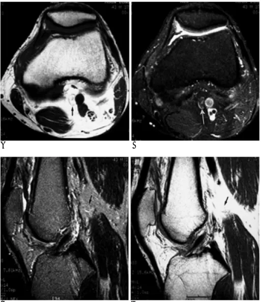
A B
Fig. 2. A 43 year-old-man with knee pain.
A. On T1-weigthed axial images, the popliteal lymph node with fatty change is identified as discrete, round or oval structures, which were well-demarcat- ed high signal structures surrounded by a low signal rim.
B, C. On T2-weighted axial and sagittal images, lymph nodes were identified as central low signal intensity with pe- ripheral high signal rim on fat-sup- pressed imaging.
D. On proton weighted sagittal images, lymph nodes showed high signal inten- sity.
C D
void, and their tubular branching shape (2, 3).
In terms of localization, lymph nodes were classified as anteromedial, anterolateral, posteromedial or pos- terolateral, depending upon their relationship with the popliteal vein (Fig. 4). In addition, the precise location of a node was expressed as the shortest distance from the line tangential to the most distal femoral articular sur- face to the center of the node (Fig. 5).
The size of a node was expressed as its shortest diame- ter, as measured on an axial image (5-8).
The relationship between number, fatty change, and the size of popliteal lymph nodes with aging was deter- mined by statistical analysis employing univariate logis- tic regression.
Results
A total of 158 (mean, 1.36) popliteal lymph nodes were present in 116 (52.3%) of 222 patients included in
this study. Their number correlated negatively with age (R square=0.826): they were found in all four subjects in the 1st decade of life, (100%), 15 of 23 in the 2nd (65.2%), 27 of 42 in the 3rd (64.3%), 22 of 34 in the 4th (64.7%), 22 of 40 in the 5th (55%), 12 of 40 in the 6th (30%), 11 of 28 in the 7th (39.3%), and 3 of 11 in the 8th (27.3%) (Fig. 6).
The number of lymph nodes showing fatty change correlated positively with age (R square=0.840). Fatty change was observed in none of the four subjects in the 1st decade (0%), 4 of 15 in the 2nd (26.7%), 12 of 27 in the 3rd (44.4%), 17 of 22 in the 4th (77.3%), 21 of 22 in the 5th (95.5%), 12 of 12 in the 6th (100%), 11 of 11 in the 7th (100%), and 3 of 3 in the 8th (100%) (Fig. 7).
The number of lymph nodes present in each of the four regions varied: they were most common in the an- teromedial region (65 of 222 cases, 29.3%), followed by the posterolateral region (58 of 222, 26.1%), the antero- lateral region (27 of 222, 12.2%), and the posteromedial
A B
Fig. 3. A 43-year-old-man with patella contusion.
A. On T1 weighted images, the popliteal lymph node with fatty change shows high signal intensity with in- complete peripheral low signal rim.
B, C. On fat suppressed T2 weighted axial and sagittal images, the popliteal lymph node was not identified.
D. On proton weighted sagittal images, lymph nodes shows high signal intensi- ty.
C D
region (8 of 222, 3.6%). The distance between the lymph node and the most distal femoral articular surface also varied according to region: 5.3±2 cm (mean±SD) in the anteromedial, 4.1±1.1 cm (mean±SD) in the postero- lateral, 4.5±1.1 cm (mean±SD) in the anterolateral and 4.3±1.0 cm (mean±SD) in the posteromedial. The mean distance was 4.6±1.4 cm (mean±SD) (Table 2).
The diameter of the popliteal lymph node was 4.96±
2.4 mm (mean±SD; range, 4-8 mm), and did not corre- late with age (R square=0.029).
─ 669 ─
Table 2. Precise Location of the Popliteal Lymph Nodes Relative to the Popliteal Vein. Total 158 Poplital Lymph Nodes were Detected in 222 Patients
Location
Distance (cm) I II III IV
1-1.9 01 00 0 01
2-2.9 02 02 1 08
3-3.9 07 06 1 17
4-4.9 09 15 5 20
5-5.9 01 19 0 09
6-6.9 05 16 1 02
7-7.9 02 07 0 01
Total 27 65 8 58
I: anterolateral region, II: anteromedial region III: posteromedial region, IV: posterolateral region
Fig. 4. Four popliteal regions; anteromedial, anterolateral, pos- teromedial, and posterolateral to the popliteal vein.
Popliteal space was arbitrarily divided into four regions with respect to its relationship with the popliteal vein as; the antero- medial, anterolateral, posteromedial, and posterolateral re- gions.
Fig. 5. The precise location of the lymph node was expressed as the shortest distance from the line tangential to the most dista femoral articular surface to the center of the lymph node.
Fig. 6. The prevalence of the politeal lymph nodes.
A: Number of the popliteal lymph node detected B: Number of examination
Fig. 7. Fatty change of the popliteal lymph nodes according to aging.
A: Number of the popliteal lymph node detected B: Number of the lymph node with fatty change
Discussion
In mammals, lymph nodes constitute the major part of peripheral lymphoid tissues. Lymphatic fluid is drained from anatomically distinct regions to a sentinel lymph node, and this becomes a primary target site where in- teraction occurs between antigenic material and im- munologic cells. Hence morphologic change occurring in a regional lymph node may reflect local immune reac- tivity (1).
Peripheral lymph nodes may be of the popliteal, cu- bital, axillary, or inguinal type, and show maximum de- velopment during the first year of life. At birth, popliteal lymph nodes have, to a certain extent, already under- gone fatty change, and this reaches considerable levels by year one (1). The presence of six or seven small popliteal lymph nodes has been described in anatomy textbooks (4, 9), but, to the best of our knowledge, no imaging study has described human popliteal lymph nodes. In this MR imaging report, the mean number of lymph nodes per person was found to be 0.71, fewer than would be expected on the basis of existing anatom- ic knowledge. There are several reasons for this. First, fatty change in popliteal lymph nodes makes it difficult to differentiate them at MRI, from adjacent fat. Second, owing to the partial volume averaging artifact, the node is small enough not to be detected.
Anatomically, popliteal lymph nodes usually lie be- tween the popliteal artery and the posterior aspect of the knee joint, receiving from this joint direct vessels, or may be sited near the end of the small saphenous vein (9). As far as we know, however, their precise location, as determined by imaging studies - particularly MR imaging - has not been reported. According to the results
of our study, they are commonly found in anteromedial and posterolateral regions, 4.6±1.4 cm away from the most distal femoral articular surface.
In conclusion, our findings show that with aging, the number of popliteal lymph nodes decreased but the fre- quency of fatty change increased, and that the nodes were more common in anteromedial and posterolateral regions.
References
1. Luscieti P, Hubschmid T, Cottier H, Hess MW, Sobin LH. Human lymph node morphology as a function of age and site. J Clin Pathol 1980;33:454-461
2. Grey AC, Carrington BM, Hulse PA, Swindell R, Yates W.
Magnetic resonance appearance of normal inguinal nodes. Clin Radiol 2000;55:124-130
3. Parsons VJ, Carrington BM, Dougal M. Normal axillary lymph nodes as demonstrated by CT. Br J Radiol 1996;69S:237
4. Frank H. Netter. The Ciba collection of medical illustrations, 3rd ed.
Newjersey: Ciba-Geigy Corp, 1987: 121
5. Ingram CE, Belli AM, Lewars MD, Reznek RH, Husband JE.
Normal lymph node size in the mediastinum: a retrospective study in two groups. Clin Radiol 1989;40:35-39
6. Dooms GC, Hericak H, Crooks LE, Higgins CB. Magnetic reso- nance imaging of the lymph node: comparison with CT. Radiology 1984;153:719-728
7. Robert E, Dorfman MD, Michael B, et al. Upper abdominal lymph nodes: criteria for normal size determined with CT. Radiology 1991;180:319-322
8. Vinnicombe SJ, Norman AR, Nicholson V, Husband JE. Normal pelvic lymph nodes: evaluation with CT after bipedal lymphangio- raphy. Radiology 1995;194:349-355
9. William PL, Warwick R, Dyson M, Bannister LH. Gray’s Anatomy, 37th ed. London: Churchill Livingstone, 1989: 848-849 10. Steinkamp HJ, Hosten N, Richter C, Schedel H, Felix R. Enlarged
cervical lymph nodes at helical CT. Radiology 1994;191:795-798 11. Motulsky AG, Weinberg S, Saphir O, Rosenberg E. Lymph nodes
in rheumatoid arthritis. Arch Intern Med 1952;90:660-672
─ 671 ─
대한방사선의학회지 2002;47:665-671
자기공명영상 상 정상 오금림프절의 발현 위치 및 나이와 관련된 갯수, 지방변성, 및 크기변화1
1연세대학교 신촌세브란스병원 진단방사선과
문 희 정・신 진 석・이 상 훈
목적: 나이에 따른 오금림프절의 위치, 나이에 따른 빈도, 지방변성, 크기를 평가하고자 한다.
대상과 방법: 슬관절의 통증으로 슬관절 자기공명영상 촬영을 한 222명의 환자를 대상으로 하였다. 평균 연령은
47.1세였고 8세에서 79세의 연령분포룰 보였다. 축면, 시상면, 관상면 영상을 얻었고 축면과 시상면, 축면과 관상 면의 두 단면에서 보이는 것을 림프절로 보았다. 정확한 오금림프절의 위치를 알기위해 오금절공간을 오금정맥을 기준으로 전방내측, 전방외측, 후방내측, 후방외측으로 나누었다. 림프절의 크기는 축면에서 최단경을 측정하였다.
결과: 222명의 환자중 116명에서 158개의 오금림파절이 보였으며 평균 0.71개 였다. 나이와 오금림파절의 빈도는 음의 상관관계를 보였으나(R square-0.826) 지방변성과는 양의 상관관계를 보였다(R square-0.840). 오금림프 절은 전방내측에서 23.9%, 후방외측에서 26.1%, 전방외측에서 12.2%, 후방외측에서 3.6%의 빈도를 보였으며 슬 관절의 대퇴부 연골에서 4.6±1.4 cm(mean±SD)의 거리에 위치하였다. 평균 직경은 4.96±2.4mm(범위, 4-8 mm) 였다.
결론: 오금림프절의 빈도는 나이와 음의 상관관계 보이고 지방변성과는 양의 상관관계를 보인다. 또한 대부분의 오 금림프절은 오금정맥에 대해 전방내측과 후방외측에 대부분 위치한다.