J. Exp. Biomed. Sci. 12 (2006) 177–183
Effects of Preparation Method and Evaluations on Structural Integrity in Model Antigen-Containing Biodegradable
Microspheres for Vaccine Delivery
Seong-Wan Cho1 and Young-Kwon Kim2†
1Department of Pharmaceutical Engineering, Konyang University,
2Department of Biomedical Laboratory Science, Konyang University, Daejeon 302-718, Korea
To demonstrate the effect of formulation conditions and evaluations of structural integrity from ovalbumin containing poly lactide glycolide copolymer (PLGA) microspheres for Vaccine delivery, OVA microspheres were prepared by a W/O/W multiple emulsion solvent extraction technique. Dichloromethan (DCM) and Ethyl acetate (EA) were applied as an organic phase and poly vinyl alcohol (PVA) as a secondary emulsion stabilizer. Microspheres were characterized for particle size, morphology (optical microscopy and Scanning Electron Microscope (SEM)). Protein denaturation was evaluated by size exclusion chromatography (SEC), SDS-PAGE and isoelectric focusing (IEF). Residual organic solvent was estimated by gas chromatography (GC) and differential scanning calorimetry (DSC). Optical photomicrograph and SEM revealed that microspheres were typically spherical but various morphologies were observed. Mean particle size (dVS) of microspheres were in the range of 3~50 µm. Also, The protein stability was not affected by the formulation process and residual organic solvent was beyond the detection below 0.1 ppm. These results demonstrated that microspheres might be a good candidate for the parenteral vaccine delivery system.
Key Words: PLGA (poly lactide glycolide copolymer), Solvent extraction method, Ovalbumin, Structural integrity
INTRODUCTION
In recent years, there have been various attempts to improve the efficacy of the currently available vaccines, often by using novel adjuvants or antigen delivery systems.
One approach of considerable interest is the use of PLGA microspheres with entrapped antigens as controlled release vaccines (Reza, 1991; Jeffery et al., 1993; Claudio et al., 1996). These PLGA had an excellent tissue compatibility profiles and used for many years as surgical sutures (Wise et al., 1979). In addition, the drug delivery systems using these polymers, an example of which was commercialized and used in man both in Europe and the USA. (Furr et al., 1987). The most commonly used microencapsulation tech-
nique in these polymers are based on the concept of solvent evaporation and employs methylene chloride and water as dispersed and continuous phases, respectively.
A variety of methods that rely on this concept are very well documented in a number of publications (Sah et al., 1994; Ming et al., 1995; Ming et al., 1996). However, The use of halogenated alkanes, such as methylene chloride and chloroform is not desirable from the viewpoints of environ- mental and human safety because methylene chloride is a suspected carcinogen and mutagen. Also, solvent evapora- tion method lead to decreased efficiencies in the micro- encapsulation of medicines (Simon, 1996). By this result, we focused on the development of a microencapsulation process utilizing ethyl acetate as a dispersed solvent com- pared with dichloromethane by adapting the water-in oil-in water solvent extraction technique. We examined the phy- sical properties of microspheres because the use of different solvent and preparation methods are the investigated key process parameters that affect the characteristics of PLGA microspheres (Ogawa et al., 1988; Yan et al., 1994). The effects of the microsphere preparative process on ovalbumin
*Received: August 1, 2006
Accepted after revision: August 30, 2006
†Corresponding author: Young-Kwon Kim, Department of Biomedical Laboratory Science Konyang University, Medical Science Building 519, Gasuweon-Dong 685, Seo-Gu, Daejeon.
Tel: 042-600-6371, Fax: 042-600-6314 e-mail: [email protected]
(OVA) structural conformation and antigenicity were also investigated by SDS-PAGE, IEF and SEC. The aim of the study was to develope microparticulate systems for vaccine delivery by microencapsulation techniques.
MATERIALS AND METHODS 1. Chemicals
PLGA; molecular weight 40,000, lactide : glycolide ratio
= 50 : 50, PVA; molecular weight 13,000~23,000, 87~89%
hydrolyzed, OVA (chicken egg, grade V) was supplied by Sigma-Aldrich, USA. Sodium dodecyl sulfate (SDS) from Bio-rad. Dichloromethane (DCM) and ethyl acetate (EA) were supplied by Fischer. Micro-BCA assay reagent and isoelectric marker were supplied by Pierce. All materials were used as received.
2. Characterization of PLGA
The thermal behavior of the polymer was recorded using a differential scanning calorimetry with Setram DSC92 model. A accurately weighed polymer sample was placed in an aluminum pan, sealed and placed into the sample compartment. An empty pan was placed in the reference compartment. The sample and the reference were heated at a rate of 5℃/min from 25 to 80℃ under nitrogen. All the DSC thermograms were obtained from the first heating cycle (Crotts et al., 1995; Mehta et al., 1996).
3. Preparation of PLGA microspheres with entrapped ovalbumin.
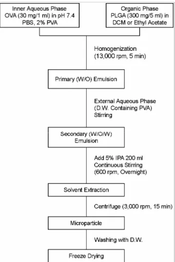
A solution of OVA in pH 7.4 phosphate buffer (internal aqueous phase) containing 2% PVA solution as a stabilizer was emulsified with 6% (W/V) polymer in DCM and EA (oil phase) using a homogenizer (X-520D, CAT, Germany) at high speed (approximately 13,000 rpm) in ice bath for 5 min. The resulting water in oil emulsion was then emulsified applying magnetic stirrer (Cole-Parmer, USA) at 600 rpm with a PVA solution to produce a W/O/W emulsion. After W/O/W emulsion was produced, emulsion maintained for 10 min agitation and the solvent was rapidly estimated by extraction 200ml of an aqueous isopropyl alcohol solution (5% V/V). Microspheres were then collected by centrifu- gation (Union 55R, Hanil, Korea), washing and freeze-dried (Labconco, USA) after microsphere formation. The final product was stored in a desiccator at below 25℃. Antigen
and polymer ratio was fixed at 1:5 and the viscosity of the external aqueous phase was varied by dissolving PVA at various concentrations. Mirospheres were prepared with the following concentrations of PVA in the external aqueous phase : 1.0, 5.0, 10.0% (W/V) (Fig. 1).
4. Characterization of particle size
The freeze dried microspheres were redispersed in PBS (pH 7.4) and sized by laser diffractometry using a Malvern sizer/E (Malvern Ins., UK). The freeze dried microspheres were dispersed by bath sonication in the presence of sur- factant to prevent aggregation and particle size is expressed as volume surface mean diameter (Vds) in micrometers.
Focal length is used with a 300 mm length lense and beam length is evaluated with a condition of 2.4 mm. Particle size is expressed as volume-surface mean diameter (dvs) in micrometers, which is expressed as belows:
Fig. 1. Preparation of biodegradable microspheres by solvent extraction method.
d: Mean diameter measured by particle size analyzer n: The number of the particle
5. Surface morphology and entrapment efficiency in microspheres
The surface appearance of the different PLGA microsp- res was analyzed by scanning electron microscopy (SEM) (JEOL 35CF, USA). For the surface analysis, freeze-dried microspheres were mounted onto metal slubs using double sided adhesive tape, dried under vacuum and coated with gold-palladium.
The protein content of microspheres were analyzed by a previously reported method (Hora et al., 1990). Briefly, 20 mg of lyophilized microspheres were digested in 5 ml of a 5% sodium dodecyl sulfate (SDS), 0.1 N NaOH solution by shaking overnight on IKA Vibrax shaker until the complete dissolution of the microspheres. The sample was centrifuged and bichinchoninic acid (BCA) protein microassay was used to determine the OVA concentration in the supernatant.
From this result, the percentage (W/W) of OVA entrapped per dry weight of microspheres were determined. Each sample was assayed in triplicate. The loading efficiency (%) was expressed as the actual OVA loading to the theoretical OVA loading.
... (2)
6. Determination of the residual organic solvent
Accurately weighed microsphere samples (30 mg) were completely dissolved in 4 ml of N-N-dimethyl formamide and samples were prepared in triplicate for each microsphere formulation (Crotts et al., 1995). A Varian-3800 gas chro- matograph system with a flame ionization detector (FID) was used in this experiment. The initial oven temperature was set at 50℃ for 2 min and then increased to a final temperature of 250℃ at the rate of 10℃/min. The injector and detector temperature were maintained at 250℃. The DB-1 cross-linked methyl siloxane capillary column (length:
30 cm, I.D: 0.25 mm) was employed as a stationary phase with nitrogen as a carrier gas.
7. SDS-PAGE
Polyacrylamide gel electrophoresis (PAGE) analysis of native OVA and OVA released from PLGA microspheres were undertaken and samples of OVA released from PLGA microspheres in phosphate buffered saline (PBS). Native OVA and molecular weight reference marker were solubi- lized with sample buffer, loaded onto a vertical slab gel (10%) and subjected to electrophoresis at 200 mV. Follo- wing electrophoresis, the gels were stained with silver stain- ing solution to visualize the protein.
8. Isoelectric focusing (IEF)
Retention of the structural integrity of the entrapped OVA was assessed by isoelectric focusing (IEF). OVA released from microspheres into PBS, native OVA and IEF standards were loaded onto a IEF ready gel and focused using a Bio- rad mini IEF cell. The gel was fixed and destained as de- scribed in the Bio-rad protocol for the mini IEF cell.
9. Size exclusion chromatography (SEC)
Significant conformational changes associated with the folding-unfolding equilibria of solvent or temperature de- pendent denaturation of proteins may be studied using analytical size exclusion chromatography. The amount of soluble OVA aggregates in each sample were quantitated by SEC. A zorbax GF250 (Hewlett packard, USA) column was operated at 1.0 ml/min using a mobile phase of 130 mM NaCl/20 mM Na2HPO4 (pH 7.0) on an HPLC system pro- tein peak detection was performed at 210 nm. The relative peak area of OVA and soluble aggregates were measured and total peak area were compared to control OVA samples of known concentration.
RESULTS
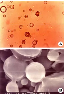
1. Photomicrographs and SEM of microspheres
The appearance of the PLGA microspheres were obser- ved by photomicroscopy and SEM. Microspheres show spherical shape in initial stage because of the well diffusion of solvent to the continuous phase by photomicroscopy (Fig.
2A). Furthermore, optical photomicrograph revealed that the extraction of solvent led to significant shrinkage of micro- actual protein loading
(w/w %) Loading efficiency (%) =
theoretical protein loading (w/w %)
×100 Σ nd3
dvs =
Σ nd2 ... (1)
spheres overnight after preparation. SEM examination also showed different morphological characteristics for micro- spheres produced by the W/O/W techniques. Immediately after preparation (Fig. 2B), the microspheres showed an
intact outer surface.
2. Loading efficiency and particle size of microspheres
There was a size reduction by the increase of PVA con- centration used in the secondary stabilizer to prepare the microspheres irrespective of preparation method, used sol- vent. In particular, the microspheres prepared by EA had a smaller size distribution and larger loading efficiency and protein entrapment than DCM microspheres in the same PVA concentration (Table 1). It was concluded that we could fabricate microspheres with different sizes by varying the concentration of poly vinyl alcohol in the external aqueous
Table 1. Physical properties of microspheres prepared by dichlo- romethane and ethyl acetate
Microspheres* Entrapment efficiency (%)
Particle size (dvs, µm)
DEX, 1% 4.19±0.01 20.11
EEX, 1% 16.53±0.10 15.42
DEX, 5% 10.48±0.40 15.16
EEX, 5% 17.39±0.26 14.36
DEX, 10% 10.72±0.21 4.38
EEX, 10% 12.34±1.14 2.33
*D or E means solvent used, dichloromethane or ethyl acetate, respectively
*EX means solvent extraction method
*1, 5, 10% means PVA concentration in W2 phase B
A
Fig. 2. Photomicrographs (×400) (A) and Scannig Electron Microscopes (SEM) (B) of PLGA (50:50) microspheres.
Fig. 3. Glass transition temperature (Tg) measured by differential scanning calorimetry (DSC). The sample was heated at a rate of 5℃
/min from 25 to 80℃ under nitrogen. The DSC thermograms were obtained from the first heating cycle.
phase and it could be a very useful for the sustained vaccine delivery.
3. Differential scanning calorimetry (DSC) of poly- mers and determination of residual organic solvents
The glass transition temperature (Tg) was determined by differential scanning calorimetry (DSC). The value was very important because the Tg could be changed when the residual amounts of organic solvent was exist. In our results, The Tg of the solvent free polymer was 45℃. Thereafter, the Tg was maintained to 45℃ after preparation of micro- spheres. It means that the polymer was not changed from glassy state to rubbery state after preparation and there is few residual organic solvent in the microspheres (Fig. 3).
This result also could be proved by gas chromatography.
Residual organic solvent was beyond the detection below 0.1 ppm after residual organic solvent was estimated by gas chromatography.
4. Stability and integrity of protein: SDS-PAGE
During microsphere preparation, the antigen was exposed to potentially harsh conditions, such as DCM and EA and mechanical agitation. SDS-PAGE analysis by silver staining revealed that the OVA released from microspheres produced from the two method above were the same as native OVA (Fig. 4). There were no additional bands to indicate the pre- sence of molecular weight aggregates or fragments greater or smaller than 45 kDa. These result pointed that OVA released initially from the PLGA microspheres had a native protein structure and the encapsulation process and polymer type do not affect the protein stability.
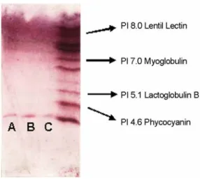
5. Stability and integrity of protein: Isoelectric focu- sing (IEF)
Also, IEF showed identical bands for the native OVA and the OVA initially released from PLGA microspheres (Fig.
5). IEF separates proteins on the basis of surface charge as a function of pH, and since a protein contains both positive and negative charge - bearing groups, the net charge of the protein will vary with pH. A significant alteration of the conformaton of OVA following entrapment into PLGA microspheres could result in a variation in the surface charge (Smardar and Howard, 1996). By comparing these bands Fig. 4. 10% Polyacrylamide gel electrophoresis (SDS-PAGE)
visualized by silver stain. Native OVA and molecular weight reference marker were solubilized with sample buffer, loaded onto a vertical slab gel (10%) and subjected to electrophoresis at 200 mV. A; native OVA, B; prepared by DCM, C; prepared by EA.
Fig. 5. Isoelectric focusing (IEF) gel electrophoresis. The gel was fixed and destained as described in the Bio-rad protocol for the mini IEF cell. A; native OVA, B; prepared by DCM, C; pre- pared by EA.
Fig. 6. Size exclusion chromatography analysis of OVA released initially from PLGA microspheres. A zorbax GF250 (Hewlett pac- kard, USA) column was operated at 1.0 ml/min using a mobile phase of 130 mM NaCl/20 mM Na2HPO4 (pH 7.0) on an HPLC system protein peak detection was performed at 210 nm. A; native OVA, B; prepared by DCM, C; prepared by EA.
with the isoelectric point marker, it could be seen that the isoelectric point of both the native OVA and the OVA rele- ased from microspheres is PI 4.5.
6. Size exclusion chromatography
The initial proteins released from the PLGA microspheres were equivalent to the control protein in the SEC. It has been possible to assess the concentration and characteristics of partially unfolded species within the same time scale as the chromatographic run. As shown in Fig. 6, the retention time of OVA as compared to the control solution was same.
This observation provides that OVA was entrapped without significantly altering the antigenic structure or conformation.
DISCUSSION
Antigens are mostly proteins that must adopt specific, folded, three-dimensional structures to be biological active products. The inactivation of proteins is often connected with a destruction of the conformation. In order to success- fully develop a protein PLGA delivery system, it is essential that sufficient biological activity of a therapeutic protein should be retained throughout encapsulation, storage, and duration of release. During the preparation of polymer part- icles using the multiple emulsion methods, one of the major obstacles faced is the contact of protein with organic solvent.
Interaction of the protein molecule with organic solvent leads to the protein's denaturatioin, resulting in loss of immuno- reactivity and/or bioactivity. Denaturation of the protein by organic solvent also leads to the protein's precipitation at the aqueous-organic interface, resulting in the protein's uneven distribution in the polymer matrix. Organic solvent-induced denaturation during the process of microencapsulation may be responsible for the unpredictable and incomplete release profile often found in many studies. The problem of protein denaturation by the organic solvent is of primary importance because this occurs at the very first step of the encapsulation process during which primary emulsification of the protein solution is carried out with organic solvent containing the polymer (Cohen and Bernstein, 1996; Raghuvanshi et al., 1998). Destabilization of protein and peptide molecules is two types: chemical, which involves changes in covalent bonds, and physical, which involves changes in the spatial three-dimensional structure (denaturation). The chemical de- gradation mechanism has been well reviewed and most
commonly include hydrolysis, oxidation, deamidation, di- sulfide exchange, and racemization. The physical or denatu- ration process has also been extensively reviewed (Manning et al., 1989). This process of unfolding molecule results in problem of aggregation, adsorption, and loss of activity. It is the tertiary structure of the protein that must be stabilized against the various disruptive forces that occur during pro- cessing and handling. Potential denaturing forces include chemical stress from factors used in purification, such as pH, ionic strength, or detergents, or physical stress during filtration and filling where surface adsorption and shear contribute to unwinding of the tertiary structure into a ran- dom coil. Solvent protein interaction strongly influences conformation. Water, their normal solvent of proteins, forms a hydration layer around the protein and contributes to the carbon side chains buried within the molecule provides additional stabilization energy. Disruption of the structure occurs when the solvent partitions the hydrocarbon side chains between a hydrocarbon side chains between a hydro- carbon phase and aqueous phase (Manning et al., 1989). In our experiment, PLGA microspheres have been successfully prepared by W/O/W emulsion solvent extraction method and protein denaturation was not found during the manu- facturing procedure. The results described in the present study showed the effect of the manufacturing method and used solvent on the microcapsule properties and protein release. Different preparation procedures produced variation of the release characteristics of microspheres. Microenca- psulation processes and used solvent worked well and fea- tured various properties. Application of a mixture of PLGA microspheres with different kinetics most likely would pro- vide a sustained/pulsed antibody response. In conclusion, PLGA microspheres are delivery systems capable of encap- sulating a wide range of materials, formulation with various entrapped antigens prepared by several techniques. The controlled release of antigens using this polymer might be useful in prolonged vaccine formulations for the parenteral carrier system. These microspheres were extensively char- acterized in vitro, before being used parenterally as drug and antigen delivery systems in small animal models.
REFERENCES
Claudio T, Giampietro C, Ying M, Hans PM, Brauno G. Tetanus toxoid and synthetic malaria antigen containing poly(lactide)/
poly(lactide-co-glycolide) microspheres.: importance of poly- mer degradation and antigen release for immune response. J Control Release 1996. 41: 131-145.
Cohen S, Bernstein H. Microparticulate systems for the delivery of proteins and vaccines. 1996. pp51-88. Marcel Dekker, Inc.
Crotts G, Park TG. Preparation of porous and nonporous biode- gradable polymeric hollow microspheres. J Control Rel. 1995.
35: 91-106.
Furr BJA. Pharmacological studies with zoladex, a novel luteini- zing hormone-releasing hormone analogue. In: Zoladex. A new treatment for prostatic cancer (ed. G.D. Chisholm). Royal society of Medicine International Congress and Symposium Series. 1987. 125. pp. 1-10.
Hora HS, Rana RK, Nunberg JH, Tice TR, Gilley RM, Hudson ME. Release of human serum albumin from poly lactide-co- glycolide microspheres. Pharm Res. 1990. 7: 1190-1194.
Jeffery H, Davis SS, O'Hagan DT. The preparation and characteri- zation of poly lactide-co-glycolide Microparticles II. The entrapment of a model protein using a (water-in-oil-in-water) emulsion solvent evaporation technique. Pharm Res. 1993.
10: 362-368.
Manning MC, Patel K, Borchandt RT. Stability of protein pharma- ceuticals. Pharm Res. 1989. 6: 903-909.
Mehta RC, Thanoo BC, DeLuca PP. peptide containing micro- spheres from low molecular weight and hydrophilic poly- lactide-co-glycolide. J Control Rel. 1996. 41: 249-257.
Ming KY, Coombes AGA, Jenkins PG, Davis SS. A novel emulsification - solvent extraction technique for production of protein loaded biodegradable microparticles for vaccine and drug delivery. J Control Release 1995. 33: 437-445.
Ming KY, Stanley SD, Allan GAC. Improving Protein Delivery from Microparticles Using Blends of Poly (DL lactide co- glycolide) and Poly (ethylene oxide)-Poly (propylene oxide)
Copolymers. Pharm Res. 1996. 13: 1693-1698.
Ogawa Y, Yamamoto M, Okada H, Yashiki T, Shimamoto T. A new technique to efficiency entrap leuprolide acetate into microcapsules of copolymer (lactic/glycolic acid). Chem Pharm Bull. 1988. 36: 1095-1098.
Raghuvanshi RS, Goyal S, Singh O, Panda AK. Stabilization of dichloromethane-induced protein denaturation during micro- encapsulation. Pharm Develop Tech. 1998. 3: 269-276.
Reza A. Preparation of biodegradable microspheres and micro- capsules: 2. Polyactides and releated polyesters. J Control Release 1991. 17: 1-22.
Sah H, Toddywala R, Chien YW. The influence of biodegradable microcapsule formulations on the controlled release of a protein. J Control Release 1994. 30: 201-211.
Smardar C, Howard B. Microparticulate systems for the delivery of proteins and vaccines. 1996. pp51-88. Marcel Dekker. Inc.
USA
Smardar C, Toshio Y, Melissa L, Lena HH, Robert L. Controlled delivery systems for proteins based on poly (lactic/glycolic acid) microsphere. Pharm Res. 1991. 8: 713-720.
Simon B. Microencapsulation; Methods and industrial applications.
1996. pp35-72. Marcel Dekker, Inc. USA
Wise DL, Fellman TD, Sanderson JE, Wentworth RL. Lactide/
glycolide polyers. Description of the polymers used as sur- gical suture material, raw material for osteosynthesis and in sustained release forms of drugs. In: Drug carriers in Medicine (ed. G. Gregoriadis), 1979. pp237-245. Academic Press, London.
Yan C, Resau JH, Hewetson J, West M, Rill WL, Kende M.
Characterization and morphological analysis of protein-loaded poly (lactide-co-glycolide) microparticles prepared by water- in-oil-in water emulsion technique. J Control Release. 1994.
32: 231-235.