Biomedical Science Letters 2019, 25(4): 407~416 https://doi.org/10.15616/BSL.2019.25.4.407 eISSN : 2288-7415
Probe-based qPCR Assay for Rapid Detection of Predominant Candida glabrata Sequence Type in Korea
Jinyoung Bae
*, Kyung Eun Lee
* *and Hyunwoo Jin
†,* *Department of Clinical Laboratory Science, College of Health Sciences, Catholic University of Pusan, Busan 46252, Korea
Recent years have seen an increase in the incidence of candidiasis caused by non-albicans Candida (NAC) species. In fact, C. glabrata is now second only to C. albicans as the most common cause of invasive candidiasis. Therefore, the rapid genotyping specifically for C. glabrata is required for early diagnosis and treatment of candidiasis. A number of genotyping assays have been developed to differentiate C. glabrata sequence types (STs), but they have several limitations.
In the previous study, multi-locus sequence typing (MLST) has performed with a total of 101 C. glabrata clinical isolates to analyze the prevalent C. glabrata STs in Korea. A total of 11 different C. glabrata STs were identified and, among them, ST-138 was the most commonly classified. Thus, a novel probe-based quantitative PCR (qPCR) assay was developed and evaluated for rapid and accurate identification of the predominant C. glabrata ST-138 in Korea. Two primer pairs and hybridization probe sets were designed for the amplification of internal transcribed spacer 1 (ITS1) region and TRP1 gene. Analytical sensitivity of the probe-based qPCR assay was 100 ng to 10 pg and 100 ng to 100 pg (per 1 μL), which target ITS1 region and TRP1 gene, respectively. This assay did not react with any other Candida species and bacteria except C. glabrata. Of the 101 clinical isolates, 99 cases (98%) were concordant with MLST results. This novel probe- based qPCR assay proved to be rapid, sensitive, highly specific, reproducible, and cost-effective than other genotyping assay for C. glabrata ST-138 identification.
Key Words: Candida glabrata, Genotyping, Quantitative PCR (qPCR), Sequence types (STs), TRP1
INTRODUCTION
Candidiasis is a widespread fungal infection caused by various Candida species, especially in acquired immune deficiency syndrome (AIDS) patients, immunocompromised individuals, patients with hematopoietic stem cells or organ transplantations, and those who present underlying valvular heart diseases or have received long-term antibiotic therapy (Bineshian et al., 2015; Rezazadeh et al., 2016; Ortiz et al.,
2018).
Although C. albicans is still the major cause of candi- diasis, in recent years the number of infections caused by non-albicans Candida (NAC) species has increased (Ho and Haynes, 2015). Among them, C. glabrata has received considerable attention as one of the most common cause of invasive candidiasis (Shahrokhi et al., 2017). The mortality rate associated with candidemia by infection with C. glabrata is also becoming higher than with any other NAC species because of its resistance to azole antifungal agents (Gohar et
Original Article
Received: October 28, 2019 / Revised: November 26, 2019 / Accepted: November 28, 2019
*Graduate student, **Professor.
†Corresponding author: Hyunwoo Jin. Department of Clinical Laboratory Science, College of Health Science, Catholic University of Pusan, Busan 46252, Korea.
Tel: +82-51-510-0567, Fax: +82-51-510-0568, e-mail: [email protected]
○CThe Korean Society for Biomedical Laboratory Sciences. All rights reserved.
○CCThis is an Open Access article distributed under the terms of the Creative Commons Attribution Non-Commercial License (http://creativecommons.org/licenses/by-nc/3.0/) which permits unrestricted non-commercial use, distribution, and reproduction in any medium, provided the original work is properly cited.
al., 2017). Furthermore, with rapidly expanded use of echino- candin as the primary treatment for invasive candidiasis, echinocandin resistance in C. glabrata has posed a serious clinical challenge (Zhao et al., 2016). In spite of its growing prominence, however, little is known of the population structure, epidemiology, and basic biology of C. glabrata (Dodgson et al., 2003).
It is essential to develop DNA fingerprinting methods which can assess genetic distance between independent isol- ates in broad epidemiological studies (Abbes et al., 2010).
A number of DNA fingerprinting methods have been de- veloped to differentiate C. glabrata strains, such as random amplification of polymorphic DNA (RAPD) (Becker et al., 2000), pulsed-field gel electrophoresis (PFGE) (Lin et al., 2007), multi-locus enzyme electrophoresis (MLEE) (Pujol et al., 1997), multi-locus sequence typing (MLST) (Lott et al., 2010), and multi-locus variable-number tandem-repeat analysis (MLVA, also known as microsatellite analysis) (Abbes et al., 2012). Above all, the sequence-based method, MLST analyzes five to seven selected housekeeping genes for single nucleotide polymorphisms (SNPs) (Berila and Subik, 2010; Enache-Angoulvant et al., 2010). However, these approaches have several limitations. Although these genotyping methods can discriminate between closely related strains, they are costly, time-consuming, have lower repro- ducibility, and require highly trained clinicians for proper execution (Essendoubi et al., 2007). In order to overcome these limitations, a novel C. glabrata genotyping method is required.
In the previous study (Kang, 2017), MLST was performed with a total of 101 C. glabrata clinical isolates to analyze genetic polymorphisms and investigate the most prevalent sequence types (STs) in Korea. In a total of 3,345 base-pair DNA sequences, 49 variable nucleotide sites were found. A total of 11 different C. glabrata STs were identified, and among them, C. glabrata ST-138 was the most common (53%). Each ST has a number of nucleotide differences between alleles; therefore, these polymorphic sites can be utilized for PCR-based molecular assays (Pérez-Losada et al., 2013).
Quantitative PCR (qPCR) assays have advantages over other PCR-based molecular assays, such as higher sensitivity,
less setup cost, shorter turnaround time, and reproducibility (Foongladda et al., 2014). In addition, amplification reactions and data analysis are processed in a closed-tube system, eliminating the post-amplification step and reducing chances for cross-contamination (Navarro et al., 2015).
In this study, a novel probe-based qPCR assay was de- veloped for rapid and specific identification of C. glabrata ST-138 from other STs in Korea. Its performance was eval- uated with a total of 101 C. glabrata clinical isolates provided from Korean Culture Collection of Medical Fungi (KCMF, Daejeon, Korea).
MATERIALS AND METHODS
Clinical strainsA total of 101 C. glabrata clinical isolates were provided from KCMF (Daejeon, Korea) and those isolates were col- lected from Asan Medical Center, Seoul, Yonsei University Wonju Severance Christian Hospital, Wonju, The Catholic University of Korea Seoul St. Mary's Hospital, Seoul, and Chungbuk National University Hospital, Cheongju, Korea.
Clinical isolates were isolated from a wide variety of clinical samples, including bloodstream, catheterized urine, bile, and other body fluids (Table 1).
Genomic DNA extraction
Genomic DNA (gDNA) from C. glabrata clinical isolates was extracted using a I-genomic BYF DNA Extraction Mini kit (iNtRON Inc., Seongnam, Korea) according to the manufacturer's instructions (Da Silva-Rocha et al., 2014).
Briefly, cultured yeast samples were added 200 μL of MYP Table 1. Origin of C. glabrata clinical isolates used in this study
Origin of clinical isolates Number of cases
Blood 64
Urine 14
Bile 8
Others1) 14
-2) 1
Total 101
1) Ascitic fluid, joint fluid, pleural fluid, tissue etc
2) No clinical information
buffer and 2 μL β-mercaptoethanol. After incubation at 37℃
for 15 min, the samples were centrifuged and the super- natants were removed completely. Then the samples were lysed by 100 μL of MP buffer and 2 μL lyticase solution (4.2 unit/μL). After incubation at 37℃ for 15 min, the sam- ples were centrifuged and the supernatants were discarded, and added 200 μL of MG buffer, 20 μL of proteinase K solution (20 mg/mL) and 5 μL of RNase A solution (10 mg /mL). The samples were incubated at 65℃ for 30 min for complete yeast lysis. The lysates were added 250 μL of MB buffer and 250 μL of 80% ethanol. Portions of the lysates were transferred to column in 2.0 mL collection tube and centrifuged. After washing stages, the gDNA was eluted by adding EB buffer. The concentration and purity of the gDNA was checked by 260/280 optical density using a Nanodrop 2000 Spectrophotometer (Thermo Scientific, Wilmington, DE, USA) (Da Silva-Rocha et al., 2014).
MLST analysis for identifying sequence type of C.
glabrata clinical isolates
MLST was performed using a procedure described pre- viously (Kang, 2017). The six housekeeping gene fragments including FKS, LEU2, NMT1, TRP1, UGP1, and URA3 were selected for MLST analysis. The reaction products were purified and sequenced at Macrogen Inc. (Daejeon, Korea). The obtained molecular sequences were analyzed by using the C. glabrata MLST databases (http://pubmlst.org /cglabrata/) (Dodgson et al., 2003). The allele profiles of
the strains were defined according to the six MLST loci.
Each unique allele profile was designated as a ST (Table 3).
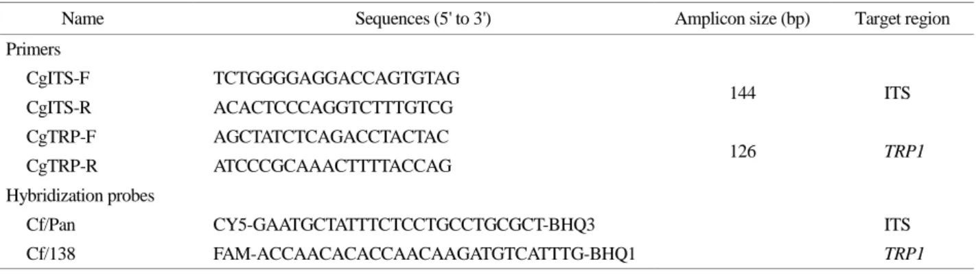
Design of primer pairs and hybridization probes
For identification of C. glabrata ST-138, the two primer pairs and hybridization probe sets were designed based on alignments of the ITS1 region, the conserved region between the 18S and 28S ribosomal RNA (rRNA) and the TRP1 gene of C. glabrata, respectively, from the National Center for Biotechnology Information (NCBI) database. Optimal primer pairs and hybridization probes were designed by using Primer 3 (v. 0. 4. 0) online software (Rozen et al., 2012). The characteristics and the sequences of two primer pairs and probe sets are shown in Table 4. Two primer pairs (CgITS and CgTRP) recognized a 144 bp region of ITS1 and a 126 bp region of TRP1 gene of C. glabrata. Two hy- bridization probes (Cf/Pan and Cf/138) were also designed by labelling different fluorophores (FAM and CY5) at 5' end, and non-fluorescent quencher (BHQ1 and BHQ3) at 3' end, respectively.
Probe-based qPCR assay
Two-tube TaqMan probe qPCR assay was carried out on the ABI 7500 Fast Real-Time PCR system (Life- Technologies, Waltham, MA, USA). Target amplification was performed in 20 μL reaction mixture containing 10 μL Table 2. Uploaded MLST status of new C. glabrata sequence
types Assigned
ST1) FKS LEU2 NMT1 TRP1 UGP1 URA3
ST-138 3 22 4 3 3 4
ST-139 7 22 3 50 1 8
ST-140 8 22 3 5 1 2
ST-141 10 21 14 50 1 9
ST-142 7 17 11 10 5 9
ST-143 5 17 8 7 3 50
ST-144 5 17 8 7 14 6
1) Sequence type.
*All sequence types indicated in this table are novel combinations of existing database
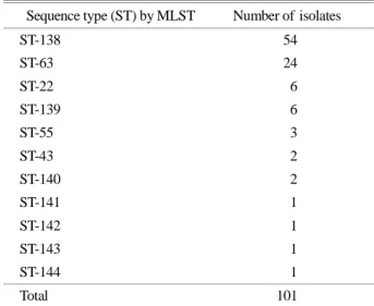
Table 3. MLST status of C. glabrata isolates
Sequence type (ST) by MLST Number of isolates
ST-138 54
ST-63 24
ST-22 6
ST-139 6
ST-55 3
ST-43 2
ST-140 2
ST-141 1
ST-142 1
ST-143 1
ST-144 1
Total 101
THUNDERBIRD® Probe qPCR Mix (Toyobo, Osaka, Japan), 4.5 μL of ultrapure water, 1 μL of each primer (10 pmol/
μL), 0.5 μL of each hybridization probe (10 pmol/μL), and 3 μL of gDNA template. The cycling conditions of qPCR were as follow: 2 min incubation at 95℃; 35 cycles of 10 sec at 95℃ and 45 sec at 63℃. A positive result was in- dicated when the cycle threshold (CT) value was less than 30 after observing signal formation of a wavelength from each channel (FAM and CY5).
Determination of analytical sensitivity and specificity
The limit of detection (LOD) of the qPCR assay of the ITS1 region and TRP1 gene was determined through the use of a 10-fold dilution [100 ng, 10 ng, 1 ng, 100 pg, 10 pg, and 1 pg (per 1 μL)] along a standard curve of gDNA isolated from C. glabrata ST-138. Each dilutions were tested in triplicate, and the LOD of the qPCR assay was determined as the dilution at which all replicates were positive.
A total of five different strains were used to determine the specificity of the qPCR assay for detection of ITS1 region:
C. albicans, C. glabrata, C. tropicalis, Staphylococcus aureus, and Escherichia coli. A total of 11 C. glabrata STs were used to determine the specificity of the qPCR assay for detection of TRP1 gene: ST-22, ST-43, ST-55, ST-63, ST-138, ST-139, ST-140, ST-141, ST-142, ST-143, and ST-144. Each gDNA samples were assayed by the same procedures for the qPCR assay application.
RESULTS
Sequence type of C. glabrata clinical isolates
With a total of 11 C. glabrata STs, seven of which were identified as new STs that were not discovered in the pre- vious study. These data of new C. glabrata STs were up- loaded and assigned at Candida glabrata MLST Databases (https://pubmlst.org/cglabrata/) (Table 2).
The data demonstrates that the ST-138 was the most pre- dominant ST in this study as a total of 54 clinical isolates were contained in this ST, and the following most pre- dominant ST was the ST-63 as a total of 24 clinical isolates Table 4. The primers and hybridization probes sequences for the qPCR assay
Name Sequences (5' to 3') Amplicon size (bp) Target region
Primers
CgITS-F TCTGGGGAGGACCAGTGTAG
144 ITS
CgITS-R ACACTCCCAGGTCTTTGTCG
CgTRP-F AGCTATCTCAGACCTACTAC
126 TRP1
CgTRP-R ATCCCGCAAACTTTTACCAG
Hybridization probes
Cf/Pan CY5-GAATGCTATTTCTCCTGCCTGCGCT-BHQ3 ITS
Cf/138 FAM-ACCAACACACCAACAAGATGTCATTTG-BHQ1 TRP1
Fig. 1. C. glabrata ST-138 specific sequences of TRP1 locus in Multi-align analysis.
were contained in this ST. In addition, this study obtained the ST-22, ST-55, and ST-43 were as a total of 6, 3, and 2 clinical isolates were contained in respective ST and the ST- 139 was identified in 6 isolates. The ST-140 was identified in 2 isolates and the remaining STs (141, 142, 143, and 144) were classified only once each (Table 3).
Analytical sensitivity and specificity of probe-based qPCR assay
The LOD of the qPCR assay of the ITS1 region and TRP1
gene was determined through the use of a 10-fold dilution [100 ng, 10 ng, 1 ng, 100 pg, 10 pg, and 1 pg (per 1 μL)]
along a standard curve of gDNA isolated from C. glabrata ST-138. The LOD of the qPCR assay of the ITS1 region was 10 pg (per 1 μL) (Fig. 4A). The CT values for each sample concentrate ranged from 15.898 to 30.043. The LOD of the qPCR assay of the TRP1 gene was 100 pg (per 1 μL) (Fig. 4B). The CT values for each sample concentrate ranged from 21.358 to 32.382. The average CT values for each sample concentration are shown in Table 5.
Fig. 3. Multi-alignment analysis of forward primers, reverse primers, and hybridization probes based on reference TRP1 gene sequences.
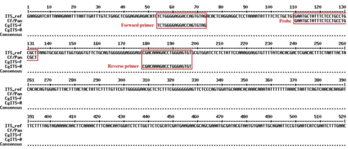
Fig. 2. Multi-alignment analysis of forward primers, reverse primers, and hybridization probes based on reference ITS1 gene sequences.
A
B
Fig. 4. Limit of detection of real-time PCR assay. The LOD of real-time PCR assay of the ITS1 region and TRP1 gene was determined through the use of a 10-fold dilution [100 ng, 10 ng, 1 ng, 100 pg, 10 pg, and 1 pg (per 1 μL)] along a standard curve of gDNA isolated from C. glabrata ST-138. (A) the LOD of real-time PCR assay of the ITS1 region was 10 pg (per 1 μL). (B) the LOD of real-time PCR assay of the TRP1 gene was 100 pg (per 1 μL).
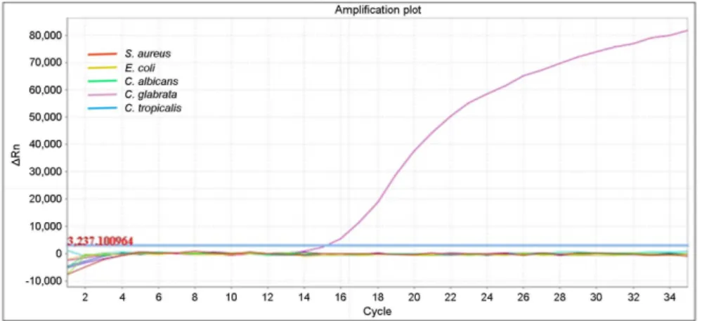
Fig 5. Specificity of real-time PCR assay for detection of ITS1 region.
A total of five different strains were used to determine the specificity of the real-time PCR assay for detection of ITS1 region: C. albicans, C. gla- brata, C. tropicalis, S. aureus, and E.
coli. The ITS1 region-specific primers detected C. glabrata accurately with- out cross-reaction.
A total of five different strains were used to determine the specificity of the qPCR assay for detection of ITS1 region:
C. albicans, C. glabrata, C. tropicalis, S. aureus, and E.
coli. The ITS1 region-specific primers detected C. glabrata accurately without cross-reaction (Fig. 5). A total of 11 C.
glabrata STs were used to determine the specificity of the qPCR assay for detection of TRP1 gene: ST-22, ST-43, ST- 55, ST-63, ST-138, ST-139, ST-140, ST-141, ST-142, ST-143, and ST-144. The TRP1 gene-specific primers detected C.
glabrata ST-138 accurately without cross-reaction (Fig. 6).
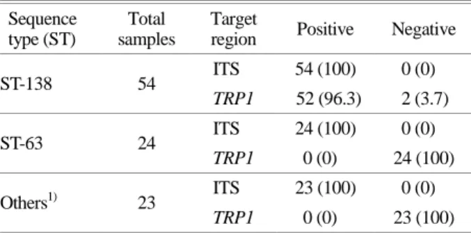
Performance of probe-based qPCR assay on C. glabrata clinical isolates
A total of 101 gDNA samples from C. glabrata clinical isolates were used for the qPCR assay. In a total of 54 C.
glabrata ST-138 cases, all were positive in targeting the ITS1 region, and 52 cases (96.3%) were positive and two cases (3.7%) negative in targeting the TRP1 gene. In a total of 47 cases of other STs (22, 43, 55, 63, 139, 140, 141, 142,
143, and 144), all assays were positive in targeting the ITS1 region and were negative in targeting the TRP1 gene (Table 6).
DISCUSSION
Candidiasis is a fungal infection caused by Candida species that features various clinical expressions, including superficial, mucocutaneous, and invasive infection (Camacho- Cardoso et al., 2017). Although Candida species are com- mensal organisms that presented in healthy individuals and in the natural environment, some of these fungi can become opportunistic pathogens induced by alteration in the host environment, especially in immunocompromised patients (Sadeghi et al., 2018). C. albicans remains the major cause of candidiasis; however, infections by NAC species have also increased, recently. Among them, the incidence of C.
glabrata infection ranks second in Candida species with a high mortality rate, and it is resistant to antifungal agents Table 5. Analytical sensitivity of the qPCR assay
Concentration (ng/μL)
Ct Mean
ITS region TRP1 gene
100 15.898±0.51 21.358±0.26
10 19.441±0.42 24.865±0.26
1 22.825±0.21 28.244±0.19
0.1 25.739±1.10 32.382±1.10
0.01 30.043±0.32 n.d1)
0.001 n.d1) n.d1)
1) Not detected
Table 6. Result of the qPCR assay
Sequence type (ST)
Total samples
Target
region Positive Negative
ST-138 54 ITS 54 (100) 0 (0)
TRP1 52 (96.3) 2 (3.7)
ST-63 24 ITS 24 (100) 0 (0)
TRP1 0 (0) 24 (100)
Others1) 23 ITS 23 (100) 0 (0)
TRP1 0 (0) 23 (100)
1) 22, 43, 55, 139, 140, 141, 142, 143, and 144
Fig 6. Specificity of real-time PCR assay for detection of TRP1 gene.
A total of 11 C. glabrata STs were used to determine the specificity of real-time PCR assay for detection of TRP1 gene: ST-22, ST-43, ST-55, ST- 63, ST-138, ST-139, ST-140, ST-141, ST-142, ST-143, and ST-144. The TRP1 gene-specific primers detected C. glabrata ST-138 accurately without cross-reaction.
(Becker et al., 2000). Notably, C. glabrata has a wide min- imum inhibitory concentration (MIC) range (≤ 32 μg/mL) for fluconazole susceptible-dose dependence (SDD) with no MIC cut-off for the susceptible range, according to the Clinical and Laboratory Standards Institute (CLSI). However, despite fluconazole's wide MIC range, C. glabrata has dem- onstrated reduced susceptibility and high-level resistance to fluconazole therapy (Whaley et al., 2018). The mortality rate of candidemia caused by C. glabrata is also increasing, becoming higher than other NAC species due to resistance to azole antifungal agents (Gohar et al., 2017). In spite of its growing prominence, little is known of the population structure, epidemiology, and basic biology of C. glabrata (Dodgson et al., 2003).
In order to investigate the epidemiology of these patho- gens, it is crucial to perform genotype analysis of C. glabrata (Paluchowska et al., 2014). Several genotyping assays have been used to differentiate C. glabrata. PFGE compares total DNA banding patterns with or without restriction enzyme digestion. MLEE discriminates genetic variations of the DNA products for a number of loci. RAPD compares band- ing patterns following PCR with a non-specific primer. The sequence-based methods, MLST analyzes five to seven selected housekeeping genes for SNPs, whereas MLVA ex- amines length variation markers in six to nine PCR-amplified sequences that contain polymorphic tandem repeats (Abbes et al., 2010; Berila and Subik, 2010; Enache-Angoulvant et al., 2010; Katiyar et al., 2016). But these methods have sev- eral limitations, in that they are high-priced, time-consuming, and low reproducibility.
In the previous study (Kang, 2017), MLST was perfor- med to analyze prevalent C. glabrata STs in Korea. Of a total of 101 C. glabrata clinical isolates, 11 different STs were identified and, among them, ST-138 was predominant.
Several studies have assessed the C. glabrata genotype in Korea by using MLST. In a study by Byun et al. (2018), MLST analysis showed C. glabrata ST-7 (100 isolates, 47.8%) was the most common type among 209 C. glabrata bloodstream isolates. A comparison between C. glabrata ST-7 and ST-138 allele profiles showed only the LEU2 locus was different in their overall allelic profiles. Due to its haploid nature, C. glabrata may undergo rapid genotypic change
during infection in humans, unlike the usually diploid Can- dida species. However, little is known about the relation- ship between the genotypic variation and antifungal therapy among isolates of C. glabrata (Shin et al., 2007). It is im- portant to discriminate the most prevalent ST because the investigation of its virulence factor, resistance for antifungal agents or possible routes of transmission can assist in the prevention of pathogenic fungi (Paluchowska et al., 2014).
For this reason, we developed a novel genotyping method for rapid and accurate identification of C. glabrata ST-138 using a probe-based qPCR assay. qPCR assays have advan- tages such as rapidity, high sensitivity, less setup cost and reproducibility (Takahashi et al., 2017). The rRNA gene operon, encoding the 18S, 5.8S, and 28S rRNA gene sub- units, ITS1, ITS2, and ITS4 are commonly used for specific identification of Candida species in PCR-based assay (Silva et al., 2012). One of the housekeeping genes used for analysis C. glabrata ST in MLST, the TRP1 gene has unique poly- morphic sites that can identify ST-138 accurately. Of 101 samples, 99 (98%) were concordant compared with MLST results. The results of sequence analysis of two samples were uncertain which specifically targeted the TRP1 region.
Because the TRP1 gene specific primer targets only two different polymorphic sites in TRP1 gene, these two incon- sistent samples require further sequence analysis for accurate identification.
In conclusion, the novel probe-based qPCR assay pro- ved rapid, sensitive and highly specific identification of C.
glabrata ST-138. This novel genotyping assay can be used for investigation of epidemiology, virulence factor or re- sistance to antifungal agents of predominant C. glabrata STs in Korea.
ACKNOWLEDGEMENT
This study was supported by Basic Science Research Program through the National Research Foundation of Korea (NRF) funded by the Ministry of Education (NRF- 2017R1C1B5076822) and Brain Busan 21 Plus project.
CONFLICT OF INTEREST
No potential conflict of interest relevant to this article was reported.
REFERENCES
Abbes S, Amouri I, Sellami H, Sellami A, Makni F, Ayadi A. A review of molecular techniques to type Candida glabrata isolates. Mycoses. 2010. 53: 463-467.
Abbes S, Sellami H, Sellami A, Hadrich I, Amouri I, Mahfoudh N, Neji S, Makni F, Makni H, Ayadi A. Candida glabrata strain relatedness by new microsatellite markers. Eur J Clin Microbiol Infect Dis. 2012. 31: 83-91.
Becker K, Badehorn D, Deiwick S, Peters G, Fegeler W. Molecular genotyping of Candida species with special respect to Candida (Torulopsis) glabrata strains by arbitrarily primed PCR. J Med Microbiol. 2000. 49: 575-581.
Berila N, Subik J. Molecular analysis of Candida glabrata clinical isolates. Mycopathologia. 2010. 170: 99-105.
Bineshian F, Yadegari MH, Sharifi Z, Akbari Eidgahi M, Nasr R.
Identification of Candida Species Using MP65 Gene and Evaluation of the Candida albicans MP65 Gene Expression in BALB/C Mice. Jundishapur J Microbiol. 2015. 8: e18984.
Byun SA, Won EJ, Kim MN, Lee WG, Lee K, Lee HS, Uh Y, Healey KR, Perlin DS, Choi MJ, Kim SH, Shin JH. Multilocus Sequence Typing (MLST) Genotypes of Candida glabrata Bloodstream Isolates in Korea: Association With Antifungal Resistance, Mutations in Mismatch Repair Gene (Msh2), and Clinical Outcomes. Front Microbiol. 2018. 9: 1523.
Camacho-Cardoso JL, Martínez-Rivera MÁ, Manzano-Gayosso P, Méndez-Tovar LJ, López-Martínez R, Hernández-Hernández F. Molecular detection of Candida species from hospitalized patient's specimens. Gac Med Mex. 2017. 153: 581-589.
Da Silva-Rocha WP, Lemos VL, Svidizisnki TI, Milan EP, Chaves GM. Candida species distribution, genotyping and virulence factors of Candida albicans isolated from the oral cavity of kidney transplant recipients of two geographic regions of Brazil. MC Oral Health. 2014. 14: 20.
Dodgson AR, Pujol C, Denning DW, Soll DR, Fox AJ. Multilocus sequence typing of Candida glabrata reveals geographically enriched clades. J Clin Microbiol. 2003. 41: 5709-5717.
Enache-Angoulvant A, Bourget M, Brisse S, Stockman-Pannier C, Diancourt L, François N, Rimek D, Fairhead C, Poulain D, Hennequin C. Multilocus microsatellite markers for molecular typing of Candida glabrata: application to analysis of genetic relationships between bloodstream and digestive system isolates.
J Clin Microbiol. 2010. 48: 4028-4034.
Essendoubi M, Toubas D, Lepouse C, Leon A, Bourgeade F, Pinon JM, Manfait M, Sockalingum GD. Epidemiological investigation and typing of Candida glabrata clinical isolates by FTIR spectroscopy. J Microbiol Methods. 2007. 71: 325 -331.
Foongladda, S., Mongkol, N., Petlum, P, Chayakulkeeree M. Multi- probe Real-Time PCR Identification of Four Common Candida Species in Blood Culture Broth. Mycopathologia. 2014. 177:
251-261.
Gohar AA, Badali H, Shokohi T, Nabili M, Amirrajab N, Moazeni M.
Expression Patterns of ABC Transporter Genes in Fluconazole- Resistant Candida glabrata. Mycopathologia. 2017. 182: 273 -284.
Ho HL, Haynes K. Candida glabrata: new tools and technologies- expanding the toolkit. FEMS Yeast Res. 2015. pii: fov066.
Kang MJ. Sequence Type Analysis of Candida glabrata Clinical Isolates using Multi-locus Sequence Typing in Korea. Catholic University of Pusan. 2017. (Dissertation)
Katiyar S, Shiffrin E, Shelton C, Healey K, Vermitsky J-P, Edlind T. Evaluation of Polymorphic Locus Sequence Typing for Candida glabrata Epidemiology. J Clin Microbiol. 2016. 54:
1042-1050.
Lin CY, Chen YC, Lo HJ, Chen KW, Li SY. Assessment of Candida glabrata strain relatedness by pulsed-field gel electro- phoresis and multilocus sequence typing. J Clin Microbiol.
2007. 45: 2452-2459.
Lott TJ, Frade JP, Lockhart SR. Multilocus sequence type analysis reveals both clonality and recombination in populations of Candida glabrata bloodstream isolates from U.S. surveillance studies. Eukaryot Cell. 2010. 9: 619-625.
Navarro E, Serrano-Heras G, Castaño MJ, Solera J. Real-time PCR detection chemistry. Clin Chim Acta. 2015. 439: 231-250.
Ortiz B, Perez-A E, Galo C, Fontecha G. Molecular identification of Candida species from urinary infections in Honduras. Rev Iberoam Micol. 2018. 35: 73-77.
Paluchowska P, Tokarczyk M, Bogusz B, Skiba I, Budak A.
Molecular epidemiology of Candida albicans and Candida glabrata strains isolated from intensive care unit patients in Poland. Mem Inst Oswaldo Cruz. 2014. 109: 436-441.
Pérez-Losada M, Cabezas P, Castro-Nallar E, Crandall KA. Patho- gen typing in the genomics era: MLST and the future of molecular epidemiology. Infect Genet Evol. 2013. 16: 38-53.
Pujol C, Joly S, Lockhart SR, Noel S, Tibayrenc M, Soll DR. Parity among the randomly amplified polymorphic DNA method, multilocus enzyme electrophoresis, and Southern blot hybridi-
zation with the moderately repetitive DNA probe Ca3 for fingerprinting Candida albicans. J Clin Microbiol. 1997. 35:
2348-2358.
Rezazadeh E, Moazeni M, Sabokbar A. Use of cost effective and rapid molecular tools for identification of Candida species, opportunistic pathogens. Curr Med Mycol. 2016. 2: 1-4.
Rozen SG, Untergasser A, Cutcutache I, Koressaar T, Ye J, Faircloth BC, Remm M, Rozen SG. Primer3 - new capabilities and interfaces. Nucleic Acids Research. 2012. 40: e115.
Sadeghi G, Ebrahimi-Rad M, Mousavi SF, Shams-Ghahfarokhi M, Razzaghi-Abyaneh M. Emergence of non-Candida albicans species: Epidemiology, phylogeny and fluconazole suscepti- bility profile. J Mycol Med. 2018. 28: 51-58.
Shahrokhi S, Noorbakhsh F, Rezaie S. Quantification of CDR1 Gene Expression in Fluconazole Resistant Candida glabrata Strains Using Real-time PCR. Iran J Public Health. 2017. 46:
1118-1122.
Shin JH, Chae MJ, Song JW, Jung SI, Cho D, Kee SJ, Kim SH, Shin MG, Suh SP, Ryang DW. Changes in karyotype and azole susceptibility of sequential bloodstream isolates from patients with Candida glabrata candidemia. J Clin Microbiol.
2007. 45: 2385-2391.
Silva S, Negri M, Henriques M, Oliveira R, Williams DW, Azeredo
J. Candida glabrata, Candida parapsilosis and Candida tro- picalis: biology, epidemiology, pathogenicity and antifungal resistance. FEMS Microbiol Rev. 2012. 36: 288-305.
Takahashi H, Saito R, Miya S, Tanaka Y, Miyamura N, Kuda T, Kimura B. Development of quantitative real-time PCR for detection and enumeration of Enterobacteriaceae. Int J Food Microbiol. 2017. 246: 92-97.
Whaley SG, Caudle KE, Simonicova L, Zhang Q, Moye-Rowley WS, Rogers PD. Jjj1 Is a Negative Regulator of Pdr1-Mediated Fluconazole Resistance in Candida glabrata. mSphere. 2018.
pii: e00466-17.
Zhao Y, Nagasaki Y, Kordalewska M, Press EG, Shields RK, Nguyen MH, Clancy CJ, Perlin DS. Rapid Detection of FKS- Associated Echinocandin Resistance in Candida glabrata.
Antimicrob Agents Chemother. 2016. 60: 6573-6577.
https://doi.org/10.15616/BSL.2019.25.4.407
Cite this article as: Bae J, Lee KE, Jin H. Probe-based qPCR Assay for Rapid Detection of Predominant Candida glabrata Sequence Type in Korea. Biomedical Science Letters. 2019. 25: 407-416.