Functional and Histologic Changes After Repeated Transcranial Direct Current Stimulation in Rat Stroke Model
Transcranial direct current stimulation (tDCS) is associated with enhancement or weakening of the NMDA receptor activity and change of the cortical blood flow. Therefore, repeated tDCS of the brain with cerebrovascular injury will induce the functional and histologic changes. Sixty-one Sprague-Dawley rats with cerebrovascular injury were used. Twenty rats died during the experimental course. The 41 rats that survived were allocated to the exercise group, the anodal stimulation group, the cathodal stimulation group, or the control group according to the initial motor function. Two-week treatment schedules started from 2 days postoperatively. Garcia, modified foot fault, and rota-rod performance scores were checked at 2, 9, and 16 days postoperatively. After the experiments, rats were sacrificed for the evaluation of histologic changes (changes of the white matter axon and infarct volume). The anodal stimulation and exercise groups showed improvement of Garcia’s and modified foot fault scores at 16 days postoperatively. No significant change of the infarct volume happened after exercise and tDCS. Neuronal axons at the internal capsule of infarct hemispheres showed better preserved axons in the anodal stimulation group. From these results, repeated tDCS might have a neuroprotective effect on neuronal axons in rat stroke model.
Key Words: Cerebrovascular Trauma; Electrical Stimulation; Neuroprotection; Exercise;
White Matter Sang Jun Kim1, Byeong Kwon Kim2,
Young Jin Ko3, Moon Suk Bang1, Man Ho Kim4, and Tai Ryoon Han1 Departments of Rehabilitation Medicine1 and Pathology2, Seoul National University, College of Medicine, Seoul; Department of Rehabilitation Medicine3, Seoul St. Mary’s Hospital, Seoul;
Department of Neurology4, Seoul National University, College of Medicine, Seoul, Korea Received: 15 May 2010
Accepted: 6 August 2010 Address for Correspondence:
Tai Ryoon Han, M.D.
Department of Rehabilitation Medicine, Seoul National University, College of Medicine, 103 Daehang-no, Jongno-gu, Seoul 110-799, Korea
Tel: +82.2-2072-3940, Fax: +82.2-743-7473 E-mail: [email protected]
This study was supported by the research fund from the Clinical Research Institute of Seoul National University Hospital (Study Number: 06-2007-2259).
DOI: 10.3346/jkms.2010.25.10.1499 • J Korean Med Sci 2010; 25: 1499-1505
INTRODUCTION
Many trials have been performed to find potential stroke treat- ments, such as, intravenous tissue plasminogen activator, neu- roprotective agents, and stem cell transplantation (1, 2). Recent- ly, noninvasive brain stimulation techniques, such as, repetitive transcranial magnetic stimulation and transcranial direct cur- rent stimulation (tDCS) have been introduced and studied in many clinical settings (3-5).
Transcranial direct current stimulation (tDCS) is used to po- larize local brain regions by the non-invasive application of weak direct currents. The procedure elicits focal reversible shifts in cortical excitability, modulates brain function (6), and can en- hance motor function in stroke patients temporarily. Repeated tDCS has been associated with a significant motor function im- provement in stroke patients and the effect lasted for 2 weeks after the treatment (7). However, the events made in the injured brain are unknown and the mechanism of the motor recovery is uncertain.
Recently, several mechanisms have been proposed. Ardolino et al. (8) suggested that after-effects of tDCS had a non-synaptic mechanism of action based upon changes in neural membrane function. Nitsche et al. (9) revealed that long-lasting after-effects
of tDCS were associated with enhancement or weakening of the NMDA receptor activity depending on the polarity of the stimulation (modification of NMDA receptor efficacy). Direct current stimulation alters the resting membrane potential of re- ceptors and therefore, repeated tDCS may affect the efficacy of the NMDA receptor agonist or antagonist. Recent studies (10, 11) have revealed that several channels including glutamate and NMDA receptors are related to the cell death and that NMDA receptor blockade attenuates white matter injury in rat stroke model. Considering that repeated tDCS may affect the efficacy of the NMDA receptor agonist or antagonist, tDCS may affect the white matter protection from the ischemic injury in the acute phase of stroke through the modulation of NMDA receptors.
Another mechanism of tDCS to modify brain function is the change of regional cerebral blood flow in many cortical and sub- cortical regions, which was proven by PET study (12). A recent study (13) also supported this change using the functional near- infrared spectroscopy. Therefore, repeated tDCS will affect the infarct size by changing blood flow in cortical and subcortical regions.
In view of the above, it appears that repeated tDCS may have an effect on functional and histologic changes from ischemic in- jury. However, no study has been conducted about these chang-
es. The purpose of this study is to reveal the histologic changes of the injured brain and the changes of the motor function after repeated tDCS in rat stroke model.
MATERIALS AND METHODS
All experimental protocols were approved by the Seoul Nation- al University Animal Research Committee.
Experimental design
Sixty-one 5-week-old Sprague-Dawley rats were allowed to adapt for a week at our Clinical Research Institute. Permanent left isch- emic middle cerebral artery occlusion (MCAO) rat models were produced using a Longa method (14). At postoperative day (POD) 2, Garcia’s motor behavioral test (15), the modified foot fault test (16), and the rota rod performance test (16) were performed to evaluate motor function. Animals were then divided into 4 groups (Garcia’s motor behavior scores were matched to within 2 points);
the exercise group (n=15), the anodal stimulation group (n=15), the cathodal stimulation group (n=16), and the control group (n=15). Animals underwent tDCS, exercise, or no treatment dai- ly for 2 weeks. We anesthetized rats with 1% ketamine (15 mL/
kg) before the tDCS. The reason for the use of anesthetics was that the movement of an awakened rat would change the elec- trode position and this study focused on the neuroprotection and the change of the ischemic size after the repeated tDCS, not to investigate the movement during the tDCS. We also used the same dosage of anesthetics in the control and exercise groups to equalize anesthetic effect on motor recovery.
To examine the negative effect of repeated anesthesia on mo- tor recovery, we included the exercise group, because exercise was found to improve motor function and reduce infarct volume in rat stroke model (17). Motor function tests, as described above, were repeated at POD 9 and 16. After completing the experiments, animals were euthanized, decapitated, and brains were extract- ed to determine the pathologic changes and infarct volumes.
Twenty rats died during the experiment (control 4; anodal 5; cath- odal 6; exercise 5) and 41 rats completed the study protocol.
Transcranial direct current stimulation (tDCS)
Cathodal and anodal tDCS were applied using a constant-cur- rent stimulator developed in cooperation with Cybermedic Corp.
(Cybermedic Corp., Iksan, Korea) to deliver a current of 0.1 mA for 30 min. To simulate clinical studies in humans, the active electrode was attached transcranially to a rat and fixed with a molded plastic cup (Fig. 1). The active electrode was positioned 3 mm to the left and 2 mm in front of the interaural line. How- ever, in contrast to human studies, the counter electrode was attached to the trunk and surrounded by gauze to avoid displace- ment. The active electrode was 1 cm diameter, cup-shaped and filled with gel. The contact area of active electrode was 0.785 cm2.
The counter electrode was square with 3×3 cm2 rubber pad.
Treadmill exercise
The exercise group was trained on a rat treadmill from POD 2 to 13 for a total of 10 days (except POD 8, 9) at 30 min per day. Tread- mill velocity was set at 8 m/min on the first exercise day and 16 m/min on following days. The treadmill tilting angle was 0°. To encourage running, a heating bar was set at the end of the tread- mill.
Evaluation of motor performance
All animals were evaluated for motor performance. Three eval- uation tools were used to assess motor function (as detailed above). Testing was performed by a single experienced research- er (LJE) unaware of group allocations to eliminate the bias.
Garcia’s motor behavior test (15), composed of 6 items, was performed at 10 a.m. to account for diurnal variation. Allocated scores ranged from 3 to 18, and higher scores indicate better motor performance.
In the modified foot fault test (16), a rat was placed on the grid and encouraged to traverse the grid surface for 1 min. The foot placing apparatus was made as the same size (10×110 cm2, with a square opening of 9 cm2) of that used in the previous study (18). When a rat traversed the grid, occasionally a forelimb was misplaced and fell through the grid. These mistakes were con- sidered foot faults and their numbers per meter were counted and considered as the modified foot fault scores.
The rota rod performance test was performed using a 9 cm diameter rod. After 2 min of adaptation at 2 rpm, rotational speed was gradually increased by 1 rpm per minute. When a rat fell off the rod or rotated with the rod without moving, its performance score was determined to be the rotational speed set at the time.
To reduce failures due to lack of familiarity, 3 trials were perform- ed and the fastest rotational speed was taken.
Change of white matter axon from ischemic injury
Pathologic changes in ischemic models are visualized by loss of Fig. 1. Transcranial direct current stimulation in rat stroke model. We used a cup electrode for the active and a rubber electrode for the reference. To reduce the skin impedance, the cup electrode was fixed by a molded plastic cup and the rubber electrode by gauzes.
white matter axonal stainability using Bielschowsky’s silver im- pregnation. This technique demonstrates hypoxic injury, not at the cellular level, but at the white matter axonal level (19).
Twenty rats (control 5; anodal 5; cathodal 5; exercise 5) were anesthetized with sodium pentobarbital 100 mg intraperitone- ally. Brains were removed, fixed in 10% neutral buffered forma- lin, dehydrated in a graded ethanol series, and embedded in paraffin. Coronal blocks were taken from both sides of the sen- sorimotor cortex in intact brains and from the cortex contralat- eral to infarcts in infarcted brains. Brain tissue was sectioned serially perpendicular to the pial surface using an Oxford vibra- tome at 200 μm. The staining procedure was performed in the same manner in the previous study (19).
To determine white matter axonal integrity, optical densities were measured in bilateral internal capsules. Optical densities were measured using images collected by a video-imaging mi- croscope system running BIO-1D software (v. 11.05; Vilber Lour- mat Imaging System). White matter axonal integrities were ex- pressed as ratios of the density of ipsilesional areas to those of their contralateral counterparts. A decreased optical density value indirectly reflects destruction of white matter axon.
Infarct volume estimation
Twenty-one rats (control 6; anodal 4; cathodal 6; exercise 5) were euthanized to determine infarct volumes at POD 16. Brains were extracted and fixed by intracardiac perfusion with heparinized 0.9% saline followed by 10% formalin in a 0.1 M phosphate buf- fer (pH 7.4), removed and stored in fixative. They were then di- vided into 5 coronal sections (2 mm each), and immersed in 2%
2, 3, 5–triphenyl-tetrazolium chloride (TTC) at 37°C for 30 min.
Infarct volumes were measured using a Scion image program (Scion Corp., NIH, USA), and calculated as follows; infarct vol- ume=(the sum of 5 unaffected hemispheric areas – the sum of 5
affected hemispheric areas)/the sum of 5 unaffected hemispher- ic areas ×50 (%).
Statistical analysis
Analysis of variance (ANOVA) was done to confirm motor per- formance score homogeneities (Garcia’s, modified foot fault, and rota-rod performance scores) before treatment in the four groups. Improvement in motor performance scores were com- pared between groups using repeated measures analysis of co- variance (ANCOVARM) using pre-treatment (POD 2) scores as covariates. The Kruskal-Wallis test was used to determine the differences of the optical density ratios and of infarct volumes between groups. SPSS ver. 12.0 was used for the statistical anal- yses and P values of <0.05 were considered statistically signifi- cant. Post-hoc Mann-Whitney U testing with Bonferroni cor- rection was conducted to identify which group showed the dif- ference from the other groups if the Kruskal-Wallis test revealed the significant difference.
RESULTS
Motor performance score
Motor performance scores before treatment were not signifi- cantly different between groups. Garcia’s motor behavior scores are presented in Fig. 2. Repeated measures analysis of covari- ance revealed significant difference in terms of INTERVENTION [F(3, 36)=14.460, P<0.001] but no difference in terms of TIME [F(1, 36)=3.935, P=0.055] and TIME×INTERVENTION [F(3, 36)=
1.755, P=0.173]. Post-hoc test showed that cathodal stimulation and control groups were significantly different from anodal stim- ulation and exercise groups. However, there was no significant difference between cathodal stimulation and control groups and between anodal and exercise groups.
Control Anodal Cathodal Exercise
16.5
16.0
15.5
15.0
14.5
14.0
*
* * *
Fig. 2. Garcia’s motor behavior scores between control, anodal stimulation, cathodal stimulation, and exercise groups at postoperative 2, 9, and 16 days (white, gray, and black bars). The values in the Y-axis represent the Garcia’s scores (3-18). Asterisks represent P values less than 0.05.
Control Anodal Cathodal Exercise
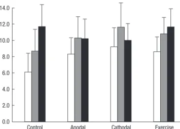
14.0 12.0 10.0 8.0 6.0 4.0 2.0 0.0
Fig. 3. Rota-rod performance scores between control, anodal stimulation, cathodal stimulation, and exercise groups at postoperative 2, 9, and 16 days (white, gray, and black bars). The values in the Y-axis represent the rotational speed (rpm) when a rat fell off the rod or rotated with the rod without moving.
For rota-rod test results (Fig. 3), ANCOVARM revealed no signif- icant effect in terms of INTERVENTION [F(3, 36)=0.570, P=0.638], TIME [F(1, 36)=0.769, P=0.386], and TIME×INTERVENTION [F(3, 36)=2.659, P=0.063].
For modified foot fault test results (Fig. 4), a significant differ- ence between groups was found [F(3, 36)=13.665, P<0.001] with- out any difference in the time factor [F(1, 376)=2.359, P=0.133].
No interaction between time and group factors was observed [F(1, 36)=4.108, P=0.050]. Cathodal stimulation and control groups were significantly different from the anodal stimulation and ex- ercise groups by post-hoc test (all the corrected P values <0.001).
Paradoxical deterioration of Garcia’s and modified foot fault scores at POD 16 were exhibited by the cathodal stimulation group.
Infarct size
Mean infarct volume (±standard deviation) was 7.5±2.7% in the control group (n=6), 11.3±6.3% in the anodal stimulation group (n=4), 7.7±4.2% in the cathodal stimulation group (n=6), and
Control Anodal Cathodal Exercise
8.0 7.0 6.0 5.0 4.0 3.0 2.0 1.0 0.0
* *
* *
Fig. 4. Modified foot fault scores between control, anodal stimulation, cathodal stimu- lation, and exercise groups at postoperative 2, 9, and 16 days (white, gray, and black bars). The values in the Y-axis represent the number of the forelimb misplacements over 1 minute period when a rat traversed the grid. Asterisks represent P values less than 0.05.
A
C
B
D right side
left side
2.0 mm
5.0 mm
Optic density
Group
Control Cathodal Exercise Anodal
0.9000
0.8000
0.7000
0.6000
0.5000
0.4000
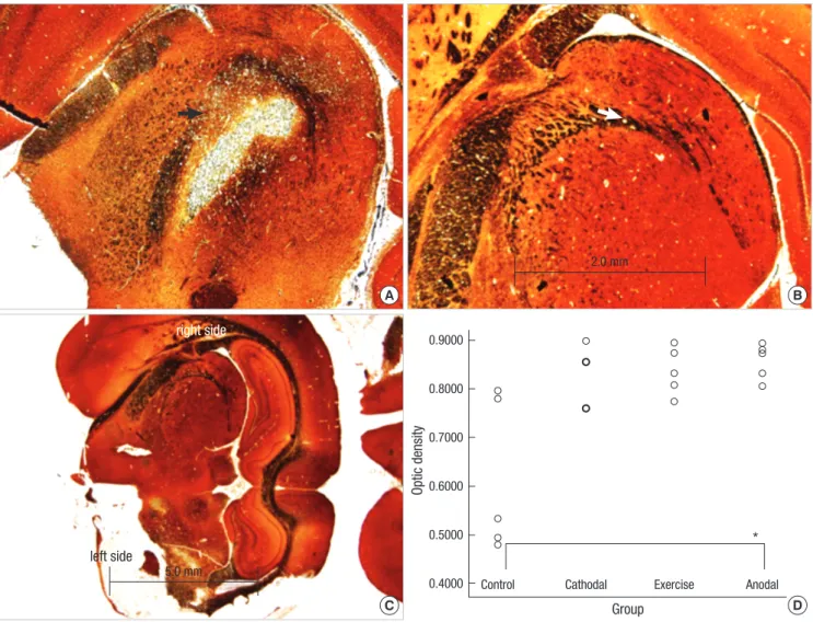
Fig. 5. Representative photomicrographs from bilateral internal capsules showing neural axons staining with Bielschowsky’s method in rat stroke model (original magnification
×40 and ×12.5). (A) and (B) represent infarct and intact hemispheres at the level of internal capsules. The arrows in figures A and B represent the left and right internal capsules.
(C) is the dorsal side of a rat brain section. (D) is a graph to show the optical density ratios in all groups. Optical density ratios of the infarct hemisphere to the intact one are measured and compared between them. Infarct areas in the anodal stimulation group show less neuronal axon and stain intensity changes than those in the control group.
*
15.4±12.4% in the exercise group (n=5). No significant difference was found between the 4 groups [χ2=1.884, P=0.597].
Integrity of white matter axon
Neuronal axons at the internal capsule of infarct hemispheres in the control group were stained poorly (Fig. 5). However, neu- ronal axons in the same region in the anodal stimulation and exercise groups contained axons that were better preserved. Op- tical density ratios of ischemic areas versus contralateral hemi- sphere were significantly different between groups [χ2df=3=8.211, P=0.042]. The anodal stimulation group had well-stained axons in the infarct areas when compared to the control group (Bon- ferroni corrected P value=0.009).
DISCUSSION
This is the first study to reveal the functional and histological changes after tDCS using the rat stroke model. Repeated tran- scranial anodal stimulation improved motor function (accord- ing to Garcia’s test and the modified foot fault test) in rat stroke model. Histologically, this had no effect on infarct size, but re- duced neuronal axon deterioration.
Transcranial direct current stimulation has not been widely studied in rats, although it has been examined with respect to anticonvulsant effects (20) and the propagation velocity of cor- tical spreading depression, which represents cortical excitabili- ty (21). In these previous studies, a plastic body jacket and a coat- ing of glass ionomer cement were used to attach the electrodes.
In the present study, we used a molded plastic cup and gauze to attach them firmly, which reduced skin resistance to several hundred kilo ohms. Although current modeling in rats during tDCS has not been developed, separation between anodal and cathodal electrodes is known to prevent current shunting effects (22). We did not use implanted subcutaneous electrodes because they might have caused infection and stress, and thus, affected the results. Since the stimulation method was noninvasive, we did not include a sham stimulation group and instead used a group that received no stimulation as a control.
Since there was no significant difference in the time factor, this suggests that anodal stimulation for 2 weeks did not show superiority over stimulation for a week in terms of the motor re- covery. These results are contrary to the findings of Boggio’s study (7). On the other hand, cathodal stimulation for 2 weeks was associated with deterioration at POD 16, which is probably attributable to a cumulative effect caused by decreased excit- ability of the infarcted brain. Improvements in Garcia’s test and the modified foot fault test, but not in the rota-rod test might be explained by the results of the previous study (18) which stated that improvement of simple movement did not always accom- pany coordinated and balanced movement.
Transcranial direct current stimulation (tDCS) and exercise
failed to reduce infarct size in our model, which contradicts the findings of previous studies (23-25). We attribute this discrepan- cy to the repeated anesthetic insults, different injury types, and exercise intensities. As mentioned in the Methods, we could not rule out false negative results caused by repeated anesthetic in- sults of ketamine (26). In addition, we used a permanent infarct model and not a temporary model, as was used in previous stud- ies. Moreover, in the present study, the treadmill speed was set at 16 m/min lower than those used in previous studies, because our rats could not endure a speed over 20 m/min. It could be said that this reduced level of exercise was insufficient to reduce in- farct size as compared with forced exercise at 30 m/min (23).
White matter axons in the infarcted brain were well preserved after transcranial anodal stimulation compared to those in the control group. The results obtained are of importance because they demonstrate that white matter axonal damage after isch- emia can be reduced by transcranial anodal stimulation. The mechanism of the protective effect of repeated transcranial an- odal stimulation may involve modulation of the activities of cal- cium channel and NMDA receptor, activations of which cause white matter injury due to excessive glutamate release. Howev- er, Nitsche’s study (27) showed that transcranial anodal stimu- lation might enhance NMDA receptor efficacy through the high- frequency presynaptic activity and postsynaptic subthreshold membrane depolarization. This is contradictory to our results.
Some other factors might involve this phenomenon and further animal studies must be performed to clarify this.
We selected the stimulus intensity as 0.1 mA because this in- tensity and total charges would not injure the rat’s brain accord- ing to the Liebetanz’ study (28). The amounts of electrical charge delivered in this study could not be calculated because a math- ematical model has not been developed in the rat brain. This makes it difficult to apply our results to clinical studies.
For the anesthesia, we used ketamine which was a non-com- petitive NMDA receptor antagonist and might affect the results in this experiment. However, ketamine is a weak glutamate an- tagonist and continuous infusion of ketamine (1.25 mg/kg/min during 55 min) failed to show the protective effect over the glu- tamate in the temporary middle cerebral artery occlusion rat model (29). Therefore, we do not think this induction dosage of ketamine would interfere with anodal and cathodal tDCS after- effects. Although ketamine has been found consistently to ame- liorate neuronal death in vitro, results from in vivo studies have proved more conflicting (30).
During this experiment, many rats (20 rats) died. The causes of death could not be found but we hypothesize that they were due to the stressful condition after the ischemic insult. Although death rates were not different between the study groups by the Chi square test [Pearson χ2=0.542, P=0.910], they may have caused selection bias. As a consequence, type 2 error increased in this study because of small sample size. Further studies using
larger sample sizes must be done to prove the negative results found in this study.
From our results, repeated transcranial anodal stimulation may have a neuroprotective effect of white matter in the cere- brovasular injured brain. However, we only observed histopath- ologic changes in axons of white matter, not in myelins using the Luxol fast blue-periodic acid Schiff stains. The addition of other stains would reveal the protection of white matter injury more accurately.
Transcranial anodal stimulation was found to make a func- tional improvement and well-preserved white matter axons in our rat stroke model. These findings may be useful for studies about the therapeutic mechanism of tDCS.
ACKNOWLEDGMENTS
We appreciate the Cybermedic Corporation (Iksan, Korea) for the support of the direct current stimulator. However, the con- tent of this report is not related to the company’s interest. We specially thank to Professor “Chung Chun Kee” for the guidance of this study. We thank to Dr. John Roberts and Shaina for assis- tance with the English and to our medical research collaborat- ing center for assistance with statistics.
REFERENCES
1. Ding DC, Shyu WC, Lin SZ, Li H. Current concepts in adult stem cell therapy for stroke. Curr Med Chem 2006; 13: 3565-74.
2. Liu DD, Shyu WC, Lin SZ. Stem cell therapy in stroke: strategies in basic study and clinical application. Acta Neurochir Suppl 2006; 99: 137-9.
3. Boggio PS, Alonso-Alonso M, Mansur CG, Rigonatti SP, Schlaug G, Pas- cual-Leone A, Fregni F. Hand function improvement with low-frequency repetitive transcranial magnetic stimulation of the unaffected hemisphere in a severe case of stroke. Am J Phys Med Rehabil 2006; 85: 927-30.
4. Fregni F, Boggio PS, Mansur CG, Wagner T, Ferreira MJ, Lima MC, Ri- gonatti SP, Marcolin MA, Freedman SD, Nitsche MA, Pascual-Leone A.
Transcranial direct current stimulation of the unaffected hemisphere in stroke patients. Neuroreport 2005; 16: 1551-5.
5. Fregni F, Boggio PS, Nitsche MA, Marcolin MA, Rigonatti SP, Pascual- Leone A. Treatment of major depression with transcranial direct current stimulation. Bipolar Disord 2006; 8: 203-4.
6. Hummel FC, Cohen LG. Non-invasive brain stimulation: a new strategy to improve neurorehabilitation after stroke? Lancet Neurol 2006; 5: 708-12.
7. Boggio PS, Nunes A, Rigonatti SP, Nitsche MA, Pascual-Leone A, Fregni F. Repeated sessions of noninvasive brain DC stimulation is associated with motor function improvement in stroke patients. Restor Neurol Neu- rosci 2007; 25: 123-9.
8. Ardolino G, Bossi B, Barbieri S, Priori A. Non-synaptic mechanisms un- derlie the after-effects of cathodal transcutaneous direct current stimula- tion of the human brain. J Physiol 2005; 568: 653-63.
9. Nitsche MA, Seeber A, Frommann K, Klein CC, Rochford C, Nitsche MS, Fricke K, Liebetanz D, Lang N, Antal A, Paulus W, Tergau F. Modulating parameters of excitability during and after transcranial direct current
stimulation of the human motor cortex. J Physiol 2005; 568: 291-303.
10. Besancon E, Guo S, Lok J, Tymianski M, Lo EH. Beyond NMDA and AMPA glutamate receptors: emerging mechanisms for ionic imbalance and cell death in stroke. Trends Pharmacol Sci 2008; 29: 268-75.
11. Manning SM, Talos DM, Zhou C, Selip DB, Park HK, Park CJ, Volpe JJ, Jensen FE. NMDA receptor blockade with memantine attenuates white matter injury in a rat model of periventricular leukomalacia. J Neurosci 2008; 28: 6670-8.
12. Lang N, Siebner HR, Ward NS, Lee L, Nitsche MA, Paulus W, Rothwell JC, Lemon RN, Frackowiak RS. How does transcranial DC stimulation of the primary motor cortex alter regional neuronal activity in the human brain? Eur J Neurosci 2005; 22: 495-504.
13. Merzagora AC, Foffani G, Panyavin I, Mordillo-Mateos L, Aguilar J, Onaral B, Oliviero A. Prefrontal hemodynamic changes produced by anodal direct current stimulation. Neuroimage 2010; 49: 2304-10.
14. Longa EZ, Weinstein PR, Carlson S, Cummins R. Reversible middle cere- bral artery occlusion without craniectomy in rats. Stroke 1989; 20: 84-91.
15. Garcia JH, Wagner S, Liu KF, Hu XJ. Neurological deficit and extent of neuronal necrosis attributable to middle cerebral artery occlusion in rats.
Statistical validation. Stroke 1995; 26: 627-34.
16. Rogers DC, Campbell CA, Stretton JL, Mackay KB. Correlation between motor impairment and infarct volume after permanent and transient middle cerebral artery occlusion in the rat. Stroke 1997; 28: 2060-5.
17. Kim MW, Bang MS, Han TR, Ko YJ, Yoon BW, Kim JH, Kang LM, Lee KM, Kim MH. Exercise increased BDNF and trkB in the contralateral hemisphere of the ischemic rat brain. Brain Res 2005; 1052: 16-21.
18. Ding Y, Li J, Lai Q, Rafols JA, Luan X, Clark J, Diaz FG. Motor balance and coordination training enhances functional outcome in rat with tran- sient middle cerebral artery occlusion. Neuroscience 2004; 123: 667-74.
19. Schabitz WR, Li F, Fisher M. The N-methyl-D-aspartate antagonist CNS 1102 protects cerebral gray and white matter from ischemic injury follow- ing temporary focal ischemia in rats. Stroke 2000; 31: 1709-14.
20. Liebetanz D, Klinker F, Hering D, Koch R, Nitsche MA, Potschka H, Loscher W, Paulus W, Tergau F. Anticonvulsant effects of transcranial direct-current stimulation (tDCS) in the rat cortical ramp model of focal epilepsy. Epilepsia 2006; 47: 1216-24.
21. Liebetanz D, Fregni F, Monte-Silva KK, Oliveira MB, Amancio-dos-San- tos A, Nitsche MA, Guedes RC. After-effects of transcranial direct current stimulation (tDCS) on cortical spreading depression. Neurosci Lett 2006;
398: 85-90.
22. Miranda PC, Lomarev M, Hallett M. Modeling the current distribution during transcranial direct current stimulation. Clin Neurophysiol 2006;
117: 1623-9.
23. Hayes K, Sprague S, Guo M, Davis W, Friedman A, Kumar A, Jimenez DF, Ding Y. Forced, not voluntary, exercise effectively induces neuropro- tection in stroke. Acta Neuropathol 2008; 115: 289-96.
24. Kim DY, Park SH, Lee SU, Choi DH, Park HW, Paek SH, Shin HY, Kim EY, Park SP, Lim JH. Effect of human embryonic stem cell-derived neuro- nal precursor cell transplantation into the cerebral infarct model of rat with exercise. Neurosci Res 2007; 58: 164-75.
25. Wang RY, Yu SM, Yang YR. Treadmill training effects in different age groups following middle cerebral artery occlusion in rats. Gerontology 2005; 51: 161-5.
26. Church J, Zeman S. Ketamine promotes hippocampal CA1 pyramidal neuron loss after a short-duration ischemic insult in rats. Neurosci Lett
1991; 123: 65-8.
27. Nitsche MA, Fricke K, Henschke U, Schlitterlau A, Liebetanz D, Lang N, Henning S, Tergau F, Paulus W. Pharmacological modulation of cortical excitability shifts induced by transcranial direct current stimulation in humans. J Physiol 2003; 553: 293-301.
28. Liebetanz D, Koch R, Mayenfels S, Konig F, Paulus W, Nitsche MA. Safe- ty limits of cathodal transcranial direct current stimulation in rats. Clin
Neurophysiol 2009; 120: 1161-7.
29. Ridenour TR, Warner DS, Todd MM, Baker MT. Effects of ketamine on outcome from temporary middle cerebral artery occlusion in the sponta- neously hypertensive rat. Brain Res 1991; 565: 116-22.
30. Church J, Zeman S. Ketamine promotes hippocampal CA1 pyramidal neuron loss after a short-duration ischemic insult in rats. Neurosci Lett 1991; 123: 65-8.