INTRODUCTION
The peroxisome proliferator activated receptor (PPAR)gis a ligand specific transcription factor. It plays an important role in the regulation of diabetes mellitus (DM). It modulates insulin sensitivity, cell growth, inflammation, and adipocyte differentiation and lipid metabolism (1). PPARgagonist is used worldwide for treatment of DM. In addition, PPARg agonist has antifibrotic effects in both diabetic nephropathy and non-diabetic nephropathy. Animal models of diabetic nephropathy treated with PPARgagonist have demonstrat- ed decreased proteinuria and glomerular matrix deposition and glomerulosclerosis; these results appear to be indepen- dent of the antidiabetic effect of PPARg(2, 3). In the 5/6- nephrectomy model, activation of PPARg has been shown to reduce glomerulosclerosis (4). In addition, in vitro exper- iments have demonstrated that PPAR-gactivation had anti- proliferative (5) and antifibrotic effects on the mesangial cells (6). Furthermore PPAR-gactivation reduced type I collagen synthesis and secretion. Its action probably was linked to a TGF-b1-dependent mechanisms (7). Based on these results,
we speculate that the antifibrotic effects of PPAR-gare linked to TGF-b. However, the mechanism involved in the antifi- brotic effects has been little known.
Tubulointerstitial lesion containing tubular atrophies and interstitial fibrosis in the kidney are the final common pathways that lead to progressive renal failure regardless of the prima- ry cause of renal disease (8). The unilateral ureteral obstruc- tion (UUO) model induces interstitial inflammation and fibro- sis; it has been used as a model for tubulointerstitial fibrosis.
The expressions of tissue growth factor (TGF)-b and plas- minogen activator inhibitor-1 (PAI-1) are increased in the UUO model (9, 10) and with decreased fibrosis, their expres- sion decreases.
In this study, we evaluated whether the PPARgagonist, pioglitazone, had antifibrotic and antiinflammatory effects in the UUO mouse model. In addition, we studied whether there are synergistic effects with angiotensin receptor blocker (ARB), widely used for anti-inflammatory and antifibrotic treatment in kidney diseases. In addition, we evaluated the mechanism involved in the antifibrotic effects of the PPARg agonist that correlate with TGF-band PAI-1.
35
Jee-Young Han1, Ye-Ji Kim1, Lucia Kim1, Suk-Jin Choi1, In-Suh Park1, Joon-Mee Kim1, Young Chae Chu1, and Dae-Ryong Cha2
Department of Pathology1, Inha University Hospital, Inha University Medical College, Incheon; Division of Nephrology2, Ansan Hospital, Korea University Medical College, Ansan, Korea
Address for Correspondence Jee-Young Han, M.D.
Department of Pathology, Inha University Hospital, 17 Biryong-gil, Jung-gu, Incheon 400-711, Korea Tel : +82.32-890-3983, Fax : +82.32-890-3464 E-mail : jeeyhan@inha.ac.kr
This study was supported by Inha University Research Grant.
PPAR g Agonist and Angiotensin II Receptor Antagonist Ameliorate Renal Tubulointerstitial Fibrosis
The peroxisome proliferator activated receptor (PPAR)gagonist is used as antidia- betic agent with antihyperglycemic and antihyperinsulinemic actions. Beyond these actions, antifibrotic effects have been reported. We examined antifibrotic effects of PPARgagonist and interaction with angiotensin receptor antagonist in the unilater- al ureteral obstruction (UUO) model. After UUO, mice were divided to four groups:
no treatment (CONT), pioglitazone treatment, L158809 treatment, and L158809+
pioglitazone treatment. On day 14, CONT mice showed severe fibrosis and all treated mice showed decreased fibrosis. The immunohistochmistry of PAI-1, F4/80 and p- Smad2 demonstrated that their expressions were increased in CONT group and decreased in the all treated groups compared to CONT. PAI-1 and p-Smad2 deter- mined from Western blotting, among treated groups, was decreased compared to CONT group. The expression of TGF-b1 from real time RT PCR showed markedly increased in the CONT group and decreased in all treated groups compared to CONT.
These data suggest the pioglitazone inhibited tubulointerstitial fibrosis, however, the synergism between pioglitazone and L158809 is not clear. Considering decreased expression of PAI-1 and TGF-b/Smad2 in the treated groups, PAI-1 and TGF-bare likely linked to the decreased renal tubulointerstitial fibrosis. According to these results, the PPARgagonist might be used in the treatment of renal fibrotic disease.
Key Words : PPAR gamma; Receptors, Angiotensin; Fibrosis; Plasminogen Activator Inhibitor 1; Transform- ing Growth Factor Beta
ⓒ 2010 The Korean Academy of Medical Sciences.
This is an Open Access article distributed under the terms of the Creative Commons Attribution Non-Commercial License (http://creativecommons.org/licenses/by-nc/3.0) which permits unrestricted non-commercial use, distribution, and reproduction in any medium, provided the original work is properly cited.
Received : 5 November 2008 Accepted : 6 March 2009
MATERIALS AND METHODS Animals
Adult male wild type C57BL/6 mice, 10 to 12 weeks of age were used for the experiments. The mice were housed in microisolator cages in a pathogen-free barrier facility. The mice had free access to tap water and standard mouse chow (RP5015; PMI feeds Inc, St. Louis, MO, USA) or prepared chow containing the PPARgagonist, pioglitazone. The mice were maintained in a temperature-controlled facility with 12- hr light/12-hr dark cycles. All procedures were followed the rules of the Inha University animal experiment committee.
Experimental protocol
The mice underwent UUO (n=40) under general anesthe- sia and sterile conditions (11). After the UUO, they were divided into 4 groups. The first group (n=10) did not receive any treatment (CONT). The second group (n=10) received the PPAR-gagonist, pioglitazone, p.o. (30 mg/kg/day) (pio- glitazone treated group). The third group (n=10) received the angiotensin receptor antagonist, L158809 (1.5 mg/kg/
day, drinking water [DW]) (L158809 treated group). The fourth group (n=10) received combined therapy including pioglitazone and L158809 of the same dose (combined treat- ment group). On day five or 14 days after the surgery, the mice were sacrificed and both the obstructed and nonobstructed contralateral kidneys were collected for morphological, im- munostaining, and molecular assessment. Four mice were underwent sham operation as normal controls.
Histological examination
For light microscopy, the tissue fixed in 4% paraformalde- hyde was embedded in paraffin and 4 μm sections were pre- pared. After Masson’s trichrome staining, the degree of tubu- lointerstitial fibrosis and injury were scored from 0 to 4+ on the day 5 specimens. Fibrosis and injury were assessed as 0 for 0%, 1 for <25%, 2 for 25 to 50%, 3 for >50 to 75%, and 4 for >75% for each field with tubulointerstitial fibrosis. Inter- stitial fibrosis, in the cortex, was assessed by point counting (12) in the cortical area on the day 14 specimens. All sections were examined without knowledge of the treatment protocol.
Immunohistochemistry
For immunostaining for PAI-1, antigen retrieval was per- formed using microwaves for 10 min. The slides were expos- ed to Power Block (BioGenex Laboratories, San Ramon, CA, USA) for 45 min and incubated with rabbit anti-rat PAI-1 antibody (1:50, American Diagnostica, Inc., Greenwich, CT, USA) at 4℃overnight followed by biotinylated goat anti- rabbit immunoglobulin (BioGenex Laboratories) for 45 min
and peroxidase-conjugated streptavidin for 45 min. Diami- nobenzidine was added as a chromogen. Positive PAI-1 stain- ing was graded from 0 to 4+ (grade 0, No significant increase, diffuse very weak stain similar to nonobstructed kidney; grade 1, very weak focal staining, <5% increase staining; grade 2, mild/moderate intensity in <20% of areas; grade 3, moderate intensity in 20-50%; grade 4, moderate intensity >50%).
Infiltration of macrophages was detected by rat anti-mouse F4/80 antibody (1:2,000; Serotec Inc., Raleigh, NC, USA) incubated at room temperature for 1 hr, followed by Envision kit (Dako, Carpinteria, CA, USA). Infiltrating macrophages, in the interstitium, were counted and expressed as the num- ber of macrophages per high-power field.
For immunostaining of phosphorylated Smad2 (p-Smad2), antigen retrieval was performed by microwave for 10 min.
Rabbit anti-human p-Smad 2 (Cell Signaling Technology, Beverly, MA, USA) was used at room temperature overnight followed by Envision kit (Dako). Positive p-Smad2 staining was graded from 0 to 3+ (grade 0, No significant increase, diffuse very weak nuclear stain similar to nonobstructed kid- ney; grade 1, nuclear staining with mild intensity <20%
increase staining; grade 2, nuclear staining with moderate intensity in ≤50% of areas; grade 3, nuclear staining with moderate intensity in >50%).
Western blot analysis
1) p-Smad 2, b-actin and PAI-1: The proteins were extracted using PRO-PREP protein extraction solution (Intron, Seong- nam, Korea) from the frozen kidney tissues. The protein con- centration was measured using Dc Protein Assay kit (Bio- Rad Laboratories, Hercules, CA, USA). Remaining proce- dures was performed to an established procedure (13). The primary antibodies used were as follows: rabbit anti-p-Smad2 polyclonal antibody, 1:500 (Upstate Biotechnology, Lake Placid, NY, USA), mouse anti-b-actin monoclonal antibody, 1:5,000, (Sigma, St. Louis, MO, USA), rabbit anti-PAI-1 antibody, 1:100 (American Diagnostica). 2) The intensities of individual bands were semiquantified using the TINA 2.0 program.
Real time RT-PCR for TGF-b1
Total RNA was obtained from frozen mouse kidney tissues (normal and obstructed kidney) using the TRIZOL (Invit- rogen, San Diego, CA, USA) according to the manufacturer’s protocols. Mouse b-actin mRNA was used as an internal con- trol and also used to assess RNA integrity.
One micrograms of total RNA was reverse transcribed into cDNA using RT system kit (Promega, Madison, WI, USA).
Reverse transcription was carried out in a total volume of 25
mL containing 10 mM Tris-HCl, (pH 8.8), 50 mM KCl, 5 mM MgCl2, 1 mM each dNTPs, 0.5 mg oligo (dT) primers, 25 U RNase inhibitor, and 15 U AMV reverse transcriptase
for 1 hr at 37℃, followed by 10 min at 95℃. The polymerase chain reaction (PCR) was performed using 1 mL cDNA in a total volume of 25 mL in the presence of 12.5 mL 2×iQ Super- mix (Bio-rad), 200 nM TGF-b primer set, 100 nM TGF-b probe. The PCR condition was 40 cycles at 95℃for 30 sec, 60℃for 1 min using iCycler iQ real-time PCR detection system (Bio-Rad). Amplification of b-actin was carried out in parallel with the genes of interest. Each assay included a no template control and all measurements were performed in duplicate. All samples with a coefficient of variation >10%
were retested. Primers and probes used for Real-Time RT- PCR were purchased from TIB MOLBIOL (Berlin, Germany) and their sequences were represented as following.
1) TGF-b1; forward; 5′-TTCAGGACTATCACCTACC- TTTCC-3′
Reverse; 5′-CGGGAACCCTCGGCAAAG-3′
Probe; 5′-6FAM-AGACCCCACCCCACAAGCCTGC- BHQ1-3′
2) b-actin; forward; 5′-CTTCTTTGCAGCTCCTTCG- TTG-3′
Reverse; 5′-CGACCAGCGCAGCGATATC-3′
Probe; 5′-HEX-TCCACACCCGCCACCAGTTCGC- BHQ2-3′
The value of TGF-bmeasured relative to Ct value.
Statistics
Results are expressed as mean±SEM. Statistical differ- ence was assessed by analysis of variance followed by Mann- Whitney U-test. A P value <0.05 was considered to be sig- nificant.
RESULTS Tubulointerstitial fibrosis
The trichrome staining revealed interstitial fibrosis and tubular atrophy at 5 days in all of the groups. There was no significant statistical difference among the groups. On the day 14, the interstitial fibrosis was markedly increased in the CONT group (0.27±0.03). The pioglitazone treated group revealed decreased fibrosis compared to CONT (0.15±0.02, P<0.05 vs. CONT). The interstitial fibrosis was more dec- reased in the L158809 treated group and combined treatment group compared to CONT and pioglitazone treated group (L158809 treated group; 0.06±0.004, combined treatment group; 0.09±0.01, P<0.05 vs. CONT and pioglitazone treat- ed group) (Fig. 1).
The number of interstitial macrophages
The number of macrophages was increased 5 days after UUO compared to contralateral non- obstructed kidney. How- ever, there was no significant difference among treated and non-treatment groups. By 14-day, the number of macrophages was significantly increased in the CONT group (117.9±9.5/
HPF). The all treated groups showed significantly small mac- rophage numbers compared to CONT (pioglitazone treated group 63.9±5.4/HPF, L158809 treated group 80.9±10.2/
HPF, combined treatment group 76.5±4.4/HPF, P<0.05 vs. CONT) (Fig. 2).
PAI-1 expression
PAI-1 expression of IHC was slightly increased at 5-day.
Fig. 1. Tubulointerstititial changes in obstructed kidneys. (A) At day 5, tubulointerstitial fibrosis is slightly increased at all groups with no significant difference among groups. At day 14, CONT mice show severe progressive fibrosis and three treated groups show decreased fibrosis compared to CONT. a, sham operated kidney; b, CONT at day 5; c, pioglitazone treated group at day 5; d, L158809 treated group at day 5; e, combined treatment group at day 5; f, CONT at day 14; g, pioglitazone treated group at day 14; h, L158809 treated group at day 14; i, combined treatment group at day 14 (Masson-trichrome stain, ×200). (B) Point counting scoring of fibrosis at day 14 shows less fibrosis in all treated groups compared to CONT (Comb; combined treatment group) (*P<0.05 vs. CONT).
A B
a b c
d e f
g h i
Tubulointerstitial fibrosis
0.35 0.30 0.25 0.20 0.15 0.10 0.05 0
Sham CONT Pioglitazone L158809 Comb
*
*
*
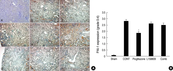
However, there was no statistical difference between the treat- ed groups. The PAI-1 was detected at the cytoplasm of dam- aged or atrophic tubular epithelial cells, interstitial inflam- matory cells and parietal epithelial cells. Their expression was significantly increased at 14-day in the CONT group (2.85
±0.12). PAI-1 expression cells were similar to the CONT but PAI-1 intensity was decreased compared to CONT in the pioglitazone treated groups (1.91±0.08, P<0.05 vs. CONT).
For the L158809 and in the combined treatment groups, the PAI-1 expression patterns were similar to CONT. Its inten- sity was decreased but there was no statistically significant difference (L158809; 2.65±0.10, P=0.3 vs. CONT, com-
bined treated group; 2.52±0.16, P=0.06 vs. CONT) (Fig. 3).
The PAI-1 determined from Western blotting, among the day 14 all treated groups (pioglitazone treated group, L158809 treated group, and combined treatment group), was decreased compared to CONT group (P<0.05); the L158809 treated group showed most decreased expression, the combined treat- ment group had the second most decreased expression and pioglitazone treated group had the smallest decrease in expres- sion compared to CONT (Fig. 4).
Macrophage number/HPF
140 120 100 80 60 40 20 0
Sham CONT Pioglitazone L158809 Comb
*
*
*
Fig. 2. Interstitial macrophage numbers in obstructed kidneys at day 14. CONT mice show markedly increased number of intersti- tial macrophages and all treated groups show decreased num- ber of macrophages compared to CONT (*P<0.05 vs. CONT).
Fig. 3. PAI-1 protein expression by Immunohistochemical staining. (A) PAI-1 expression is identified in the cytoplasm of atrophic tubular epithelial cells, interstitial inflammatory cells and parietal epithelial cells of glomerulus. At day 5, PAI-1 expression is minimally increased with no significant difference among groups. At day 14, PAI-1 expression is increased at CONT. At pioglitazone treated group, the PAI-1 expres- sions is decreased comparing to CONT. a, sham operated kidney; b, CONT at day 5; c, pioglitazone treated group at day 5; d, L158809 treated group at day 5; e, combined treatment group at day 5; f, CONT at day 14; g, pioglitazone treated group at day 14; h, L158809 treat- ed group at day 14; i, combined treatment group at day 14 (immunohistochemical staining for PAI-1, ×200). (B) Semiquantative grading of PAI-1 expression at day 14 shows significantly decreased PAI-1 expression only at the pioglitazone treated groups compared to CONT (*P<0.05 vs. CONT).
A B
a b c
d e f
g h i
PAI-1 expression (grade 0-4)
3.5 3.0 2.5 2.0 1.5 1.0 0.5
0 Sham CONT Pioglitazone L158809 Comb
*
Fig. 4. PAI-1 protein expression by western blotting at day 14. PAI- 1 expression is decreased at all treated groups compared to CONT (*P<0.05 vs. CONT).
PAI-1/b-actin ratio
2.5
2.0
1.5
1.0
0.5
0
Sham CONT Pioglitazone L158809 Comb Sham CONT Pioglitazone L158809 Comb
*
* *
PAI-1 b-actin
p-Smad 2 expression
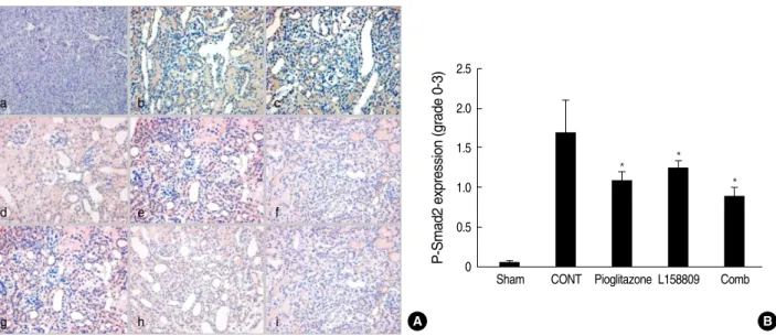
P-Smad2 expression of IHC, a marker of TGF-bsignaling activation, was detected at the nuclei of damaged or atrophied tubular epithelial cells and interstitial inflammatory cells. Its expression was not increased at day 5 compared to normal con- trol. At day 14, their intensity was increased in the CONT group. In the all of treated groups (pioglitazone treated group, L158809 treated group, combined treatment group), the p- Smad2 expressions were decreased compared to CONT (Fig. 5).
For the day 14 specimens, the p-Smad2 from Western blot- ting analysis showed that the markedly increased in the CO- NT group compared to normal control and decreased in the combined treatment group compare to CONT (Fig. 6).
TGF-b1 expression
On the day 5, TGF-b1 expression was increased in all groups.
However, there was no statistically significant difference among the groups. For the day 14 samples, the TGF-b1 expression was increased more in the CONT group and decreased in all of the treated groups (pioglitazone treated group, L158809 treated group, combined treatment group, P<0.05 vs. CONT).
Fig. 5. P-Smad2 expression by Immunohistochemical staining. P-Smad2 is detected at the nuclei of tubular epithelial cells and interstitial inflammatory cells. (A) At day 5, p-Smad2 expression of IHC at treated groups is not increased compared to normal control. Weak cyto- plasm stainings are present at all groups. At day 14, p-Smad2 expression is increased at CONT. At all treated groups, the p-Smad2 expres- sions are decreased compared to CONT. a, sham operated kidney; b, CONT at day 5; c, pioglitazone treated group at day 5; d, L158809 treated group at day 5; e, combined treatment group at day 5; f, CONT at day 14; g, pioglitazone treated group at day 14; h, L158809 treated group at day 14; i, combined treatment group at day 14 (immunohistochemical staining for p-Smad 2, ×200). (B) Semiquantative grad- ing of p-Smad2 expression at day 14 shows significantly decreased p-Smad2 expression at the all treated groups compared to CONT (*P<0.05 vs. CONT) .
A B
a b c
d e f
g h i
P-Smad2 expression (grade 0-3)
2.5
2.0
1.5
1.0
0.5
0
Sham CONT Pioglitazone L158809 Comb
* *
*
Relative TGF-b1 radio 2.5
2.0
1.5
1.0
0.5
0
Sham CONT Pioglitazone L158809 Comb
*
*
*
Fig. 7. Real time RT PCR for TGF-b1 at day 14. TGF-b1 expression shows markedly increased at the CONT and significantly decreases at the all treated groups compared to CONT (*P<0.05 vs. CONT).
Fig. 6. P-smad 2 expression by western blotting at day 14. P-Smad2 expression blotting at combined treatment group is decreased compared to CONT (*P<0.05 vs. CONT).
p-Smad2/b-actin ratio 0.7 0.6 0.5 0.4 0.3 0.2 0.1 0
Sham CONT Comb
Sham CONT Comb
* p-Smad2
b-actin
The relative value for the real time RT PCR of TGF-b1 was the following: CONT, 1.52±0.53; pioglitazone treated group, 0.83±0.12; L158809 treated group, 0.63±0.13; and com- bined treated group, 0.54±0.06 (Fig. 7).
DISCUSSION
PPARgagonists are used worldwide for their antidiabetic activity including the regulation of insulin sensitivity and lipid metabolism. In addition to these effects, PPARghas antifibrotic and anti-inflammatory effects. Clinical and ani- mal studies have shown that PPARgagonists not only regu- late insulin sensitivity but also may have a protective effect against kidney damage, such as reduced microalbuminuria and delaying progression to diabetic nephropathy in humans with type 2 diabetes and diabetic animal model (2, 14-16).
These renoprotective effects appear to be independent from blood glucose control activity of PPARg. In vitro studies also have revealed that PPAR-gagonists can inhibit cell pro- liferation and suppress the expression of extracellular matrix components (7, 17). UUO is a well established experimen- tal model of progressive tubulointerstitial fibrosis. In this study, we observed the antifibrotic and anti-inflammatory effects of PPARgagonist, pioglitazone. Pioglitazone had an antifibrotic effect on the UUO kidney. The interstitial fibro- sis was decreased in the pioglitazone-treated group compared the CONT group.
The mechanism underlying the antifibrotic effects of the PPARgagonist has little known. TGF-bhas been known to be increased in progressive renal disease and PAI-1 plays an im- portant role in the progression of renal fibrosis. Angiotensin, aldosterone, and TGF-binduce PAI-1 expression, and inhibi- tion of these compounds resulted in decreased PAI-1 and scle- rosis in experimental models of chronic kidney disease (18- 20). In this connection, we tried to evaluate that TGF-band PAI-1 were linked to the antifibrotic effects of pioglitazone.
The results of our study showed that PAI-1 and TGF-b expression were increased in the UUO kidneys, and decreased in all of the treatment groups including the PPARgagonist treated group. Based on these results, the mechanism under- lying the antifibrotic effects of pioglitazone appeared to be correlated with the decrease of TGF-band PAI-1. There are several prior studies that support these findings. In vitro, pioglitazone inhibited fibronectin production induced by TGF-bin mesangial cells (7). PPARgagonists had an antifi- brotic effect on human proximal tubular cells in a high glu- cose environment by inhibiting the increase in AP-1 and TGF-b1, and decreasing the production of the fibronectin (21). The PPARgagonist also inhibited TGF-b1 mediated alpha smooth muscle actin, fibronectin, and PAI-1 expression in the mesangial cells (22). In the 5/6 nephrectomy model, PPARg ameliorated the development of sclerosis and this effect was linked to decreased TGF-band PAI-1 (4).
Our findings showed that the p-Smad2 expression was also decreased in the PPARgagonist treated group. The Smad proteins are essential mediators and regulators of the intra- cellular signaling pathways, acting as transcription factors of TGF-b-mediated responses, including the fibrotic process (23).
After TGF-bbinds to the TGF-b receptor II, phosphoryla- tion of the TGF-breceptor I, and the intracellular substrates, Smad2 and Smad3, occurs. When activated, these Smads with their common partner, Smad4 complexes, are translocated to the nucleus, where they regulate transcriptional responses together with additional DNA binding cofactors. An in vitro study showed that PPARgagonists inhibited the Smad sig- naling pathway in the renal fibroblasts and blocked TGF- beta/Smad-mediated gene transcription in mesangial cells (22). Therefore, PPARgappears to have antifibrotic effects correlated with PAI-1, and TGF-b/Smad2 signaling.
The PPARgagonist also has anti-inflammatory effects. Our results showed that the number of infiltrated interstitial mac- rophage was markedly decreased in the pioglitazone treated groups. PAI-1 affects recruitment of interstitial macrophages and myofibroblasts, a potential source of both PAI-1, TGF-b and other profibrotic cytokines such as PDGFs, macrophage chemoattractant protein-1 (MCP-1), osteopontin, and inte- grins (24). Persistent infiltration of activated macrophages is profibrotic and closely correlated with progressive fibrosis in the kidney (25). Therefore inhibition of macrophage accu- mulation can produce not only antiinflammatory but also antifibrotic effects.
Angiotensin II is involved in the pathogenesis of renal dis- ease, through the regulation of two key processes inflamma- tion and fibrosis. Treatment of angiotensin II receptor antag- onist ameliorates the renal interstitial fibrosis caused by UUO by decreasing the TGF-band the number of interstitial mac- rophages (26). We also evaluated the antifibrotic effects of ARB, L158809. Renal interstitial fibrosis and the number of infiltrating macrophage were decreased in the L158809 treated group. Interstitial fibrosis was decreased more in the L158809 treated group compared to the pioglitazone treat- ed group. However, the number of interstitial macrophages was greater in the L158809 treated group than in the piogli- tazone treated group. The PAI-1 and TGF-bexpression were also decreased in the L158809 treated group. However, the synergistic effects of pioglitazone and L158809 were not clear;
a synergistic effect was only observed with TGF-bexpression.
Combination of pioglitazone and L158809 is almost equal- ly effective compared to their single applications concerning attenuation of fibrosis, infiltration of macrophages, PAI-1 and TGF-b/p-Smad2 expression after UUO. The synergistic effect between pioglitazone and L158809 was not clear in our study.
The studies about synergism of these two drugs are few and the results are still controversial. In vitro, combination ther- apy of a PPARgagonist and an ARB suppresses proinflamma- tory signaling and stimulates expression of Smad7 in human peritoneal mesothelial cells (27). However, in vivo examina-
tion of ischemia induced brain injury of rat, there was no syn- ergistic effect between candesartan and pioglitazone (28). The synergistic effect with ARB is not clear. It needs further inves- tigation.
In the conclusion, the PPARgagonist inhibited tubuloin- terstitial fibrosis and infiltration of interstitial macrophages in UUO model. The findings of decreased expression of PAI- 1, TGF-b, and p-Smad2 in the treated groups, suggest that PAI-1 and TGF-b/Smad2 were linked to the decreased renal tubulointerstitial fibrosis observed. Therefore, PPARgago- nists might be used for the treatment of renal fibrotic dis- eases other than those associated with diabetes mellitus.
REFERENCES
1. Guan Y, Zhang Y, Breyer MD. The role of PPARs in the transcrip- tion control of the cellular processes. Drug News Prospect 2002;
15: 147-54.
2. McCarthy KJ, Routh RE, Shaw W, Walsh K, Welbourne TC, John- son JH. Troglitazone halts diabetic glomerulosclerosis by blockade of mesangial expansion. Kiney Int 2000; 58: 2341-50.
3. Smith SA, Lister CA, Toseland CD, Buckingham RE. Rosiglitazone prevents the onset of hyperglycemia and proteinuria in the Zucker diabetic fatty rats. Daibetes Obes Metab 2000; 2: 363-72.
4. Ma LJ, Marcantoni C, Linton MF, Fazio S, Fogo AB. Peroxisome proliferator-activated receptor-gamma agonist troglitazone protects against non-diabetic glomerulosclerosis in rats. Kidney Int 2001; 59:
1899-910.
5. Ghosh SS, Gehr TW, Ghosh S, Fakhry I, Sica DA, Lyall V, School- werth AC. PPARgamma ligand attenuates PDGF-induced mesangial cell proliferation: role of MAP kinase. Kidney Int 2003; 64: 52-62.
6. Routh RE, Johnson JH, McCarthy KJ. Troglitazone suppresses the secretion of type I collagen by mesangial cells in vitro. Kidney Int 2002; 61: 1365-76.
7. Guo B, Koya D, Isono M, Sugimoto T, Kashiwagi A, Haneda M.
Peroxisome proliferator-activated receptor-gamma ligands inhibit TGF-beta 1-induced fibronectin expression in glomerulomesangial cells. Diabetes 2004; 53: 200-8.
8. Eddy AA. Experimental insights into the tubulointerstitial disease accompanying primary glomerular lesions. J Am Soc Nephrol 1994;
5: 1273-87.
9. Miyajima A, Chen J, Lawrence C, Ledbetter S, Soslow RA, Stern J, Jha S, Pigato J, Lemer ML, Poppas DP, Vaughan ED, Felsen D. Anti- body to transforming growth factor-beta ameliorates tubular apop- tosis in unilateral ureteral obstruction. Kidney Int 2000; 58: 2301-13.
10. Duymelinck C, Dauwe SE, De Greef KE, Ysebaert DK, Verpooten GA, De Broe ME. TIMP-1 gene expression and PAI-1 antigen after unilateral ureteral obstruction in adult male rat. Kidney Int 2000;
58: 1186-201.
11. Ma J, Nishimura H, Fogo A, Kon V, Inagami T, Ichikawa I. Accel- erated fibrosis and collagen deposition develop in the renal intersti- tium of angiotensin type 2 receptor null mutant mice during ureteral obstruction. Kidney Int 1998; 53: 937-44.
12. Bonegio RG, Fuhro R, Wang Z, Valeri CR, Andry C, Salant DJ, Lieberthal W. Rapamycin ameliorates proteinuria associated tubu- lointerstitial fibrosis inflammation and fibrosis in experimental mem- branous nephropathy. J Am Soc Nephrol 2005; 16: 2063-72.
13. Yang J, Liu Y. Dissection of key events in tubular epithelial to myofi- broblast transition and its implications in renal interstitial fibrosis.
Am J Pathol 2001; 159: 1465-75.
14. Buckinham RE, Al-Barazanji KA, Toseland CD, Slaughter M, Con- nor SC, West A, Bond B, Turner NC, Clapham JC. Peroxisom pro- liferator-activated receptor gamma agonist, rosiglitazone, protects against nephropathy and pancreatic islet abnormalities in Zucker fatty rats. Diabets 1998; 47: 1326-34.
15. Fujii M, Takemura R, Yamaguchi M, Hasegawa G, Shigeta H, Na- kano K, Kondo M. Troglitazone (CS-045) ameliorates albuminuria in streptozotocin-induced diabetic rats. Metabolism 1997; 46: 981-3.
16. Imano E, Kanda T, Nakatani Y, Nishida T, Arai K, Motomura M, Kajimoto Y, Yamasaki Y, Hori M. Effect of troglitazone halts dia- betic glomerulosclerosis by blockade of mesangial expansion. Dia- betes Care 1998; 21: 2135-9.
17. Zafirious S, Stanners SR, Saad S, Polhill TS, Poronnik P, Pollock CA.
Pioglitazone inhibits cell growth and reduces matrix production in human kidney fibroblasts. J Am Soc Nephrol 2005; 16: 638-45.
18. Eddy AA. Plasminogen activator inhibitor-1 and the kidney. Am J Physiol Renal Physiol 2002; 283: F209-20.
19. Fogo AB. The role of angiotensin II and plasminogen activator in- hibitor-1 in progressive glomerulosclerosis. Am J Kidney Dis 2000;
35: 179-88.
20. Brown NJ, Vaughan DE, Fogo AB. Aldosterone and PAI-1: impli- cations for renal injury. J Nephrol 2002; 15: 230-5.
21. Panchapakesan U, Sumual S, Pollock CA, Chen X. PPARgamma agonists exert antifibrotic effects in renal tubular cells exposed to high glucose. Am J Physiol Renal Physiol 2005; 289: F1153-8.
22. Li Y, Wen X, Spataro BC, Hu K, Dai C, Liu Y. Hepatocyte growth factor is a downstream effector that mediates the antifibrotic action of peroxisome proliferator-activated receptor-gamma agonists. J Am Soc Nephrol 2006; 17: 54-65.
23. Massague J, Chen YG. Controlling TGF-beta signaling. Genes Dev 2000; 14: 627-44.
24. Eddy AA. Molecular basis of renal fibrosis. Pediatr Nephrol 2000;
15: 290-301.
25. Diamond JR. Macrophages and progressive renal disease in exper- imental hydronephrosis. Am J Kidney Dis 1995; 26: 133-40.
26. Ishidoya S, Morrissey J, McCracken R, Reyes A, Klahr S. Angiotensin II receptor antagonist ameliorates renal tubulointerstitial fibrosis caused by unilateral ureteral obstruction. Kidney Int 1995; 47: 1285-94.
27. Yao Q, Ayala ER, Qian JQ, Stenvinkel P, Axelsson J, Lindholm B.
A combination of PPAR gamma agonist and an angiotensin recep- tor blocker attenuates proinflammatory signaling and stimulates ex- pression of Smad7 in human peritoneal mesothelial cells. Clin Nephrol 2007; 68: 295-301.
28. Schmerbach K, Schefe JH, Krikov M, Mu_ller S, Villringer A, Kint- scher U, Unger T, Thoene-Reineke C. Comparison between single and combined treatment with candesartan and pioglitazone follow- ing transient focal ischemia rat brain. Brain Res 2008; 7: 225-33.