* Corresponding author: Hye Hyun Cho
Department of Biomedical Laboratory Science, Daejeon Institute of Science and Technology, 100 Hyechon-ro, Seo-gu, Daejeon 35408, Korea
E-mail: [email protected]
* ORCID: https://orcid.org/0000-0002-0471-4938
ORIGINAL ARTICLE
Molecular Detection of Virulence Factors in
Carbapenem-Resistant Pseudomonas aeruginosa Isolated from a Tertiary Hospital in Daejeon
Hye Hyun Cho
Department of Biomedical Laboratory Science, Daejeon Institute of Science and Technology, Daejeon, Korea
대전지역의 3차 병원에서 분리된 Carbapenem 내성 Pseudomonas aeruginosa 의 병독성 인자 검출
조혜현
대전과학기술대학교 임상병리과
ARTICLE INFO ABSTRACT
Received August 15, 2019 Revised 1st August 23, 2019 Revised 2nd August 27, 2019 Accepted August 27, 2019
The emergence and spread of multidrug resistant (MDR) Pseudomonas aeruginosa is a critical problem worldwide. The pathogenesis of P. aeruginosa is due partly to the production of several cell-associated and extracellular virulence factors. This study examined the distribution of virulence factors and antimicrobial resistance patterns of carbapenem-resistant P. aeruginosa (CRPA) isolated from a tertiary hospital in Daejeon, Korea. Antimicrobial susceptibility testing was performed using the disk diffusion method, and PCR and DNA sequencing were performed to determine for the presence of virulence genes. In addition, the sequence type (ST) of MDR P. aeruginosa was investigated by multilocus sequence typing (MLST). Among 32 CRPA isolates, 14 (43.8%) were MDR and the major ST was ST235 (10 isolates, 71.4%). All isolates were positive for the presence of virulence genes and the most prevalent virulence genes were toxA, plcN, and phzM (100%). All isolates carried at least eight or more different virulence genes and nine (28.1%) isolates had 15 virulence genes. The presence of the exoU gene was detected in 71.4% of the MDR P. aeruginosa isolates. These results indicate that the presence of the exoU gene can be a predictive marker for the persistence of MDR P. aeruginosa isolates.
Copyright © 2019 The Korean Society for Clinical Laboratory Science. All rights reserved.
Key words ExoU gene Multidrug-resistant Virulence factor
서 론
Pseudomonas aeruginosa는 만성 감염과 심각한 급성 감염 을 일으키는 광범위한 기회감염균이다. 또한 P. aeruginosa는 폐렴, 창상감염, 심각한 화상, 욕창, 면역력이 저하된 환자의 다 양한 전신감염을 일으키는 원내감염균의 10∼15%를 차지하고
있다. 최근 원내감염에 의한 사망율이 18∼61%로 보고되고 있 으며, 특히 다제내성 P. aeruginosa에 의한 원내감염은 전 세계 적으로 증가하고 있는 실정이다[1-3].
최근 P. aeruginosa에 의한 발병기전 중 하나로, 세포 관련 및 세포외 병독성 인자의 생성은 광범위 조직 손상, 혈류 침입 및 확 산을 일으키는 것으로 보고되고 있다[4, 5]. 세포관련 병독성 인 자로는 flagella, lipopolysaccharide, pili (pilA, pilB), type III system effector proteins, type III secretion system (exoS, exoT, exoU, exoY)과 alginate가 있으며, 세포외 병독성 인자 로는 exotoxin A (toxA), phospholipases (plcH, plcN),
Korean Society for Clinical Laboratory Science
phenazine (phzI, phzII, phzH, phzM, phzS), elastase (lasA), zinc metalloprotease (lasB), alkaline protease (aprA), pyoverdin (pvdA)이 있다[6-9]. 이러한 세포 관련 및 세포외 병 독성 인자에 의한 침입성 P. aeruginosa 감염은 carbapenem 과 같은 항균제의 사용에도 불구하고 높은 사망률을 나타내고 있다. 최근 carbapenem에 대한 높은 내성이 발달함에 따라 다 제내성 P. aeruginosa의 출현은 전 세계적으로 심각한 문제로 대두되고 있으며 일본, 대만, 인도, 이란 등 아시아 지역 국가에 서 항균제의 선택과 사용의 제한으로 인해 치료에 많은 어려움 이 보고되고 있다[10-13]. 특히, 우리나라의 경우 carbapenem 에 대한 내성이 지속적으로 증가하고 있으며, 대부분의 car- bapenem 내성 P. aeruginosa가 다제내성으로 보고되어 큰 우 려를 낳고 있다[14]. 현재 우리나라에서는 다제내성 P. aeru- ginosa에 대한 많은 연구가 보고되고 있으나, 세포 관련 및 세포 외 병독성 인자와 관련된 연구는 매우 미비한 실정이다.
이에 본 연구에서는 대전지역의 3차 병원에서 분리된 carbapenem 내성 P. aeruginosa를 대상으로 주요 세포 관련 및 세포외 병독성 인자의 분포를 확인하고, 항균제 내성과의 관 계에 대해 조사하고자 한다.
재료 및 방법
1. 균주의 수집과 동정
본 연구는 2011년 3월부터 2012년 12월까지 대전지역의 3 차 병원에 의뢰된 객담(21), 소변(5), 창상(3), 농(1), 담즙(1), 혈 액(1)로부터 분리된 carbapenem 내성 P. aeruginosa 32균주 를 대상으로 하였다. 이 중 동일 환자에서 반복 분리된 균주는 수 집대상에서 제외하였다. 임상검체로부터 분리 배양된 균주는 Vitek II automated ID system (BioMerieux, Hazelwood, MO, USA)을 이용하여 동정하였고, carbapenem 내성 P. aeruginosa 는 imipenem과 meropenem에 내성인 균주로 선별하였다.
2. 항균제 감수성 검사
Clinical and Laboratory Standards Institute (CLSI) 지침에 따라, amikacin, gentamicin, imipenem, meropenem 및 ciprofloxacin, levofloxacin, ceftazidime, cefepime (BBL, Cockeysville, MI, USA)에 대한 감수성 검사는 Mueller-Hinton 한천배지 (Difco, Cockeysville, MD, USA)를 사용하여 디스크 확산법으로 확인하였다[15]. 정도관리를 위해 Escherichia coli ATCC 25922와 Pseudomonas aeruginosa ATCC 27853을 동 시에 시험하여 허용범위 내에 있는지 확인하였다. Magiorakos
등[16]의 연구를 참고하여, aminoglycosides, carbapenems 및 fluoroquinolones 계열에 내성을 보인 균주를 다제내성 P.
aeruginosa로 하였다.
3. 다제내성 P. aeruginosa의 multilocus sequencing typing (MLST) 분석
항균제 감수성 시험 결과, 다제내성 P. aeruginosa로 확인된 균주는 brain heart infusion broth (Difco)에 접종하여 37°C 에서 24시간 배양한 후, Genomic DNA Prep kit (Solgent, Daejeon, Korea)을 사용하여 DNA를 추출하였다. DNA 추출 액(5 L), 10× Taq buffer (2.5 L), 10 mM dNTP mix (0.5
L), primer 각 10 pmol, 0.7 U Taq DNA polymerase (Solgent) 및 증류수를 혼합하여 총 부피 25 L의 반응용액을 만들었다. 7개의 housekeeping gene (acsA, aroE, guaA, mutL, nuoD, ppsA, trpE)은 Gene Amp PCR System 9600 (Perkin-Elmer, Norwalk, CT, USA)을 사용하여 96°C에서 1 분간 반응시킨 후, 96°C에서 1분, 55°C에서 1분, 72°C에서 1분 으로 30회 증폭 반응시키고, 72°C에서 10분간 연장 반응시켰 다. 각각의 PCR 반응산물은 ethidium bromide가 포함된 1%
agarose gel에서 30분간 전기영동하여 밴드를 확인하였다. 증 폭산물은 PCR purification kit (Solgent)로 분리한 후, BigDye Terminator cycle sequencing kit (PE Applied Biosystems, Foster City, CA, USA)와 ABI PRISM 3730xl DNA analyzer (PE Applied Biosystems)를 이용하여 염기서열을 분석하였다.
MLST는 P. aeruginosa MLST database website (http://
pubmlst.org/ paeruginosa/)에 설명된 방법에 따라 분석하였 다. 7개의 housekeeping gene에 대한 각각의 염기서열 분석 결과는 MLST database에 입력하여 allelic number와 sequence type (ST)를 확인하였다.
4. 세포 관련 및 세포외 병독성 인자의 검출
P. aeruginosa의 주요 병독성 인자인 toxA, exoS, exoT, exoU, exoY, plcH, plcN, phzI, phzII, phzH, phzM, phzS, lasA, lasB, pilA, pilB, aprA, pvdA 유전자를 검출하기 위해 이 전 연구에서 사용된 primer (Table 1)를 이용하여 PCR을 진행 하였다[7, 17]. 먼저, toxA, exoS, plcH, plcN, lasB 유전자를 확인하기 위해, MLST 분석에서와 동일한 방법으로 DNA 추출 액(5 L), 10× Taq buffer (2.5 L), 10 mM dNTP mix (0.5
L), primer 각 10 pmol, 0.7 U Taq DNA polymerase
(Solgent) 및 증류수를 혼합하여 총 부피 25 L의 반응용액을
만들었다. PCR 과정은 94°C에서 3분간 반응시킨 후, 94°C에
Table 1. Oligonucleotides primers used for virulence genes amplification
Gene Sequence (5′-3′)
Size of product
(bp)
References
toxA F: CTGCGCGGGTCTATGTGCC 270 7
R: GATGCTGGACGGGTCGAG
exoS F: CGTCGTGTTCAAGCAGATGGTGCTG 444 7 R: CCGAACCGCTTCACCAGGC
exoT F: CAATCATCTCAGCAGAACCC 1159 17
R: TGTCGTAGAGGATCTCCTG
exoU F: GATTCCATCACAGGCTCG 3308 17
R: CTAGCAATGGCACTAATCG
exoY F: TATCGACGGTCATCGTCAGGT 1035 17 R: TTGATGCACTCGACCAGCAAG
plcH F: GCACGTGGTCATCCTGATGC 608 7
R: TCCGTAGGCGTCGACGTAC
plcN F: TCCGTTATCGCAACCAGCCCTACG 481 7 R: TCGCTGTCGAGCAGGTCGAAC
phzI F: CATCAGCTTAGCAATCCC 392 17
R: CGGAGAAACTTTTCCCTC
phzII F: GCCAAGGTTTGTTGTCGG 435 17
R: CGCATTGACGATATGGAAC
phzH F: GGGTTGGGTGGATTACAC 1752 17
R: CTCACCTGGGTGTTGAAG
phzM F: ATGGAGAGCGGGATCGACAG 875 17
R: ATGCGGGTTTCCATCGGCAG
phzS F: TCGCCATGACCGATACGCTC 1752 17
R: ACAACCTGAGCCAGCCTTCC
lasA F: GCAGCACAAAAGATCCC 1075 17
R: GAAATGCAGGTGCGGTC
lasB F: GGAATGAACGAAGCGTTCTCCGAC 284 7 R: TGGCGTCGACGAACACCTCG
pilA F: ACAGCATCCAACTGAGCG 1675 17
R: TTGACTTCCTCCAGGCTG
pilB F: TCGAACTGATGATCGTGG 408 17
R: CTTTCGGAGTGAACATCG
aprA F: TGTCCAGCAATTCTCTTGC 1017 17
R: CGTTTTCCACGGTGACC
pvdA F: GACTCAGGCAACTGCAAC 1281 17
R: TTCAGGTGCTGGTACAGG
Table 2. Antimicrobial susceptibility of 32 CRPA isolates
Antimicrobial agent No. of isolates (%)
Resistant Susceptible Intermediate
Amikacin 14 (43.8%) 17 (53.1%) 1 (3.1%)
Gentamicin 16 (50.0%) 16 (50.0%) 0 (0.0%)
Imipenem 32 (100.0%) 0 (0.0%) 0 (0.0%)
Meropenem 32 (100.0%) 0 (0.0%) 0 (0.0%)
Ciprofloxacin 22 (68.8%) 10 (31.3%) 0 (0.0%)
Levofloxacin 22 (68.8%) 8 (25.0%) 2 (6.3%)
Ceftazidime 13 (40.6%) 13 (40.6%) 6 (18.8%)
Cefepime 16 (50.0%) 12 (37.5%) 4 (12.5%)
서 30초, 55°C에서 1분, 72°C에서 1분 30초로 30회 증폭 반응 시키고, 72°C에서 5분간 연장 반응시켰다. 또한, exoT, exoU, exoY, phzI, phzII, phzH, phzM, phzS, lasA, pilA, pilB, aprA, pvdA 유전자 검출을 위해 Finnan 등[17]의 연구에서 사 용된 primer와 반응조건을 참고하여 진행하였다. PCR 반응산 물은 ethidium bromide가 포함된 1% agarose gel에서 30분간 전기영동하여 밴드를 확인하였다. 증폭산물은 PCR purification kit (Solgent)로 분리한 후, BigDye Terminator cycle sequencing kit (PE Applied Biosystems, Foster City, CA, USA)와 ABI PRISM 3730xl DNA analyzer (PE Applied Biosystems)를 이용하여 염기서열을 분석하였다.
5. 통계분석
Microsoft Excel 2010 (Microsoft Inc., Redmond, WA, USA)를 이용한 chi-square test를 통해 P<0.05인 경우를 유의 한 것으로 판정하였다.
결 과
1. 항균제 감수성 양상과 다제내성균의 MLST 분석
총 32균주의 carbapenem 내성 P. aeruginosa를 대상으로
항균제 감수성 검사를 실시한 결과, amikacin에 14균주
(43.8%), gentamicin에 16균주(50.0%), ciprofloxacin에 22
균주(68.8%) levofloxacin에 22균주(68.8%), ceftazidime에
13균주(40.6%), cefepime에 16균주가 내성을 보였으며, imi-
penem과 meropenem에 32균주(100%) 모두 내성을 보였다
(Table 2). 이 중 14균주는 다제내성임을 확인하였고, ST235가
10균주(71.4%), ST245가 2균주(14.3%), ST589, ST654가 각
각 1균주(7.1%)를 나타내었다. 또한, 다제내성균(14균주)와 다
제내성이 아닌 균(18균주)에 대한 세포관련 및 세포외 병독성
인자의 분포 차이를 확인하였다(Table 3). 15개의 세포관련 및
Table 3. Prevalence of virulence genes between MDR and non-MDR P. aeruginosa isolates
Genes No. of isolates (%)
Total (N=32) MDR (N=14) non-MDR (N=18)
toxA 32 (100.0%) 14 (100.0%) 18 (100.0%)
exoS 20 (62.5%) 4 (28.6%) 16 (88.9%)
exoT 31 (96.9%) 13 (92.9%) 18 (100.0%)
exoU 12 (37.5%) 10 (71.4%) 2 (11.1%)
exoY 27 (84.4%) 10 (71.4%) 17 (94.4%)
plcH 27 (84.4%) 10 (71.4%) 17 (94.4%)
plcN 32 (100.0%) 14 (100.0%) 18 (100.0%)
phzI 31 (96.9%) 13 (92.9%) 18 (100.0%)
phzII 27 (84.4%) 10 (71.4%) 17 (94.4%)
phzH 30 (93.8%) 12 (85.7%) 18 (100.0%)
phzM 32 (100.0%) 14 (100.0%) 18 (100.0%)
phzS 31 (96.9%) 14 (100.0%) 17 (94.4%)
lasA 27 (84.4%) 9 (64.3%) 18 (100.0%)
lasB 31 (96.9%) 14 (100.0%) 17 (94.4%)
pilA 8 (25.0%) 3 (21.4%) 5 (27.8%)
pilB 0 (0.0%) 0 (0.0%) 0 (0.0%)
aprA 30 (93.8%) 12 (85.7%) 18 (100.0%)
pvdA 16 (50.0%) 3 (21.4%) 13 (72.2%)
Abbreviation: MDR, multidrug resistant.
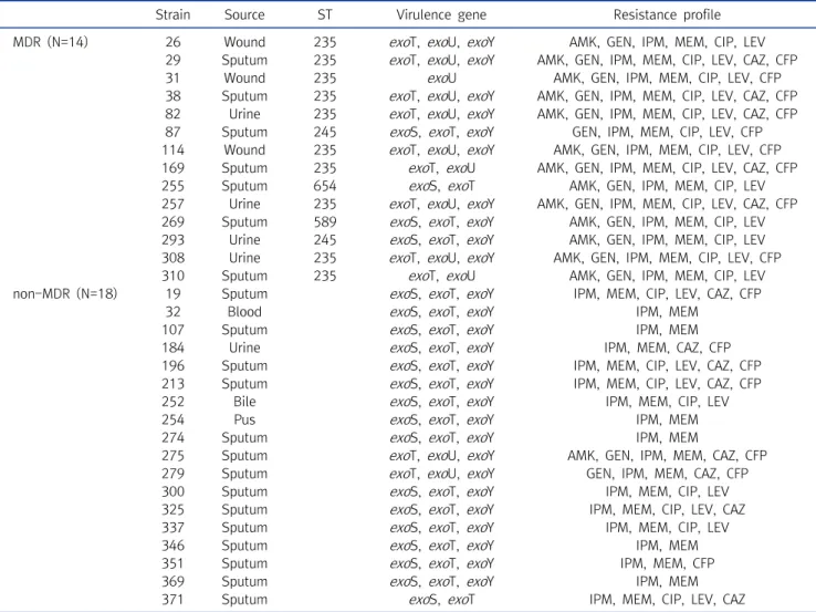
세포외 병독성 인자의 유전자의 경우, 다제내성균과 다제내성 이 아닌 균에서 큰 차이를 보이지 않았으나, exoS, exoU, pvdA 유전자의 경우, 유의한 차이를 보였다(P<0.001, P<0.001, P=0.004). 특히, exoU 유전자의 경우, 다제내성균에서 71.4%
(10/14)의 높은 비율을 보인 반면, 다제내성이 아닌 균에서는 11.1% (2/18)의 낮은 비율로 확인되었으며, exoS 유전자의 경 우, 다제내성균에서 28.6% (4/14)의 낮은 비율을 보였으나, 다 제내성이 아닌 균에서는 88.9% (16/18)의 높은 비율이 확인되 었다(Table 4).
2. 세포 관련 및 세포외 병독성 인자의 확인
18개의 세포 관련 및 세포외 병독성 인자의 유전자 확인을 위 해 PCR과 염기서열을 분석한 결과, 총 32균주의 carbapenem 내성 P. aeruginosa 중 32균주(100.0%) 모두에서 확인되었다 (Table 3). 18개의 세포 관련 및 세포외 병독성 인자 유전자 중 toxA, plcN, phzM 유전자(100%, 32/32)가 가장 높은 비율로 확인되었으며, exoT, phzI, phzS, lasB 유전자가 각각 96.9%
(31/32), phzH, aprA 유전자가 각각 93.8% (30/32)의 높은 비 율로 확인되었다. 반면, pilB 유전자는 32균주 모두에서 확인되 지 않았으며, exoU와 pilA 유전자는 각각 37.5% (12/32)와 25.0% (8/32)로 비교적 낮은 비율로 확인되었다. 또한, 32균주 는 각각 8∼16개의 유전자를 동시에 가지고 있는 것으로 확인 되었으며, 그 중 15개의 유전자를 가지고 있는 균주가 9균주
(28.1%)로 가장 높은 비율을 보였다(Table 5). 그 다음으로 14 개의 유전자를 가지고 있는 균주가 8균주(25.0%), 13개와 16개 의 유전자를 가지고 있는 균주가 각각 5균주(15.6%)로 확인되 었으며, 12개의 유전자를 가지고 있는 균주가 2균주(6.3%), 8개 와 9개, 11개의 유전자를 가지고 있는 균주가 각각 1균주(3.1%) 로 확인되었다.
고 찰
Carbapenem 내성 P. aeruginosa의 증가는 전세계적으로 높은 사망율의 원인이 되고 있으며, 다제내성으로 발달하고 있 어 매우 심각한 실정이다[9, 14, 18]. 본 연구에서도 32균주의 carbapenem 내성 P. aeruginosa 중 14균주(43.8%)가 다제내 성균이었으며, 이 중 71.4% (10균주)가 ST235로 확인되었다.
이전 많은 연구에서 다제내성으로 보고되고 있는 ST235는 CC235의 대표적인 클론으로, 유럽, 아시아, 남아메리카 등에 서 보고되고 있다[19, 20]. 우리나라의 경우, IMP-6를 생성하 는 ST235의 출현과 확산에 대해 보고하고 있으며, 최근에는 IMP-10을 생성하는 ST235가 보고되어 carbapenemases의 다양화에 대한 우려를 낳고 있다[21, 22].
또한, 항균제 감수성 결과, 8개의 항균제에 대해 모두 40%이
상의 높은 내성율을 보임에 따라, 향후 다제내성으로의 발달 가
능성과 항균제의 선택과 사용에 대한 어려움이 대두되고 있다
Table 4. Distribution of exo genes between MDR and non-MDR P. aeruginosa isolates
Strain Source ST Virulence gene Resistance profile
MDR (N=14) 26 Wound 235 exoT, exoU, exoY AMK, GEN, IPM, MEM, CIP, LEV 29 Sputum 235 exoT, exoU, exoY AMK, GEN, IPM, MEM, CIP, LEV, CAZ, CFP
31 Wound 235 exoU AMK, GEN, IPM, MEM, CIP, LEV, CFP
38 Sputum 235 exoT, exoU, exoY AMK, GEN, IPM, MEM, CIP, LEV, CAZ, CFP 82 Urine 235 exoT, exoU, exoY AMK, GEN, IPM, MEM, CIP, LEV, CAZ, CFP 87 Sputum 245 exoS, exoT, exoY GEN, IPM, MEM, CIP, LEV, CFP 114 Wound 235 exoT, exoU, exoY AMK, GEN, IPM, MEM, CIP, LEV, CFP 169 Sputum 235 exoT, exoU AMK, GEN, IPM, MEM, CIP, LEV, CAZ, CFP
255 Sputum 654 exoS, exoT AMK, GEN, IPM, MEM, CIP, LEV
257 Urine 235 exoT, exoU, exoY AMK, GEN, IPM, MEM, CIP, LEV, CAZ, CFP 269 Sputum 589 exoS, exoT, exoY AMK, GEN, IPM, MEM, CIP, LEV 293 Urine 245 exoS, exoT, exoY AMK, GEN, IPM, MEM, CIP, LEV 308 Urine 235 exoT, exoU, exoY AMK, GEN, IPM, MEM, CIP, LEV, CFP
310 Sputum 235 exoT, exoU AMK, GEN, IPM, MEM, CIP, LEV
non-MDR (N=18) 19 Sputum exoS, exoT, exoY IPM, MEM, CIP, LEV, CAZ, CFP
32 Blood exoS, exoT, exoY IPM, MEM
107 Sputum exoS, exoT, exoY IPM, MEM
184 Urine exoS, exoT, exoY IPM, MEM, CAZ, CFP
196 Sputum exoS, exoT, exoY IPM, MEM, CIP, LEV, CAZ, CFP 213 Sputum exoS, exoT, exoY IPM, MEM, CIP, LEV, CAZ, CFP
252 Bile exoS, exoT, exoY IPM, MEM, CIP, LEV
254 Pus exoS, exoT, exoY IPM, MEM
274 Sputum exoS, exoT, exoY IPM, MEM
275 Sputum exoT, exoU, exoY AMK, GEN, IPM, MEM, CAZ, CFP
279 Sputum exoT, exoU, exoY GEN, IPM, MEM, CAZ, CFP
300 Sputum exoS, exoT, exoY IPM, MEM, CIP, LEV
325 Sputum exoS, exoT, exoY IPM, MEM, CIP, LEV, CAZ
337 Sputum exoS, exoT, exoY IPM, MEM, CIP, LEV
346 Sputum exoS, exoT, exoY IPM, MEM
351 Sputum exoS, exoT, exoY IPM, MEM, CFP
369 Sputum exoS, exoT, exoY IPM, MEM
371 Sputum exoS, exoT IPM, MEM, CIP, LEV, CAZ
Abbreviations: MDR, multidrug resistan; ST, sequence type; AMK, amikacin; GEN, gentamicin; IPM, imipenem; MEM, meropenem; CIP, ciprofloxacin; LEV, levofloxacin; CAZ, ceftazidime; CFP, cefepime.
[23]. 최근 P. aeruginosa는 많은 항균제에 내재된 또는 획득된 내성 관련 연구 뿐만 아니라, 세포 관련 및 세포외 병독성 인자에 대한 많은 연구가 보고되고 있다[2, 9]. 이러한 병독성 인자들은 심각한 조직 손상, 혈액 감염 전파 및 질병의 진행에 관여하며, 현재 사용 가능한 항균제의 치료를 제한하고 있어 그 심각성이 중요시되고 있다[24, 25]. 이전 연구에서 특정 항균제인 fluoroquinolne과 일부 병독성 유전자인 exoS와 exoU의 관계 를 확인한 결과를 토대로, 이번 연구에서는 동일 지역의 병원에 서 분리한 carbapenem 내성 P. aeruginosa를 대상으로 다양 한 세포 관련 및 세포외 병독성 인자의 분포 및 항균제 내성과의 관계를 확인하였다. 18개의 세포 관련 및 세포외 병독성 인자를 확인한 결과, 32균주 모두에서 유전자가 확인되었으며, toxA, plcN, phzM 유전자(100%, 32/32)가 가장 높은 비율로 확인되 었다. 앞서 발표된 많은 연구에서도 toxA 유전자는 높은 빈도로
확인되었는데, Badr RI 등[26]의 연구에 의하면, 화상환자로부
터 분리된 P. aeruginosa에서 89%의 높은 비율로 확인되었으
며, Haghi 등[9]의 연구에서도 총 93균주의 P. aeruginosa 중
97.8%에서 확인되었다. toxA 유전자에 의해 암호화된
exotoxin A는 type II secretion system (T2SS)의 주요 구성 성
분으로, 인체에서 세포 사멸, 심각한 조직손상과 괴사를 일으키
고 있다. 이러한 exotoxin A는 ADP-ribosyl 부분을 신장인자 2
로 전달하는 ADP-ribosyl transferase로서, 포유동물 세포에
서 단백질 합성을 억제하고 있다[7, 8]. 또한, P. aeruginosa에
서 rhamnolipid와 2종류의 phospholipases C (용혈성
phospholipase C (PlcH)와 비용혈성 phospholipase C
(PlcN))는 3가지 용해성 단백질로써, 숙주세포의 침입에 관여
하고 있다. PlcH는 적혈구 막의 분해를 촉진하는데 외막의 인지
질 성분인 phosphatidylcholine과 sphingomyelin을 분해하
Table 5. Distribution of virulence genes among 32 CRPA isolates
Genes Virulence genes profile No. of
isolates (%)
Total No.
(%) 8 genes toxA, exoU, plcN, phzI, phzM, phzS, lasA, lasB 1 (3.1%) 1 (3.1%) 9 genes toxA, exoT, exoU, plcH, plcN, phzM, phzS, lasB, aprA 1 (3.1%) 1 (3.1%) 11 genes toxA, exoT, exoU, plcN, phzI, phzII, phzH, phzM, phzS, lasA, lasB 1 (3.1%) 1 (3.1%) 12 genes toxA, exoT, exoU, exoY, plcH, plcN, phzI, phzH, phzM, phzS, lasB, aprA 1 (3.1%) 2 (6.3%)
toxA, exoT, exoU, exoY, plcN, phzI, phzH, phzM, phzS, lasA, lasB, aprA 1 (3.1%)
13 genes toxA, exoS, exoT, exoY, plcH, plcN, phzI, phzII, phzH, phzM, phzS, lasA, aprA 1 (3.1%) 5 (15.6%) toxA, exoS, exoT, exoY, plcH, plcN, phzI, phzII, phzH, phzM, phzS, lasB, aprA 1 (3.1%)
toxA, exoS, exoT, plcH, plcN, phzI, phzH, phzM, phzS, lasA, lasB, aprA, pvdA 1 (3.1%) toxA, exoT, exoU, exoY, plcH, plcN, phzI, phzII, phzH, phzM, lasA, lasB, aprA 1 (3.1%) toxA, exoT, exoU, exoY, plcN, phzI, phzII, phzH, phzM, phzS, lasA, lasB, aprA 1 (3.1%)
14 genes toxA, exoT, exoU, exoY, plcH, plcN, phzI, phzII, phzH, phzM, phzS, lasA, lasB, aprA 5 (15.6%) 8 (25.0%) toxA, exoS, exoT, exoY, plcH, plcN, phzI, phzII, phzH, phzM, phzS, lasA, lasB, aprA 2 (6.3%)
toxA, exoS, exoT, plcH, plcN, phzI, phzII, phzH, phzM, phzS, lasB, pilA, aprA, pvdA 1 (3.1%)
15 genes toxA, exoS, exoT, exoY, plcH, plcN, phzI, phzII, phzH, phzM, phzS, lasA, lasB, aprA, pvdA 7 (21.9%) 9 (28.1%) toxA, exoS, exoT, exoY, plcH, plcN, phzI, phzII, phzH, phzM, phzS, lasB, pilA, aprA, pvdA 1 (3.1%)
toxA, exoS, exoT, exoY, plcN, phzI, phzII, phzH, phzM, phzS, lasA, lasB, pilA, aprA, pvdA 1 (3.1%)
16 genes toxA, exoS, exoT, exoY, plcH, plcN, phzI, phzII, phzH, phzM, phzS, lasA, lasB, pilA, aprA, pvdA 5 (15.6%) 5 (15.6%)
여 내막을 노출시키고, PlcN은 내막에 존재하는 phospha- tidylserine을 가수분해함으로써, 2종류의 phospholipases는 상승작용을 하고 있다[27]. Georgescu 등[28]은 plcH 유전자 만 확인되었던 루마니아의 이전 연구 결과와 달리, 혈액배양과 상처부위로부터 분리된 P. aeruginosa에서 plcH와 plcN 유전 자 모두 84.6%의 높은 빈도로 확인된 결과를 보고하였다. 2016 년 이란에서 발표한 Faraji 등[29]은 낭포성 섬유증과 화상감염 을 가진 소아에서 P. aeruginosa의 병독성 인자를 각각 확인한 결과, plcH (87.7%, 79.0%)와 plcN (60.0%, 63.1%) 유전자 모 두 검출되었으며, 본 연구에서도 plcN와 plcH 유전자가 100%
와 84.4%로 확인되어, 이전 연구보다 높은 결과를 보였다.
Phenazine 오페론(phzI, phzII) 및 유전자(phzH, phzM, phzS)는 P. aeruginosa에 의해 수동적으로 분비되는 3 가지 phenazine 화합물의 형성에 관여하는 전구 단백질을 암호화한 다[30]. 이러한 3가지 phenazine 화합물은 pyocyanin, 1-hyd- roxyphenazine, phenazine-1-carboxamide로써, 세포 내 산화 스트레스를 증가시키는 원인이 되고 있으며, 2015년 미국 에서 발표한 Goldufsky 등[6]은 P. aeruginosa 균주의 90% 이 상이 pyocyanin을 생성함으로써 만성 폐 감염에서 관찰된 폐 조직 손상에 중요한 역할을 하는 것으로 보고하였다. 본 연구에 서는 5개의 phenazine 오페론 및 유전자가 84% 이상의 높은 빈 도를 보였으며, phzM 유전자가 32균주 모두에서 확인되었다.
P. aeruginosa 감염에 의한 조직손상의 또 다른 원인 중 하나는 type III secretion proteins로써, exo 유전자(exoS, exoT, exoU, exoY)는 type III secretion systems의 발현과 분비에
관여하고 있다[31, 32]. Allydice-Francis 등[33]의 연구에 의
하면, 대부분의 많은 P. aeruginosa 균주들이 여러 exo 유전자
를 동시에 가지고 있는 것을 보고하였다. 이와 유사하게, 본 연
구에서는 exoT 유전자가 96.9%로 가장 높은 빈도로 확인되었
고, 그 다음으로 exoY 유전자가 84.4%, exoS 유전자가 62.5%,
exoU 유전자가 37.5%를 보였으며, 1균주를 제외한 31균주
(96.9%)가 2개 이상의 exo 유전자를 가지고 있었다. 이 중
exoU 유전자와 exoS 유전자는 유의한 관계를 보이는데, exoU
유전자가 확인된 12균주 중 10균주는 다제내성균(83.3%)인 반
면, exoS 유전자가 확인된 20균주 중 16균주는 다제내성이 아
닌 균(80.0%)으로 확인되었으며, 두 유전자를 모두 가지고 있는
균주는 한 균주도 확인되지 않았다. 2019년 이란에서 발표한
Khodayary 등[18]은 exoU 유전자의 획득과 항균제의 높은 내
성에 대한 정확한 기전은 알려지지 않았으나, exoU 유전자의
존재는 exoS 유전자와 비교하여 항균제에 대한 내성이 높다는
점을 확인하였고, 이러한 결과는 이란과 한국, 인도, 호주 등 다
른 국가에서 연구한 결과와 일치하였다. 그 밖에 P. aeruginosa
는 lasA와 lasB 유전자가 암호화하는 elastase 효소를 가지고 있
는데, 이 효소는 사이토카인, 백혈구 및 방어세포를 분해할 수
있으며, 폐와 혈관 조직의 분해에 중요한 역할을 하고 있다[34,
35]. 또 다른 병독성 인자 중 하나로, aprA 유전자가 암호화하는
alkaline protease는 각막염, 중이염, 낭포성 섬유증 및 균혈증
에서 생성되며, 사이토카인, 보체, 인터페론 및 종양괴사인자와
같이 생물학적으로 중요한 단백질의 가수분해에 관여하고 있다
[36]. 본 연구에서 lasB와 lasA, aprA 유전자는 각각 96.9%와
84.4%, 93.8%의 높은 빈도로 확인되었다. 또한, pyoverdine 생합성에 관여하는 pvdA 유전자는 50.0%가 확인되었으며, type IV pili의 생합성에 관여하는 pilA 유전자는 25.0%를 보였 고 pilB 유전자는 한 균주에서도 검출되지 않았다. 또한, 이전 연구에서 Fazeli N 등[2]은 다제내성 P. aeruginosa 균주에서 병독성 관련 유전자의 다양한 범위와 조합을 확인하였다. 이와 유사하게 32균주에서 확인된 17개의 세포 관련 및 세포외 병독 성 인자는 8개 이상의 유전자가 다양한 조합으로 확인되었으며, 이 중 15개의 유전자를 가지고 있는 9균주(28.1%)는 가장 높은 빈도로 확인되었다.
2011년 3월부터 2012년 12월까지 대전지역의 3차 병원에 서 분리된 carbapenem 내성 P. aeruginosa를 대상으로 한 항 균제 감수성 결과, 43.8%가 다제내성균으로 확인되었으며, 각 항균제에 대한 내성율이 40%이상을 보임에 따라 다제내성으로 의 발달 및 확산, 항균제의 선택과 제한이 심각한 실정이다. 또 한, 세포 관련 및 세포외 병독성 인자의 생성을 확인한 결과, 32 균주 모두에서 확인되었고 8개 이상의 유전자를 가진 다양한 조 합으로 존재하는 것을 확인하였으며, 이 중 exoU 유전자와 다 제내성과의 유의한 관계를 확인하였다(P<0.001). 향후 다제내 성균에 대한 중요한 예측 마커로써 활용되기 위해 exoU 유전자 와 다제내성균과의 연관성에 대한 연구가 지속적으로 진행되어 야 할 것으로 사료된다.
요 약
다제내성 P. aeruginosa의 출현과 확산은 전 세계적으로 중 요한 문제가 되고 있다. P. aeruginosa에 의한 발병은 일부 몇몇 세포 관련 및 세포외 병독성 인자의 생성에 기인한다. 본 연구에 서는 대전지역의 3차 병원에서 분리된 carbapenem 내성 P.
aeruginosa를 대상으로 병독성 인자의 분포와 항균제 내성 양 상을 조사하였다. 항균제 감수성 시험은 디스크 확산법으로 확 인하였고, 병독성 유전자의 분석을 위해 PCR과 염기서열분석 을 수행하였다. 또한, 다제내성 P. aeruginosa의 sequence type (ST)은 multilocus sequence typing (MLST)을 통해 확인 하였다. 32균주의 carbapenem 내성 P. aeruginosa 중, 14균 주(43.8%)가 다제내성이었으며, 주요 ST는 ST235 (10균주, 71.4.%)임을 확인하였다. 병독성 유전자는 32균주 모두에서 확 인되었고, 이 중 가장 높은 빈도로 확인된 병독성 유전자는 toxA, plcN, phzM (100.%)이었다. 또한, 32균주는 모두 8개 이 상의 병독성 유전자를 가지고 있었으며, 9균주(28.1%)가 15개 의 병독성 유전자를 가지고 있었다. exoU 유전자는 다제내성 P.
aeruginosa 균주의 71.4%에서 확인되었다. 이러한 결과는 exoU 유전자가 다제내성 P. aeruginosa 균주의 지속성에 대한 예측 표지자가 될 수 있을 것으로 사료된다.
Acknowledgements: None Conflict of interest: None
Author’s information (Position): Cho HH, Professor.
REFERENCES
1. Kumari H, Balasubramanian D, Zincke D, Mathee K. Role of
Pseudomonas aeruginosa
AmpR on -lactam and non--lac- tam transient cross-resistance upon pre-exposure to sub- inhibitory concentrations of antibiotics. J Med Microbiol.2014;63:544-555. https://doi.org/10.1099/jmm.0.070185-0.
2. Fazeli N, Momtaz H. Virulence gene profiles of multi- drug-resistant
Pseudomonas aeruginosa
isolated from Iranian hospital infections. Iran Red Crescent Med J. 2014;16:E15722.http://dx.doi.org/10.5812/ircmj.15722.
3. Strateva T, Yordanov D.
Pseudomonas aeruginosa
- a phenom- enon of bacterial resistance. J Med Microbiol. 2009;58:1133- 1148. https://doi.org/10.1099/jmm.0.009142-0.4. Lanotte P, Watt S, Mereghetti L, Dartiguelongue N, Rastegar- Lari A, Goudeau A, et al. Genetic features of
Pseudomonas aer- uginosa
isolates from cystic fibrosis patients compared with those of isolates from other origins. J Med Microbiol. 2004;53:73-81.https://doi.org/10.1099/jmm.0.05324-0.
5. Choy MH, Stapleton F, Willcox MD, Zhu H. Comparison of viru- lence factors in
Pseudomonas aeruginosa
strains isolated from contact lens- and non-contact lens-related keratitis. J Med Microbiol. 2008;57:1539-1546. https://doi.org/10.1099/jmm.0.2008/003723-0.
6. Goldufsky J, Wood S, Hajihossainlou B, Rehman T, Majdobeh O, Kaufman HL, et al.
Pseudomonas aeruginosa
exotoxin T in- duces potent cytotoxicity against a variety of murine and hu- man cancer celllines. J Med Microbiol. 2015;64:164-173.https://doi.org/10.1099/jmm.0.000003.
7. Wolska K, Szweda P. Genetic features of clinical
Pseudomonas aeruginosa
strains. Pol J Microbiol. 2009;58:255-260.8. Kipnis E, Sawa T, Wiener-Kronish J. Targeting mechanisms of
Pseudomonas aeruginosa
pathogenesis. Med Mal Infect.2006;36:78-91. https://doi.org/10.1016/j.medmal.2005.10.007.
9. Haghi F, Zeighami H, Monazami A, Toutouchi F, Nazaralian S, Naderi G. Diversity of virulenc egenes in multidrug resistant
Pseudomonas aeruginosa
isolated from burn wound infections.Microb Pathog. 2018;115:251-256. https://doi.org/10.1016/
j.micpath.2017.12.052.
10. Makedou KG, Tsiakiri EP, Bisiklis AG, Chatzidimitriou M, Halvantzis AA, Ntoutsou K, et al. Changes in antibiotic resist- ance of the most common Gram-negative bacteria isolated in intensive care units. J Hosp Infect. 2005;60:245-248.
https://doi.org/10.1016/j.jhin.2005.01.013.
11. Tsukayama DT, van Loon HJ, Cartwright C, Chmielewski B, Fluit
AC, van der Werken C, et al. The evolution of
Pseudomonas aer- uginosa
during antibiotic rotation in a medical intensive care unit: the RADAR-trial. Int J Antimicrob Agents. 2004;24:339-345. https://doi.org/10.1016/j.ijantimicag.2004.04.011.
12. Rossi Gonçalves I, Dantas RCC, Ferreira ML, Batistão DWDF, Gontijo-Filho PP, Ribas RM. Carbapenem-resistant
Pseudomonas aeruginosa
: association with virulence genes and biofilm formation. Braz J Microbiol. 2017;48:211-217. https://do- i.org/10.1016/j.bjm.2016.11.004.13. Khosravi Y, Tay ST, Vadivelu J. Analysis of integrons and asso- ciated gene cassettes of metallo--lactamase-positive
Pseudomonas aeruginosa
in Malaysia. Med Microbiol.2011;60:988-994. https://doi.org/10.1099/jmm.0.029868-0.
14. Cho HH. Molecular analysis of carbapenem-resistant
Pseudo- monas aeruginosa
isolated from patients hospitalized in Daejeon between 2008 and 2014 year. Korean J Clin Lab Sci.2018;50:406-413. https://doi.org/10.15324/kjcls.2018.50.4.406.
15. Clinical and Laboratory Standards Institute. Performance standards for antimicrobial susceptibility testing; twentieth in- formational supplement, M100-S20. Wayne, PA: Clinical and Laboratory Standards Institute; 2010.
16. Magiorakos AP, Srinivasan A, Carey RB, Carmeli Y, Falagas ME, Giske CG, et al. Multidrug-resistant, extensively drug-resistant and pandrug-resistant bacteria: an international expert pro- posal for interim standard definitions for acquired resistance.
Clin Microbiol Infect. 2012;18:268-281. https://doi.org/10.
1111/j.1469-0691.2011.03570.x.
17. Finnan S, Morrissey JP, O'Gara F, Boyd EF. Genome diversity of
Pseudomonas aeruginosa
isolates from cystic fibrosis patients and the hospital environment. J Clin Microbiol. 2004;42:5783-5792. https://doi.org/10.1128/JCM.42.12.5783-5792.2004.
18. Khodayary R, Nikokar I, Mobayen MR, Afrasiabi F, Araghian A, Elmi A, et al. High incidence of type III secretion system asso- ciated virulence factors (exoenzymes) in
Pseudomonas aerugi- nosa
isolated from Iranian burn patients. BMC Res Notes.2019;12:28. https://doi.org/ 10.1186/s13104-019-4071-0.
19. Samuelsen O, Toleman MA, Sundsfjord A, Rydberg J, Leegaard TM, Walder M, et al. Molecular epidemiology of metal- lo-beta-lactamase-producing
Pseudomonas aeruginosa
iso- lates from Norway and Sweden shows import of international clones and local clonal expansion. Antimicrob Agents Chemother.2010;54:346-352. http://doi.org/10.1128/AAC.00824-09.
20. Cholley P, Thouverez M, Hocquet D, van der Mee-Marquet N, Talon D, Bertrand X. Most multidrug-resistant
Pseudomonas aeruginosa
isolates from hospitals in eastern France belong to a few clonal types. J Clin Microbiol. 2011;49:2578-2583.http://doi.org/10.1128/JCM.00102-11.
21. Hong JS, Yoon EJ, Lee H, Jeong SH, Lee K. Clonal dissemination of
Pseudomonas aeruginosa
sequence type 235 isolates carry- ingbla
IMP-6 and emergence ofbla
GES-24 andbla
IMP-10 on novel ge- nomic islands PAGI-15 and –16 in South Korea. Antimicrob Agents Chemother. 2016;60:7216-7223. http://doi.org/10.1128/AAC.01601-16.
22. Wi YM, Choi JY, Lee JY, Kang CI, Chung DR, Peck KR, et al.
Emergence of colistin resistance in
Pseudomonas aeruginosa
ST235 clone in South Korea. Int J Antimicrob Agents. 2017;49:767-769. http://doi.org/10.1016/j.ijantimicag.2017.01.023.
23. Kim D, Ahn JY, Lee CH, Jang SJ, Lee H, Yong D, et al. Increasing resistance to extended-spectrum cephalosporins, fluo- roquinolone, and carbapenem in Gram-negative bacilli and the emergence of carbapenem non-susceptibility in
Klebsiella pneumoniae
: Analysis of Korean Antimicrobial Resistance Monitoring System (KARMS) data from 2013 to 2015. Ann Lab Med.2017;37:231-239. https://doi.org/10.3343/alm.2017.37.3.231.
24. Breidenstein EB, de la Fuente-Núñez C, Hancock RE.
Pseudo- monas aeruginosa
: all roads lead to resistance. Trends Microbiol.2011;19:419-426. http://doi.org/10.1016/j.tim. 2011. 04.005.
25. Kerr KG, Snelling AM.
Pseudomonas aeruginosa
: a formidable and ever-present adversary. J Hosp Infect. 2009;73:338-344.http://doi.org/10.1016/j.jhin.2009.04.020.
26. Badr RI, Nagdy M, Sabagh A, Din AB.
Pseudomonas aeruginosa
exotoxin A as a virulence factor in burn wound infections.Egypt J Med Microbiol. 2008;17:125–132.
27. Cotar AI, Chifiriuc MC, Banu O, Lazar V. Molecular character- ization of virulence patterns in
Pseudomonas aeruginosa
strains isolated from respiratory and wound samples. Biointerface Res Appl Chem. 2013;3:551-558.28. Georgescu M, Gheorghe I, Curutiu C, Lazar V, Bleotu C, Chifiriuc MC. Virulence and resistance features of
Pseudomonas aeruginosa
strains isolated from chronic leg ulcers. BMC Infect Dis. 2016;16:92. http://doi.org/10.1186/s12879-016-1396-3.29. Faraji F, Mahzounieh M, Ebrahimi A, Fallah F, Teymournejad O, Lajevardi B. Molecular detection of virulence genes in
Pseudo- monas aeruginosa
isolated from children with cystic fibrosis and burn wounds in Iran. Microb Pathog. 2016;99:1-4.http://doi.org/10.1016/j.micpath.2016.07.013.
30. Jamunadevi S, Balashanmugam P, Muralitharan G, Kalaichelvan PT. Molecular characterization of pathogenic and non-patho- genic
Pseudomonas aeruginosa
with special reference to phe- nazine gene. J Mod Biotechnol. 2012;1:70-74.31. Galle M, Carpentier I, Beyaert R. Structure and function of the type III secretion system of
Pseudomonas aeruginosa
. Curr Protein Pept Sci. 2012;13:831-842.32. Hauser AR. The type III secretion system of
Pseudomonas aeru- ginosa
: infection by injection. Nat Rev Microbiol. 2009;7:654- 665. http://doi.org/10.1038/nrmicro2199.33. Allydice-Francis K, Brown PD. Diversity of antimicrobial resist- ance and virulence determinants in
Pseudomonas aeruginosa
associated with fresh vegetables. Int J Microbiol. 2012;2012:426241. http://doi.org/10.1155/2012/426241.
34. Kruczek C, Kottapalli KR, Dissanaike S, Dzvova N, Griswold JA, Colmer-Hamood JA, et al. Major transcriptome changes ac- company the growth of
Pseudomonas aeruginosa
in blood from patients with severe thermal injuries. PLoS One. 2016;11:E0149229. http://doi.org/10.1371/journal.pone.0149229.
35. Islamieh DI, Afshar D, Esmaeili D. Effect of Satureja khuzistan- ica essential oil (SKEO) extract on expression of
las
A andlas
B genes inPseudomonas aeruginosa
. Iran J Microbiol. 2019;11:55-59.
36. Andrejko M, Siemińska-Kuczer A, Janczarek M, Janik E, Bednarczyk M, Gagoś M, et al.
Pseudomonas aeruginosa
alka- line protease exhibits a high renaturation capability. Acta Biochim Pol. 2019;66:91-100. http://doi.org/10.18388/abp.2018_2741.