Korean J. Malacol. 29(2): 97-103 2013
Received: April 17, 2013; Accepted: May 29, 2013 Corresponding author : Kwang-Sik Choi
Tel: +82 (64) 754-3422 e-mail: [email protected] 1225-3480/24475
This is an Open Access article distributed under the terms of the Creative Commons Attribution Non-Commercial License with permits unrestricted non-commercial use, distribution, and reproducibility in any medium, provided the original work is properly cited.
주사전자현미경 (Scanning Electron Microscope) 을 이용한 제주 북부 연안에 서식하는 가시굴 ( Saccostrea
kegaki Torigoe & Inaba, 1981) 의 초기 유생발달관찰
이희중, 강현실, 정희도, 홍현기, 최광식
제주대학교 해양과학대학 해양의생명과학부 해양생명과학전공
First observation on the early embryonic and larval development of spiny oyster Saccostrea kegaki Torigoe & Inaba, 1981 (Bivalvial:
Ostreoida) using scanning electron microscope on the north coast of Jeju, Korea
Hee-Jung Lee, Hyun-Sil Kang, Hee-Do Jeung, Hyun-Ki Hong and Kwang-Sik Choi
School of Marine Biomedical Science, Jeju National University 102 Jejudaehakno, Jeju 690-756, Republic of Korea
ABSTRACT
In the present study, we monitored the early development of Saccostrea kegakia subtropical oyster species distributing on rocky intertidal off the northern Jeju Island using scanning electron microscope (SEM). The female oyster collected in early August, 2012 were fully mature exhibiting relatively small eggs (46.5 ± 1.4 μm in diameter) in the gonad, while testis of the mature male oysters were filled with fully developed sperms of 36.9 μm in length.
The fertilized eggs developed into 2-cell stage with polar body after 1 hr 20 min of fertilization, then followed by Morula stage (3 hr 20 min), Blastula stage (4 hr 50 min), Gastrula stage (7 hr), and trochophore larvae stage (9 hr 30 min). The observed early development of S. kegaki in this study was similar the early development of other oysters, although size of the fertilized eggs were somewhat smaller.
Keyword: Saccostrea kegaki, larval development, embryonic development, SEM, Ostreoid
서 론
굴 (Oyster) 은 연안의 조간대 및 조하대에 분포하는 고착성 이매패류로 수산 양식업 종으로서 높은 경제적 가치와 더불어 해양환경 변화모니터링의 지표 종으로 널리 활용되고 있다 (Damiensa et al. 2006). 현재까지 보고된 굴 과 (Ostreidae) 는 594종 이며 (http://www.marinespecies.org), 세계식량기
구 (Food and Agriculture Organization, FAO) 는 Crassostrea 속 8종, Ostrea 속 4종, Saccostrea 속 2종 및 Ostreola 속 1종 등, 총 15종을 상업적으로 유용하거나 현재 양식되고 있는 종으로 소개하고 있다. 우리나라 연안에는 현재 14종의 굴이 분포하고 있는 것으로 확인되고 있으며 (Min, 2004), 이중 제주연안에는 토굴 (Ostrea denselamellosa), 악 어굴 (Dendostrea folium), 톱니턱굴 (Dendostrea crenulifera), 참굴 (Crassostrea gigas), 바위굴 (Crassostrea nippona), Crassostrea echinata, Crassostrea nigromarginata, 가시굴 (Saccostrea kegaki), Saccostrea mordax, 및 태생굴 (Striostrea circumpicta) 등, 10 종이 분 포하고 있는 것으로 알려지고 있다 (과학기술부, 2005).
가시굴 (S. kegaki) 은 일본 Seto Inland Sea (Torigoe, 1999 Okutani, 2000)와 Morozaki (Lam and Morton, 2006)를 비롯하여 대만 (Taiwan malacofauna database, http://dx.doi.org/10.9710/kjm.2013.29.2.97
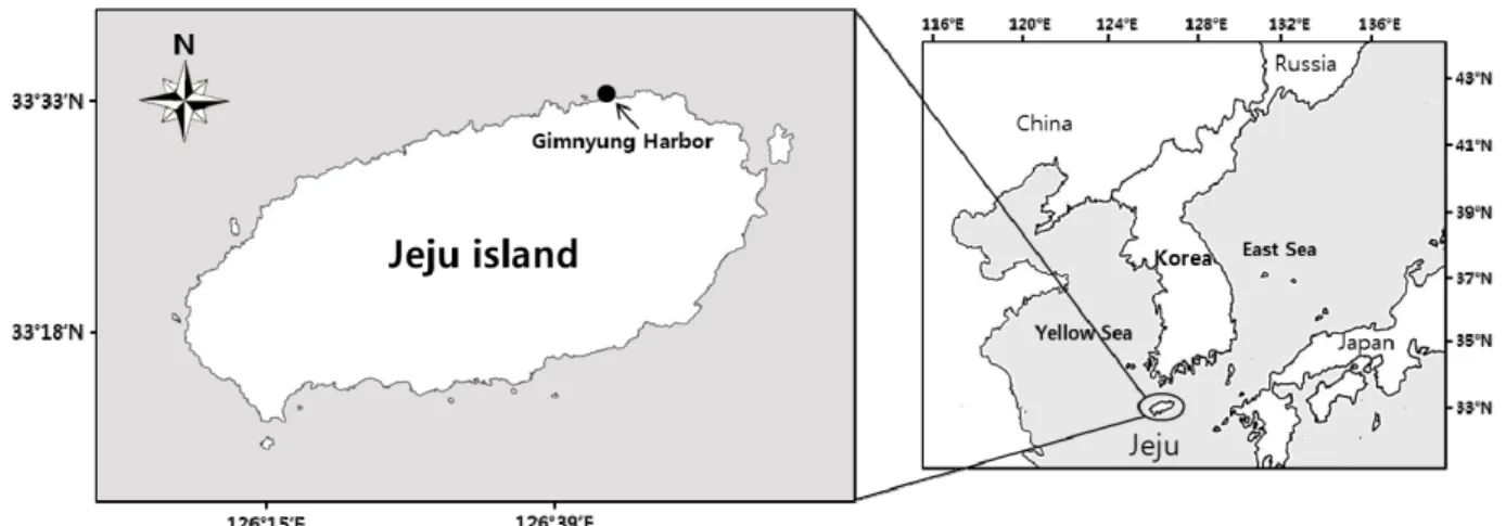
Fig. 1. Map showing the sampling site.
http://shell.sinica.edu.tw), 필리핀 (Poppe, 2010) 과 같은 아열대 지역에 서식하며 우리나라에서는 제주도에 유일하게 보고되고 있다 (Choi et al. 2000; Min, 2004; Kim et al.
2010; Yang and Choi, 2011). 가시굴 각장의 크기는 2-5 cm로 중·소형이며, 제주지역의 경우 주로 용천수가 분포하는 조간대 지역에 밀집하여 서식하고 있다. 패각은 난원형으로 표 면에는 관모양의 돌기가 가시처럼 돋아있는 것이 특징이다 (Choi et al. 2000; Min, 2004 Yang and Choi 2011). 가시 굴에 관한 생태학적 연구는 수산업적으로 가치가 높은 다른 굴 에 비하여 그 연구가 매우 미미한 실정이며, 국내의 경우 Kim et al. (2010) 이 서귀포 연안에 서식하는 가시굴의 연중 생식 주기에 관한 연구를 수행한 바 있으며, 이 지역의 가시굴은 7-8월에 산란하는 것으로 보고하였다.
이매패류 초기유생발달 연구는 양식 대상 종의 생활사 규명 하는데 중요한 분야이며, 이매패류 부유유생 식별을 통해 자연 채묘 예보에 활용될 수 있다. 유생 발달과정을 광학현미경만을 이용하여 관찰할 경우 배 발생 과정 중 나타나는 발생학적 특 징을 확인하는데 한계가 있다. 이를 보완하기 위한 주사전자현 미경(Scanning Electron Microscope, SEM) 관찰이 패류 초기유생 발달 연구에 널리 이용되어 왔다 (Moueza et al.
1999; Silberfeld and Gros, 2006). Kakoi et al. (2008) 은 광학현미경과 주사전자현미경을 이용하여 일본 와카야마와 시 즈오카 지역에 서식하는 가시굴의 초기 배 발생 (early embryogenesis) 에 관여하는 유전자들의 발현 특성을 보고한 바 있다. 우리나라의 경우, 이매패류 유생발달 연구는 대부분 의 경우 광학현미경만을 이용하여 초기유생 발달 과정을 관찰 보고하고 있으며, 주사전자현미경을 이용한 연구는 상대적으로 미비한 실정이다 (Lee et al. 2012).
이 연구는 제주연안에 서식하는 가시굴의 생활사를 이해하 는 기초연구의 일환으로 산란기간 중, 초기 유생발달 과정을
광학 현미경 및 주사전자현미경으로 관찰하여 보고하고자 하 였다.
재료 및 방법
1. 시료 채집 및 수정
가시굴의 수정 및 초기발생을 관찰하기 위하여 각장 4-6 cm 크기의 가시굴 20개체를 2012년 8월 초, 제주시 김녕항 (33°33’N, 126°44’E) 의 암반 조간대에서 채집하였다 (Fig.
1). 김녕항은 제주도 제주시 구좌읍에 위치한 포구로 상부에는 간조 시 외부로 노출되는 모래해안과 화산암으로 구성된 암반 해안이 발달해 있다.
채집된 가시굴은 실험실로 옮겨, 암·수 구분을 위해 가시굴 외투막 (mantle) 에 발달된 생식세포를 주사바늘을 이용해 일 부를 적출 후 슬라이드에 도말하여 광학현미경 (CH30, Olympus) 하에서 확인하였다. 가시굴의 채란 및 채정을 위해 성숙한 알 또는 정소가 포함된 가시굴 생식소 조직을 가위로 오려낸 후, Petri Dish에 올려놓고 1 mm간격으로 세밀히 절 개하여 정자와 알을 수집하였다. 수집된 가시굴 알은 20 μm 망목을 이용해 수세 한 뒤, 여과해수가 채워진 5 L 원형 비이 커로 옮겼다. 수정은 가시굴 정자를 알이 들어있는 비이커에 주입시켜 자연수정을 실시하였다. 수정된 가시굴 알은 바닥에 가라앉지 않도록 통기 (aeration) 하였다.
2. 수정란발달 관찰 (광학, 전자 현미경)
가시굴 수정란 초기 발달과정을 관찰하기 위해 광학 현미경 과 주사 전자현미경 (SEM) 을 이용하여 정자, 알 및 수정란의 배 발생을 관찰하였다. 광학 현미경을 이용한 수정란 발달 관 찰은 수정 후, 30분 간격으로 수행하였고, 세포분열이 관찰된 수정난은 SEM 관찰을 위하여 2% glutaraldehyde 고정액에 1시간 동안 전 고정 하였다. 고정이 완료된 수정란은 50, 70,
Korean J. Malacol. 29(2): 97-103 2013
90, 95, 100% 에탄올로 탈수 과정을 거쳤으며 치환을 위해 30, 50, 70, 100% isoamyl acetate 처리하였다. 치환이 완료 된 수정란은 진공건조 뒤 gold 입자로 코팅하였으며, 장방출 주사 전자현미경 (JSM-6700F, JEOL Korea Ltd) 을 이용하 여 관찰하였다.
결과 및 고찰
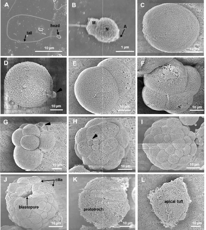
2012년 8월 제주 김녕항에서 채집된 성숙한 가시굴의 정자 는 짧은 원추형의 두부와 36.9 μm의 긴 편모 (flagellum) 로 구성되었다 (Fig. 3A). 정자 두부의 직경은 1.55 μm이며, 상 단부의 첨체 (acrosome) 와 타원형의 핵 (nuclear), 중편 (mid-piece) 의 미토콘드리아 (mitoch ondria) 로 구성되어 있었다 (Fig. 3B). 완숙한 가시굴의 알은 난막에 둘러싸인 타 원형으로, 알의 직경은 46.5 ± 1.4 μm 이었다 (Fig. 2A &
3C). 또한, 성숙한 가시굴 알은 해수에 가라앉는 침성란 (demersal egg) 의 특징을 보였다.
가시굴의 배 발생 (embryogenesis) 은 수정 40분 후 수정 란의 표면에 극체 (polar body) 가 형성되면서 시작되었다 (Fig. 2B & 3D). 수정란은 1시간 10분 후 2세포기 (2-cell stage) 를 보였으며 (Fig. 2C & 3E), 수정 1시간 40분 후에 는 4세포기 (4-cell stage) 에 도달하였으며, 이 시기에 관찰된 세포는 크기가 비슷한 3개의 난할구 (blastomere) 와 세포 크 기가 가장 큰 1개의 난할구로 구성되어 있었다 (Fig. 2D &
3F). 이후, 8세포기 (Fig. 2E & 3G), 16세포기 (Fig. 3H) 의 배 발생이 관찰되었으며, 수정 3시간 20분 후 세포분열이 절정 에 이르러 가시굴의 조직 또는 기관 분화를 위한 상실기 (morula) 로 분화하였다 (Fig. 2F & 3I). 수정 4시간 50분 후 에는 상실기 중앙에 포배강 (blastocoel) 이 형성되면서 포배 기 (blastula) 가 형성되었고 (Fig. 2G), 원구 (blastopore) 의 함입 (invagination) 과 할구 표면에 섬모 (cilia) 가 생성되는 초기 낭배기 (early gastrula) 가 수정 5시간 50분 후에 관찰 되었다 (Fig. 2H & 3J). 수정 7시간 후에는 embryo의 난할 표면 전체에 섬모가 발달하면서 활동 범위가 작은 나선 또는 구형의 회전 운동을 보이는 후기 낭배기 (late gastrula) 로 발달하였다. 특히, 이 시기의 embryo에서는 활발한 자유 유영 활동을 위한 구전섬모환 (prototroch) 이 관찰되었다 (Fig. 2I
& 3K). 활발한 운동성을 갖는 담륜자 유생 (trochophore larvae) 은 수정 9시간 30분 후에 관찰되었으며, 담륜자 유생 은 구전섬모환 (prototroch) 과 정단모 (apical tuft)를 이용하 여 유영 (swimming) 을 시작하였다 (Fig. 2J & 3L).
Kim et al. (2010) 은 제주 남부 연안에 서식하는 가시굴의 연중 생식주기를 조직학적 방법으로 구명하였으며, 이 지역에 서식하는 가시굴은 수온이 24.6-26.7℃ 범위를 보이는 7-8월 이 주 산란기이며, 산란기 때 완숙한 알의 크기는 36.9 ± 5.7
μm라고 보고하였다. 또한 Kakoi et al. (2008) 은 일본 와카 야마와 시즈오카 지역에 서식하는 가시굴을 이용하여 초기 배 발생 (early embryogenesis) 에 관여하는 유전자들의 발현 특성을 구명하였으며, 이 연구에 이용된 완숙한 가시굴 알의 크기는 40 μm으로 보고한 바 있다. 2012년 8월 제주 북부 김녕항에서 채집된 가시굴의 성 성숙 정도는 대부분의 암, 수 가 산란 직전인 것으로 판단되어, Kim et al. (2010) 이 보고 와 같이 제주의 경우 7, 8월이 가시굴의 산란기인 것으로 확인 되었다.
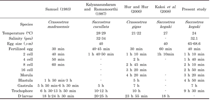
Table 1은 Crassostrea와 Saccostrea 속의 초기발생에 관 한 연구 보고로, 초기 배 발생 (early embeyogenesis) 은 12 시간 내에 완료되며, D상 유생 (D-shaped larvae) 로의 분화 는 24시간 이내에 이루어진다 하였다. 참굴 (C. gigas) 의 경 우, 21-22℃ 수온에서 수정난에서 담륜자 유생까지 10시간, D-상 유생까지 약 24시간이 소요되며 (Hur and Hur, 2000), Mangrove oyster (C. madrasensis) 의 초기 배 발생 소요 시간도 수정난에서 담륜자 유생까지 6시간 30분-13시간 50분, D-상 유생까지 18시간-24시간 정도 소요됨을 알 수 있다 (Samuel, 1983). 한편, 가시굴 (S. kegaki) 의 경우, Kakoi et al. (2008) 은 27℃ 환경에서 수정난에서 D-상 유생까지 18시간이 소요됨을 보고하였다. 또한, Kalyanasundaram and Ramamoorthi (1987) 는 가시굴과 같은 과에 속하는 S.
cucullata의 초기 유생발달에 관한 연구에서 수정에서 담륜자 유생까지 10-12시간이 소요됨을 보고하고 있다. 따라서 이 연 구에서 관찰된 가시굴의 초기 유생발생 소요 시간은 Kakoi et al. (2008) 이 일본에서 관찰한 가시굴의 초기유생발생 및 S.
cucullata의 초기 유생발생과정 관찰결과와 일치하는 것으로 사료된다.
이매패류의 정자는 형태학적으로 표준형 정자 (primitive type) 에 속하며, 두부의 뾰족한 첨체와 타원형의 핵, 미토콘 드리아가 위치하는 중편 (mid-piece) 과 한 개의 긴 편모를 가 지는 미부 (tail) 로 구성되어 있다 (Franzan, 1983). 특히, 굴 의 정자 형태는 첨체 모양, 핵의 형태 (구형, 타원형, cylinder 형) 와 중편의 미토콘드리아 수에 따라 구별된다.
Crassostrea 속에 속하는 굴의 정자 형태는 모자형의 첨체, 구형의 핵과 4개의 미토콘드리아를 갖고 있는 것으로 보고되 고 있다 (Yurchenko, 2012). 이와 달리, Saccostrea 속에 속 하는 호주굴 (Saccostrea commercialis) 의 정자 형태는 Crassostrea 속과 아주 유사하지만, 첨체의 미세구조에서 Crassostrea 속의 정자 형태와 구별된다 (Healy and Lester, 1991). 따라서 제주도 연안에 서식하는 가시굴 정자의 외부 형 태는 굴과 (Ostreidea) 에 속하는 굴류의 정자 형태와 유사하 지만, 종 특이적 특징을 결정하는 첨체의 미세구조에 대한 상 세한 연구가 필요할 것으로 사료된다.
Fig. 2. Photographs of Saccostrea kegaki in different development stages. A, ripe egg. B, fertilized egg (40 min). C, 2-cell (1 h 10 min). D, 4-cell (1 h 40 min). E, 8-cell (2 h 10 min). F, morula (3 h 20 min). G, blastula (4 h 50 min). H, early gastrula (5 h 50 min). I, late gastrula (7 h). J, trochophore larvae (9 h 30 min).
Korean J. Malacol. 29(2): 97-103 2013
Fig. 3. SEM (Scanning Electron Microscopy) ultrastructure of Saccostrea kegaki in different development stages. A, Spermatozoa (ripe sperm). B, Head part of the ripe sperm, N, Nuclear; M, Mitochondria; A, Acrosome. C, ripe egg. D, fertilized egg with polar body (arrow) (40 min). E, 2-cell (1 h 10 min). F, 4-cell (1 h 40 min). G, 8-cell with polar body (arrow) (2 h 10 min). H, 16-cell with polar body (arrow) (2 h 50 min). I, morula stage(3 h 20 min). J, early gastrula (5 h 50 min). K, late gastrula (7 h). L, trochophore larvae (9 h 30min).
Samuel (1983)
Kalyanasundaram and Ramamoorthi
(1987)
Hur and Hur (2000)
Kakoi et al.
(2008) Present study Species Crassostrea
madrasensis Saccostrea
cucullata Crassostrea
gigas Saccostrea
kegaki Saccostrea kegaki
Temperature (°C) 28-29 21-22 27 24
Salinity (psu) 32-34 - 32.1
Egg size (μm) 40 40 65-68.6
Fertilized egg 30 min 40-45 min 30 min 60 min 40 min
2 cell 40 min 1 h 40-50 min 1 h 10 min 1h 10min 1 h 10 min
4 cell 50 min - 2 h - 1 h 40 min
8 cell 60 min - 2 h 45 min 2 h 10 min
16 cell - 3 h 20 min 2 h 50 min
Morula - 4 h 20 min - 3 h 20 min
Blastula 1 h 50 min-3 h - 5 h - 4 h 50 min
Gastrula 5 h 50 min-6 h 30 min 5 h 7 h - 7 h
Trochophore 6 h 30-13 h 50 min 10-12 h 10 h - 9 h 30 min
D-larvae 18 h-24 h 30 min 20-25 h 23 h 55 min 18 h -
Table 1. Summary of early development of oysters in the Genus Saccostrea and Crassostrea
사 사
이 연구는 교육과학기술부 기초연구사업과제 "제주연안 아 열대 굴의 번식, 면역 생리 및 분자생물학적 분류, 2010-0009352"의 후원으로 수행되었습니다.
REFERENCES
과학기술부 (2005) 제주 해양생물 자원의 정보 및 추출물 은행 구축 (제주연안 무척추동물 다양성 조사연구). 과학기술부, 118p
Choi, K.S., Je, J.G., Lee, J.J. (2000) Commercially exploitable marine bivalves in Jeju island.
Underwater Science & Technology, 2: 29-38.
Damiens, G., Mouneyrac, C., Quiniou, F., His, E., Gnassia-Barelli, G., Romeeo, M. (2006) Metal bioaccumulation and metallothionein concentrations in larvae of Crassostrea gigas. Environmental Pollution, 140: 492-499.
Franzen, A. (1983) Ultrastructural studies of spermatozoa in three bivalve species with notes on evolution of elongated sperm nucleus in primitive spermatozoa. Gamete Research, 7: 199-214.
Healy, J.M., Lester, R.J.G. (1991) Sperm ultrastructure in the Australian oyster Saccostrea comer Cialis (Iredale & Roughley) (Bivalvia: Ostreoidea), Journal of Molluscan Studies, 57: 219-224.
Hur, Y.B., Hur, S.B. (2000) Development and growth of larvae of four bivalve species. Journal of Aquaculture, 13: 119-128.
Kakoi, S., Kin, K., Miyazaki, K., Wada, H. (2008) Early development of the Japanese spiny oyster (Saccostrea kegaki): Characterization of some genetic markers.
Zoological Science, 25: 455-464.
Kalyanasundaram, M., Ramamoorthi, K. (1987) Larval development of the oyster Saccostrea cucullata (Born). Mahasagar-Bulletin of the National Institute of Oceanography, 20: 53-58.
Kim, B.K., Kang, D.H., Ko, D.K., Yang, H.S., Kim, D.K., Kang, C.K., Choi, S.K. (2010) Annual reproductive cycle of the oyster, Saccostrea kegaki (Torigoe &
Inaba 1981) on the southern coast of Jeju island, Korea. Invertebrate Reproduction and Development, 54:19-26.
Lam, K., Morton, B. (2006) Morphological and mitochondrial-DNA analysis of The indo-west Pacific rock oysters (Ostreidae: Saccostreaspecies). Journal of Molluscan Studies, 72: 235-245.
Lee, H.J., Kang, H.S., Park, K.I., Choi, S.K. (2012).
Quantification of reproductive effort and microscopic observation on the larval development of Manila clam Ruditapes philippinarum (Adams and Reeve, 1850). Malacological Society of Korea, 28(2): 145-156.
Min, D.K. (2004) Mollusks in Korea. 401pp, Hangul Graphics. Busan
Moueza, M., Gros, O., Frenkiel, L. (1999) Embryonic, larval and postlarval development of the tropical clam, Anomalocardia brasiliana (Bivalvia, Veneridae).
Journal of Molluscan Strdies, 65: 73-88.
Okutani, T. (2000) Marine mollusks in Japan. 927pp, Tokai University. Japan
Poppe, G.T. (2010) Philippine marine mollusks: III.
Gastropoda Part 3 and Bivalvia Part 1, 665pp, Hackenheim. Philippine
Samuel, D. (1983) Early larval development of Edible oyster Crassostrea madrasensis (Perston). Proceedings of the symposium on coastal aquaculture, 2: 483-487.
Korean J. Malacol. 29(2): 97-103 2013
Silberfeld, T., Gros, O. (2006) Embryonic development of the tropical bivalve Tivela mactroides (Bron, 1778) (Veneridae: subfamily Meretricinae): a SEM study.
Cahiers de Biologie Marine, 27: 243-251.
Torigoe, K. (1999) The distribution of Saccostrea kegaki in the inland sea of Seto. Bulletin of the Faculty of School Education Hiroshima University, 21: 25-29.
Yang, H.S., Choi, S.K. (2011) A field guide to the Jeju seashore. 160-161pp, The Ministry of Land, Transport and Maritime Affairs and Jeju Sea Grant.
Jeju
Yurchenko, O.V. (2012) Comparative ultrastructure study of spermatozoa in some oyster species from the Asian-Pacific coast. Micron, 43: 365-373.