1063
허혈성 뇌혈관질환에서 자기공명혈관조영술의 가치에 대한 임상연구
고려대학교 의과대학 안산병원 신경외과학교실, 진단방사선과학교실*
임동준·조태형·정용구·김백현*·김근회·김세훈·권택현 정흥섭·박정율·박윤관·이훈갑·이기찬·서중근
= Abstract =
Intracranial Magnetic Resonance Angiography-Its Role in the Approach to Ischemic Stroke
Dong-Jun Lim, M.D., Tae-Hyoung Cho, M.D., Yong-Gu Chung, M.D., Baek-Hyun Kim,*
Keun-Hoe Kim, M.D., Se-Hoon Kim, M.D., Taek-Hyun Kwon, M.D., Heung-Seob Chung, M.D., Jung-Yul Park, M.D., Youn-Kwan Park, M.D., Hoon-Kap Lee, M.D.,
Ki-Chan Lee, M.D., Jung-Keun Suh, M.D.
Department of Neurosurgery, Radiology,* Ansan Hospital, Korea University, Ansan, Korea
bjectives:To determine the contribution of cranial magnetic resonance angiography(MRA) for the evaluation of patients with ischemic cerebrovascular accident.
Methods::::Magnetic resonance image(MRI) and MRA studies performed on 34 patients with ischemic stroke were retrospectively reviewed with the clinical records.
Results::::There were 9 transient ischemic attacks and 25 completed strokes in our series. Twenty-three of 34 MRA examinations(68%) were positive for stenosis or occlusion. The distribution of stenotic or occlusive vascular lesions were correlated with the location of infarction in 22 of the 23 positive cases(96%). MRA provided additional information not obtained from the MRI in 19 cases(56%).
Conclusions:::Vascular lesions demonstrated on intracranial MRA show a high correlation with infarct distribution. : MRA provided information adjunctive to conventional MRI in a majority of cases. We conclude that MRA is an impo- rtant noninvasive component of the complete evaluation of ischemic stroke.
KEY WORDS:Magnetic resonance angiography(MRA)・Ischemic cerebrovascular accident・Stroke.
서 론
허혈성 뇌혈관질환은 사망의 중요 원인이며 심한 신경학적 후유증상으로 인하여 많은 경제적, 사회적 손실을 초래하는 질환이다. 최근에는 발병초기에 허혈된 부위에 혈류의 재관류 를 시도하는 적극적인 치료방법이 시도되고 있고, 이에 따라 허혈부위와 혈관계에 대한 정확한 조기진단의 중요성이 제기 되고 있다. 뇌 자기공명영상(Magnetic Resonance Image, MRI)은 조기 뇌경색의 해부학적 영역을 정확히 진단함으로
서 초기 뇌경색의 진단에 매우 유용한 진단방법으로 보편화 되고 있고, 뇌 자기공명혈관조영술(Magnetic Resonance Angiography, MRA)은 뇌혈관계를 평가할 수 있는 비침습 적인 진단방법으로 최근 사용이 증가하고 있다. 저자들은 허 혈성 뇌혈관질환의 진단에 있어서 MRA의 유용성을 평가하 기 위하여 본 연구를 시행하였다.
대상 및 방법
1999년 3월부터 2000년 2월까지 본원 신경외과에 허혈
OOOO
성 뇌혈관질환으로 입원하여 MRI와 MRA를 시행한 34명 의 환자를 본 연구의 대상으로 하였다. 9명은 24시간이내로 지속되는 일시적 신경학적 증상을 나타낸 일과성 허혈발작 (transient ischemic attack, TIA)이었으며, 25명은 영구적 뇌경색증(completed stroke)으로 진단되었다. 모든 환자에 서 내원 직후에 뇌 전산화단층촬영(computed tomography, CT)을 시행하였고, 내원 후 3일 내에 MRI와 MRA를 동시 에 시행하였다. 모든 환자에서 1.5-tesla(Magnetom Vision plus;Siemens, Erlangen, Germany) MR기기를 이용하여 횡단면 터보 스핀에코 T2-강조영상, 스핀에코 T1-강조영 상, MRA를 시행하였다. T1-강조영상은 반복시간 및 에코 시간을 665msec, 14msec로 하였고 T2-강조영상은 9000 msec, 119msec로 하였다. MRA는 3차원 time-of-flight 기법(반복시간 35msec, 에코시간 7.2msec, 숙임각 20°, 절 편두께 36mm, 화소수 245×512)을 이용하여 시행하였다.
모든 환자의 T2-강조영상에서 고신호의 유무 및 범위를 분석하여 MRA의 소견과 비교하였다. MRA소견에서 뇌혈관 의 협착정도를‘정상’(normal),‘협착’(stenotic),‘폐색’
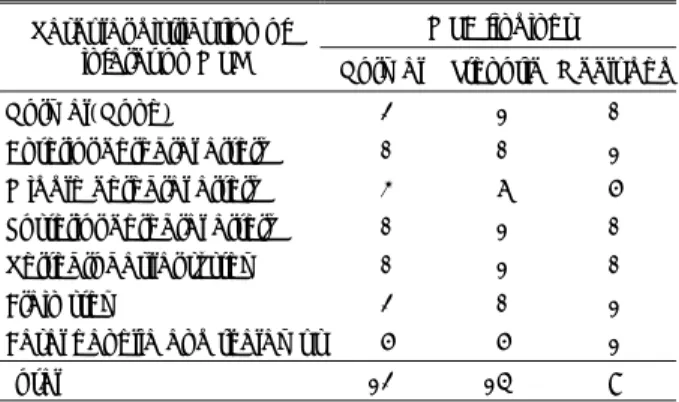
Table 1. MRI distribution of infarction versus MRA findings MRA findings Vascular distribution of
infarct on MRI Normal Stenotic Occluded
Normal(None) 2 1 0
Anterior cerebral artery 0 0 1 Middle cerebral artery 3 6 5 Posterior cerebral artery 0 1 0 Vertebrobasilar system 0 1 0
Brain stem 2 0 1
Basal ganglia and thalamus 5 5 1
Total 12 14 8
Table 2. Types of additional information from MRA
Types No. of patient
Difined a pattern of collateral flow 2 Diagnosed a major vessel occlusion 13 rather than a suspected branch occlusion
Demonstrated patency in a suspected 3 vascular occlusion
Total 18
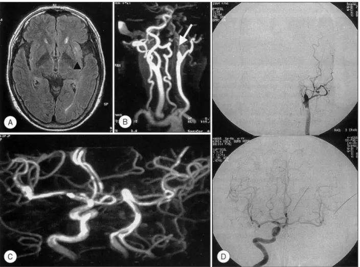
Fig. 1. A:Intermediate-weighted images showing a high signal density in right middle cerebral artery distribution with lenticul- ostriate territory. B:Magnetic resonance angiogram images demonstrating an occlusion of M1 segment of right middle cerebral artery(arrow head). A potential collateral pathway is suggested by the prominent ophthalmic artery(arrow).
J Korean Neurosurg Soc/Volume 29/August, 2000 1065 (occluded)의 3단계로 분류하였고, 협착은 국소적 신호감
소가 지름의 50%이상이거나 신호소실(singal void)과 그 원위부에 신호재생이 있는 경우로 하였다. MRA소견에서 1) 뇌경색부로의 부행혈류(collateral flow)가 관찰되는 경 우;2) MRI소견에서 중요혈관폐색이 의심되었으나 분지혈 관의 폐색으로 진단된 경우;3) MRI소견에서 분지혈관의 폐색이 의심되었으나 중요혈관의 폐색으로 진단되는 경우;
4) MRI소견에서 폐색이 의심된 혈관이 정상으로 나타나는 경우 등은 MRA가 MRI소견에서 얻을 수 없는 추가적인 정 보를 제공하는 경우로 판단하였다.
결 과
대상 환자의 연령분포는 32세에서 81세까지 평균 64세 이었고 남자가 24명, 여자가 10명이었다. 증상발생과 자기 공명촬영까지의 기간은 0에서 19일로 평균 3.4일이었다.
전체 34명의 환자 중 23명에서 MRA소견상 혈관의 협착
이나 폐색이 관찰되어 68%의 진단율을 보였고, 영구적 뇌 경색증(completed stroke)환자에서 일과성 허혈발작 환자 보다 진단율이 높았으나 통계적으로 의미있는 차이는 없었 다(z-test, p<0.05). MRA에서 혈관의 협착이나 폐색이 관 찰된 23명의 환자 중 22명(96%)에서는 혈관이상이 관찰 된 부위가 MRI에서의 뇌경색부위와 일치되는 소견이 관찰 되었다(Table 1).
전체 환자 중 19명(56%)의 환자에서 전술한 내용을 기 준으로 MRI에서 얻을 수 없는 추가적인 정보를 MRA를 시 행함으로서 얻을 수 있었다(Table 2). 2예에서는 뇌경색부 로의 부행혈류 형태가 관찰되었으며(Fig. 1), MRI소견에서 는 분지혈관의 폐색이 의심되었으나 중요혈관의 폐색으로 진단된 경우가 13례로 가장 빈도가 높았다(Fig. 2).
2명의 환자에서는 고식적인 뇌혈관조영술을 시행하여 각 각의 혈관을 정상 협착 폐색으로 분류하여 MRA소견과 비 교하여 일치되는 소견을 얻었으나, MRA로 협착의 정도를 정확히 평가할 수는 없었다(Fig. 3).
Fig. 2. A:Intermediate-weighted image showing a subtle high signal intensity in left basal ganglia(black arrow head). B, C:
Magnetic resonance angiogram(MRA) images demonstrating a left internal carotid artery occlusion(arrow). The right carotid and vertebral arteries are normal. D. Carotid angiogram images, which are well correlate with MRA findings, confirm the left internal carotid artery occlusion.
고 찰
뇌혈관질환에 대한 다양한 치료방법이 개발되고 발달함에 따라 뇌혈관질환의 신속하고 정확한 진단의 중요성이 강조 되고 있다. 뇌자기공명영상은 뇌경색을 진단하는 데 매우 유용하며 특히 질병초기에 중요한 정보를 얻을 수 있다6)20). 그러나 뇌자기공명영상만으로 복잡한 뇌혈관계를 해부학적 으로 정확하게 평가하기 어려우며, 뇌혈관질환의 위치와 정 도가 질병의 예후와 밀접한 관련을 갖고 있다는 데에서 뇌 혈관계의 정확한 해부학적 평가가 매우 중요하다1). 뇌혈관 질환의 적절한 치료방법을 결정하기 위하여 뇌혈관병소의 정도와 위치, 개수 등에 대한 정확한 진단이 중요하며3)16)19), 초기 임상양상이 뇌혈관조영술 소견과 반드시 일치하지 않 는다는 점에서 조기에 뇌혈관계를 적절히 평가할 수 있는 진단방법이 필요하다1).
뇌혈관조영술은 뇌혈관계를 평가하는 진단방법으로 널리
사용되어지고 있으며, 특히 뇌경색의 급성기에 뇌혈관계를 평가하여 적절한 치료방법의 선택과 예후를 예측하는 데에 진단적 가치가 높은 것으로 인정되고 있다19). 그러나 고식 적인 뇌혈관조영술은 시술에 따른 위험성을 내포하는 침습 적인 검사방법이며5)7), 뇌혈관질환 환자의 경우 위험성이 더 높은 것으로 보고되고 있다8). 특히 일과성허혈발작(tr- ansient ischemic attack, TIA) 환자의 경우 약 50%의 환 자에서 결국 뇌경색으로 진행되는 것으로 알려져 있어 적절 한 예방치료를 위하여 뇌혈관계의 평가가 중요하나 신경학 적 결손이 없는 상태에서 위험성을 내포하는 침습적인 뇌혈 관조영술의 시행에 있어서 유용성과 위험성에 대한 고려가 필요할 것으로 사료된다4).
자기공명혈관조영술(magnetic resonance angiography, MRA)은 내경동맥분지부의 질환이나 뇌동맥류의 진단에 효 과적인 진단방법으로 알려졌고10)12)13)15)17)
, 두개내혈관의 협 착이나 폐색의 진단에도 임상적 유용성이 보고되고 있다9)11).
허혈성 뇌혈관질환환자에서 혈관의 협착이나 폐색이 MRI
Fig. 3. A:T2-weighted images showing multiple small high signal intensities in right lenticulostriate distribution. B:Magnetic re- sonance angiogram(MRA) demonstrating focal stenosis of proximal right middle cerebral artery(M1 segment)(arrow).
C:Right carotid angiogram images confirm the stenosis(black arrow), which correlates well with MRA fingings.
J Korean Neurosurg Soc/Volume 29/August, 2000 1067 만으로 진단되는 경우도 있으나 혈관협착의 정도와 위치,
부행혈류의 형태, 다른 혈관의 질환 여부를 정확하게 진단 하기 위하여 혈관조영술이 필요하다. 본 연구에서는 MRA 소견에서 혈관의 협착이나 폐색이 관찰된 부위와 MRI에서 의 뇌경색부위가 일치되는 비율이 높게 나타났으며, MRI에 서 관찰되지 않는 혈관질환의 여부와 부행혈류의 형태가 전 체의 56%에 해당하는 환자에서 MRA로 진단되어 뇌경색 에 MRA의 진단적 가치가 높았다. MRA소견과 고식적인 혈 관조영술과의 비교에 있어서 일치되는 정도가 높은 것으로 보고 되고 있어9)11), 발병초기에 치료방법을 결정할 때나 추 적검사로 질병의 경과를 추적할 수 있는 비침습적인 진단방 법으로 침습적인 혈관조영술을 대체할 수 있는 검사로 유용 할 것으로 사료된다. 그러나 복잡한 혈류현상으로 인하여 국소적인 신호의 감소가 혈관의 협착없이 나타날 수 있고14), 지름이 작은 혈관의 경우 정확성이 떨어진다고 알려져 있으 며18), 3차원영상으로의 재합성과정에서도 MRA의 정확성에 대한 문제점이 보고되고 있어2), 내경동맥수술이나 혈관 측 로조성술과 같은 혈관수술에 고식적인 혈관조영술을 대체하 기에는 아직 한계가 있다고 사료된다.
결 론
뇌자기공명혈관조영술에서 나타난 혈관의 협착이나 폐색 은 뇌경색의 분포와 높은 상관관계를 보였으며, 대부분의 환자에서 뇌자기공명영상에서 얻을 수 없는 정보를 얻을 수 있었다. 허혈성 뇌혈관질환환자에서 발병초기의 신속한 진 단과 조기치료가 사망률과 유병률을 낮추는 데 중요하며, 이런한 관점에서 초기에 뇌자기공명영상과 뇌자기공명혈관 조영술을 동시에 시행하는 것이 뇌경색환자의 진단에 매우 유용할 것으로 사료된다. 여러 가지 제약으로 MRA가 고식 적인 뇌혈관조영술을 전적으로 대체하기 어려우나 위험성이 있는 침습적인 검사나 시술에 대한 일차적인 예검법으로 유 용할 것으로 사료된다.
•논문접수일:2000년 8월 8일
•심사완료일:2000년 11월 7일
•책임저자:조 태 형
425-050 경기도 안산시 고잔동 516 고려대학교 의과대학 안산병원 신경외과학교실 전화:031) 412-5050, 전송:031) 412-5054 E-mail:[email protected]
References
1) Acheson J, Boyd WN, Hugh AE, Hutchinson EC:Cerebral
angiography in ischemic cerebrovascular disease. Arch Neurol 20:527-532, 1969
2) Anderson CM, Saloner D, Tsuruda JS, Shapeero LG, Lee RE:
Artifacts in maximum-intensity-projection display of MR angi- ograms. AJR Am J Roentgenol 154:623-629, 1990 3) Caplan LR, Rosenbaum AE:Role of cerebral angiography in
vertebrobasilar occlusive disease. J Neurol Neurosurg Psychi- atry 38:601-612, 1975
4) Davis DO, Pressman BD:Angiography of cerebrovascular disease. in Wilkins RH, ed:Clinical neurosurgery. Baltimore: Williams & Wilkins, 1975, pp163-184
5) Dion JE, Gates PC, Fox AJ, Barnett HJ, Blom RJ:Clinical events following neuroangiography:a prospective study. Str- oke 18:997-1004, 1987
6) Elster AD, Moody DM:Early cerebral infarction:gadope- ntetate dimeglumine enhancement. Radiology 177:627-632, 1990
7) Grzyska U, Freitag J, Zeumer H:Selective cerebral intraart- erial DSA. Complication rate and control of risk factors. Neu- roradiology 32:296-299, 1990
8) Hankey GJ, Wariow CP, Sellar RJ:Cerebral angiographic risk in mild cerebrovascular disease. Stroke 21:209-222, 1990 9) Heiserman JE, Drayer BP, Keller PJ, Fram EK:Intracranial
vascular stenosis and occlusion:evaluation with three-dime- nsional time-of-flight MR angiography. Radiology 185:667- 673, 1992
10) Heiserman JE, Drayer BP, Schmalbrock P:Three-dimensional time of flight MR angiography in the evaluation of cerebral aneurysms. J Comput Assist Tomogr 14:874-881, 1990 11) Johnson BA, Heiserman JE, Drayer BP, Keller PJ:Intracra-
nial MR angiography:its role in the integrated approach to brain infarction. AJNR 15:901-908, 1994
12) Litt AW, Eidelman EM, Pinto RS, Riles TS, McLachlan SJ, Schwartzenberg S, et al:Diagnosis of carotid artery stenosis: comparison of 2DFT time-of-flight MR angiography with con- trast angiography in 50 patients. AJNR 12:149-154, 1991 13) Masaryk AM, Ross JS, DiCello MC, Modic MT, Paranandi L,
Masaryk TJ:3DFT MR angiography of the carotid bifurcat- ion:potential and limitations as a screening examination.
Radiology 179:797-804, 1991
14) Masaryk TJ, Modic MT, Ross JS, Ruggieri PM, Laub GA, Lenz GW, et al:Intracranial circulation:preliminary clinical results with three-dimensional(volume) MR angiography. Ra- diology 171:793-799, 1989
15) Masaryk TJ, Modic MT, Ruggieri PM, Ross JS, Laub G, Lenz GW, et al:Three-dimensional(volume) gradient-echo imaging of the carotic bifurcation:preliminary clinical experience. Ra- diology 171:801-806, 1989
16) Pessin MS, Hinton RC, Davis KR, Duncan GW, Roberson GH, Ackerman RH, et al:Mechanisms of acute carotid stroke. Ann Neurol 6:245-252, 1979
17) Ross JS, Masaryk TJ, Modic MT, Ruggieri PM, Haacke EM,
Selman WR:Intracranial aneurysms:evaluation with MR angiography. AJNR 11:449-456, 1990
18) Warach S, Li W, Ronthal M, Edelman RR:Acute cerebral ischemia:evaluation with dynamic contrast-enhanced MR imaging and MR angiography. Radiology 182:41-47, 1992 19) Wolpert SM, Caplan LR:Current role of cerebral angiogra-
phy in the diagnosis of cerebrovascular diseases. Am J Roent- genol 159:191-197, 1992
20) Yuh WT, Crain MR, Loes DJ, Greene GM, Ryals TJ, Sato Y:MR imaging of cerebral ischemia:Findings in the first 24 hours. AJNR 12:621-629, 1991