백나무로부터 분리된 quercetin-3- O-α- L-rhamnopyranoside 의
알도스 환원효소 및 솔비톨 억제효과
김선하*·김진규**·이연실*·배영수***·임순성*,**,****†
*한림대학교 식의약품의 효능평가 및 기능성소재개발센터, **한림대학교 천연의약연구소,
***강원대학교 산림바이오소재공학과, ****한림대학교 식품영양학과
Inhibitory Effect of quercetin-3- O-α- L-rhamnopyranoside
from Chamaecyparis obtuse on Aldose Reductase and Sorbitol Accumulation
Seon Ha Kim
*, Jin Kyu Kim
**, Yeon Sil Lee
*, Young Soo Bae
***and Soon Sung Lim
*,**,****†*Center for Efficacy Assessment and Development of Functional Foods and Drugs, Hallym University, Chuncheon 200-702, Korea.
**Institute of Natural Medicine, Hallym University, Chuncheon 200-702, Korea.
***Departement of Forest Biomaterials Engineering, Kangwon National University, Chuncheon 200-701, Korea.
****Department of Food Science and Nutrition, Hallym University, Chunchenon 200-702, Korea.
ABSTRACT : Taxifolin-3-O-β-D-xylopyranoside and quercetin-3-O-α-L-rhamnopyranoside were isolated from an EtOAc- soluble extract of the leaves of Chamaecyparis obtuse. Quercetin-3-O-α-L-rhamnopyranoside was found to possess a potent inhibitory activity of human recombinant aldose reductase in vitro, its IC50 value being 11.5µM. Kinetic analysis showed that quercetin-3-O-α-L-rhamnopyranoside exhibited uncompetitive inhibition against DL-glyceraldehyde. Also, quercetin- 3-O-α-L-rhamnopyranoside suppresses sorbitol accumulation in rat lens under high glucose conditions, demonstrating the potential to prevent sorbitol accumulation in vivo. These results suggest that this compound may be a promising agent in the prevention or treatment of diabetic complications.
Key Words :Chamaecyparis obtusa, Taxifolin-3-O-β-D-xylopyranoside, Quercetin-3-O-α-L-rhamnopyranoside, Diabetic Compli- cations, Human Recombinant Aldose Reductase, Sorbitol Accumulation
INTRODUCTION
Aldose reductase (AR, EC 1.1.1.21) catalyzesthe reduction of glucose to the corresponding sugar alcohol, sorbitol, which is subsequently metabolized to fructose by sorbitol dehydrogenase. This conversion of glucose into fructose constitutes the polyol pathway of glucose metabolism (Suzen and Buyukbingol, 2003). Under normal physiological conditions, this pathway plays a minor role in glucose metabolism for most tissues. In hyperglycemia associated with diabetes, however, cells undergoing insulin-independent glucose uptake produce significant quantities of sorbitol because sorbitol cannot penetrate cellular membranes and is metabolized by sorbitol dehydrogenase (Jedziniak et al.,
1981). The resulting hyperosmotic stress to cells is believed to be the primary cause for the development of diabetic complications such as retinopathy, cataracts, neuropathy, and nephropathy (Williamson et al., 1992). These observations suggest that the inhibition of AR may be an innovative, potentially direct pharmacological approach toward the treatment of certain diabetic complications (Kador et al., 1985; Jeon et al., 2010).
Plants are a rich source of bioactive chemicals. Because many of the biocompounds are largely free from adverse effects and have excellent pharmacological actions, they could be used in the development of new classes of safer antidiabetic agents with fewer complications. In addition, some flavonoids and polyphenols and their sugar derivatives
†Corresponding author: (Phone) +82-33-248-2133 (E-mail) [email protected]
Received 2010 August 3 / 1st Revised 2010 August 27 / 2nd Revised 2010 October 6 / Accepted 2010 October 8
are reported to be effective (Park et al., 2009). Therefore, ongoing research is focusing on the use of plants as potential agents or as lead compounds in commercial AR inhibitors.
Chamaecyparis obtusa Sieb. et Zucc., a genus in the family Cupressaceae, is native to northeast Asia. Previous phytochemical investigations of this plant isolated lignin, sesquiterpene derivatives, beyerane derivatives, and flavonoids from the leaves (Gao et al., 2008; Krauze-Baranowska et al., 2005). C. obtusareportedly exhibits neurite outgrowth- promoting activity in PC12 cells, acaricidal activity against Dermatophagides spp., and antiviral against HSV-1 replication (Jang et al., 2005; Kuo et al., 2006; Kuroyanagi et al., 2008). Reportedly, the essential oils have antimicrobial and hair-growth promoting effects (Lee et al., 2010; Yang et al., 2007). However, relatively little research has been done on the anti-diabetic effect of the compounds isolated from C.
obtusa. In the present study, repeated chromatography of the ethyl acetate-soluble fraction of the leaves of C. obtusa led to the isolation of biologically active compound. All isolates obtained in the study were tested for their potential to inhibit human recombinant aldose reductase (hrAR) and sorbitol accumulation in rat lenses.
MATERIALS AND METHODS
1. Chemicals
dl-glyceraldehyde, the reduced form of nicotinamide adenine dinucleotide phosphate (NADPH), quercetin, TL-100 medium, and antibiotics were purchased from Sigma (St.
Louis, MO, U.S.A.). Fetal bovine serum was from GenDEPOT (USA). hrAR was purchased from Wako Pure Chemical Industries (Osaka, Japan). Sephadex LH 20 (GE Healthcare Bio-Science AB, Sweden) was used as the column packing material.
2. Plant Materials
The leaves of C. obtusa were presented by Department of Forest Biomaterials Engineering, Kangwon National University. A voucher specimen (No. RIC-021) was deposited and maintained at the Herbarium of Regional Innovation Center, Hallym University, Chuncheon, Korea.
3. Extraction, fractionation, and isolation
The leaves of the dried material (2㎏) were extracted
with 70% acetone (10 L × 4) by maceration. The extracts were combined and concentrated in vacuo at 40℃ to give an acetone extract (322 g, 16.1%). The extract was suspended in water (2 L), and then successively extracted with n-hexane (2 L × 3), dichloromethane (2 L × 3), and ethylacetate (2 L × 3) to give n-hexane- (97.43 g), dichloromethane- (2.09 g), ethyl acetate- (66.83 g), and water-soluble extract (156.02 g), respectively. The ethyl acetate-soluble extract showed the greatest inhibitory activity on AR with an IC50 value of 8.52㎍/㎖. The ethyl acetate-soluble extract was chromato- graphed over Sephadex LH-20 column chromatography using a MeOH-H2O (4 : 1, v/v) to afford 3 pooled fractions (E1- E3). Fraction E2 was further fractionated using Sephadex column chromatography (50% MeOH) to give compound 1 (1.12 g). Fraction E3 was subjected to Sephadex column chromatography with MeOH:H2O (2 : 1, v/v) as the eluent to yield 3 subfractions (E3 : Fr. 1 to 3). Fraction E3 : Fr.
3was chromatographed on Sephadex column with MeOH : H2O (1 : 2, v/v) to give compound 2 (195㎎). The 1H- and
13C-NMR spectra were recorded on a Bruker DPX 400 spectrometer (Karlsrube, Germany) at 400 and 100 MHz, respectively. Chemical shifts are given in ppm using tetra- methylsilane as an internal standard.
1) Taxifolin-3-O-β-D-xylopyranoside (1)
Amorphous yellow powder; m.p. 190-195°C; + 8.5o (MeOH; c 0.3); IR (KBr) ㎝−1: 3430, 2919, 1623, 1377, 1042; UV (MeOH) λmax (ε): 269; Positive FAB MS:
[M+H]+ at m/z 437.
1H-NMR NMR (400 MHz, δ, MeOH-d4): 3.06 (2H, dd, J= 8.6 and 12.0 Hz, H-2" and H-5"β), 3.23 (2H, m, H-2"
and 3"), 3.49 (1H, m, H-4"), 3.87 (1H, d, J= 5.5 Hz, H- 1"), 3.94 (1H, dd, J= 4.6 and 12.0 Hz, H-5"α), 4.74 (1H, d, J= 9.9 Hz, H-3), 5.19 (1H, d, J= 9.9 Hz, H-2), 5.90 (1H, d, J= 2.0 Hz, H-6), 5.91 (1H, d, J= 2.0 Hz, H-8), 6.80 (2H, m, H-5' and H-6'), 6.95 (1H, d, J= 2.0 Hz, H- 2'). 13C-NMR (100 MHz, δ, MeOH-d4): 65.9 (C-5"), 70.8 (C-3"), 73.4 (C-2"), 75.8 (C-3"), 77.5 (C-3), 83.6 (C-2), 96.4 (C-6), 97.4 (C-8), 102.5 (C-1"), 103.1 (C-10), 115.7 (C-2'), 116.3 (C-5'), 120.8 (C-6'), 129.0 (C-l'), 146.5 (C-3'), 147.4 (C-4'), 164.1 (C-9), 165.5 (C-5), 169.0 (C-7), 195.6 (C-4).
2) Quercetin-3-O-α-L-rhamnopyranoside (2)
Amorphous yellow powder; m.p. 271-272℃; −178o
α [ ]D20
α [ ]D20
(MeOH; c 0.1); IR (KBr) ㎝−1: 3280, 1605, 1662, 1508, 1463, 1063; UV (MeOH) λmax (ε): 264, 314, 350; Positive FAB MS: [M+H]+ at m/z 449.
1H-NMR (400 MHz, δ, MeOH-d4): 0.94 (3H, d, J= 6.0, H-6"), 3.35 (1H, m, H-4"), 3.43 (1H, m, H-5"), 3.75 (1H, dd, J= 3.3 and 9.3, H-3"), 4.22 (1H, br d, J= 3.0, H-2"), 5.35 (1H, d, J= 1.0, H-1"), 6.19 (1H, d, J= 1.9, H-6), 6.36 (1H, d, J= 1.9, H-8), 6.85 (1H, d, J= 8.2, H-5'), 7.30 (1H, dd, J= 8.2 and 1.9, H-6'), 7.34 (1H, d, J= 1.9, H-2'). 13C- NMR (100 MHz, δ, MeOH-d4): 17.7 (C-6"), 71.9 (C-5"), 72.1 (C-3"), 72.2 (C-2"), 73.3 (C-4"), 94.8 (C-8), 99.8 (C- 6), 103.7 (C-1"), 105.9 (C-10), 116.4 (C-5'), 116.5 (C-2'), 122.9 (C-6'), 123.0 (C-1'), 136.3 (C-3), 146.4 (C-3'), 149.8 (C-4'), 158.5 (C-9), 159.3 (C-2), 163.2 (C-5), 165.9 (C-7), 179.7 (C-4).
4. Assay for hrAR inhibitory activity
hrAR activities were assayed spectrophotometrically by measuring the decrease in absorption of NADPH at 340㎚
over a 5 min period with DL-glyceraldehyde as a substrate.
Each 1.0㎕ cuvette contained equal units of enzyme, 0.10 M sodium phosphate buffer (pH 6.2), 0.3 mM NADPH, 10 mM substrate, and inhibitor. The concentration of inhibitor giving 50% inhibition of enzyme activity (IC50) was calculated from the least-squares regression line of the logarithmic concentrations plotted against the residual activity (Lee et al., 2008a and b).
5. Determination of inhibition-type of rhAR by active compound
Reaction mixtures consisted of 0.1 M potassium phosphate (pH 7.0). 0.16 mM NADPH and 2 mM of rhAR with varied concentrations of substrate DL-glyceraldehyde in a total volume of 200㎕. Concentrations were ranged from 0.02 to 0.2 mM for DL-glyceraldehyde. rhAR activity was assayed spectrophotometrically by measuring the decrease in absorption of NADPH at 340㎚ after substrate addition using BioTek Power Wave XS spectrophotometer (Lee et al., 2008a and b).
6. Lens culture and intracellular sorbitol measurement Lenses isolated form 10-week-old male Wistar rats were cultured for 6 days in TC-199 medium that contained 15%
fetal bovine serum 100 units/㎖ penicillin and 0.1㎎/㎖
streptomycin under sterile conditions in an atmosphere of 5% CO2 and 95% air at 37℃. Samples were dissolved in
dimethylsulfoxide. The lenses were divided into 3 groups and cultured in medium containing 25.5 mM glucose and sample.
Each lens was placed in a well containing 2.0㎖ of medium. Sorbitol was determined by HPLC after its derivatization by reaction with benzoic acid to a fluorescent compound (Kwang-Hyok et al., 2005; Lee et al., 2010).
7. Benzoylation
Benzoylation of processed samples and standard sugar/
sugar alcohols was performed according to the method of Shinohara with slight modification (Shinohara et al., 1998).
Briefly, to 70㎕ of the sample, 20㎕ of KH2PO4 (1㏖/L) solution, 3㎕ benzoyl chloride and 15㎕ of 8㏖/L NaOH were added in a 2.0㎖ polypropylene centrifugation tube.
The mixture was immediately vortex mixed for 5 min at 2500 vibrations/min in a vortex-mixer (REMI, India). Then the mixture was neutralized with 10㎕ of 1.4㏖/L H3PO4
and after the addition of 100㎕ ethyl acetate vortex mixed for an additional minute at the same speed. Finally, 75㎕
of ethyl acetate phase was taken in a 1.5㎖ polypropylene centrifugation tube and evaporated for 20 min in N2 gas.
The residue thus obtained was dissolved in 25㎕ of acetonitrile-water (70 : 30, v/v) mixture. 20㎕ of this sample was analyzed by HPLC.
8. Chromatographic analysis
The sample were analyzed using a Dionex HPLC System equipped with an autosampler (ASI 100), a column oven (STH 585), a UV detector (UVD 170S), quaternary pumps (P580), and Chromelone software version 4.1. The separation was achieved on a 250 4.6㎜ i.d., 4㎛ J’Sphoro ODS-H80 (YMC, Japan). The elution solvents were water (A) and acetonitrile (B) with the following gradient: 70% to 80% B from 0 to 5 min, 80% to 83% Bfrom 5 to 30 min, and 83% to 100% B from 30 to 33 min, isocratic at 100%
from 33 to 38 min, 100% to 70% B from 38 to 40 min, and isocratic at 70% B from 40 to 45 min to equilibrate the column for the next injection. UV chromatograms were monitored at 228㎚.
9. Statistical analysis
Values are expressed as means±S.D of the results of three independent experiments. Between group differences were analyzed by Student’s t-test. The level of significance was set at less than 5%.
RESULTS AND DISCUSSION
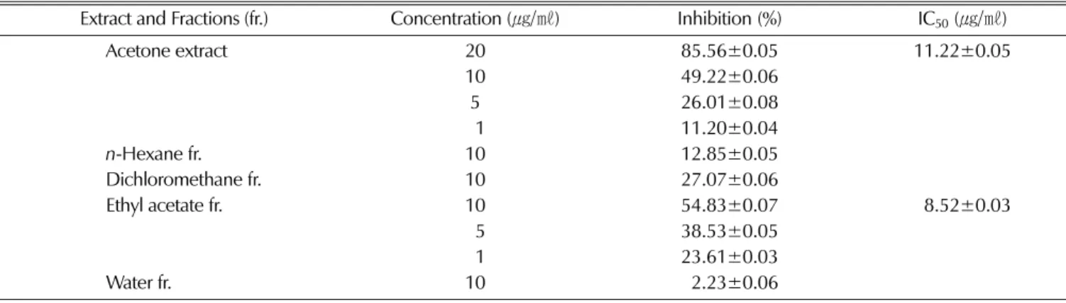
In our attempt to identify powerful, nontoxic and natural AR inhibitors (ARIs), the leaves of C. obtusa were evaluated for potential inhibitory effects on rhAR. In this study, the acetone extract of C. obtusaleaves was found to exhibit AR inhibitory activity with an IC50 value of 10.25㎍/㎖. The acetone extract was further partitioned successively with methylene chloride, ethylacetate, and water. As shown in Table 1, the ethylacetate fraction exhibited strong rhAR inhibitory activity, with an IC50 value of 8.52㎍/㎖. This finding suggested that this fraction was likely to contain many ARIs.
Therefore, this study focused specifically on the isolation of ARI compounds within this fraction. The ethyl acetate- soluble portion was subjected to repeated Sephadex LH-20 chromatography and resulted in the isolation of two compounds, quercetin-3-O-α-L-rhamnopyranoside and taxifolin- 3-O-β-D-xylopyranoside. These were identified on the basis of the 1D (1H- and 13C-NMR) and 2D NMR (HMQC and HMBC) spectral data and on comparison with published spectral data and/or by direct comparison with actual samples (Ishiguro et al., 1991a and b; Baderschneider and Winterhalter, 2001). Their chemical structures are shown in Fig. 1.
Quercetin-3-O-α-L-rhamnopyranoside exhibited strong hrAR inhibitory activity with an IC50 value of 11.5µM (Table 2).
However, taxifolin-3-O-β-D-xylopyranoside showed no inhibi- tory effect. Previous study demonstrated the possible relation- ships of structure to the inhibitory activities of flavonoids. It was reported that the glycoside of a flavonol has a higher or lower inhibitory activity than its parent aglycone, depending upon the nature of the glycosylation sugar at 3-
Table 1. Inhibitory effects of the extract and its fractions from the leaves of C. obtusa on hrAR.
Extract and Fractions (fr.) Concentration (㎍/㎖) Inhibition(%) IC50(㎍/㎖)
Acetone extract 20 85.56±0.05 11.22±0.05
10 49.22±0.06
5 26.01±0.08
91 11.20±0.04
n-Hexane fr. 10 12.85±0.05
Dichloromethane fr. 10 27.07±0.06
Ethyl acetate fr. 10 54.83±0.07 98.52±0.03
95 38.53±0.05
91 23.61±0.03
Water fr. 10 92.23±0.06
Fig. 1. Chemical structures of compounds isolated from the leaves of C. obtusa.
Table 2. Inhibitory effects of compounds isolated from C. obtusa
on hrAR.
Compounds Inhibition(IC50, µ㏖) Taxifolin-3-O-β-D-xylopyranoside (1) 187.7±0.08 Quercetin-3-O-α-L-rhamnopyranoside (2) 811.5±0.05
Quercetin 815.9±0.06
position (Matsuda et al., 2002). For example, quercetin-3-O-
α-L-rhamnopyranoside has a higher activity than its aglycone quercetin. On the other hand, the glucose and galactose of quercetin, hyperin and isoquercitrin are less active than the agylcone. Also, taxifolin is a flavanonol with the same hydroxylation pattern of quercetin but its C ring lacks the 2,3-double bond. Toxifolin, the aglycone of astilbin, has been reported to be an inhibitor of AR, which showed less activity than its rhamnoside (Haraguchi et al., 1997). From this finding, the substitution of a 3-xylosyl moiety to toxifolin was found to decrease inhibitory activity against AR. Therefore, inhibitory effect of C. obtusa on AR may be due to quercetin-3-O-α-L-rhamnopyranoside, one of major constituents of this plant.
To determine the inhibition type for quercetin-3-O-α-L- rhamnopyranoside, we conducted a kinetic study using DL- glyceraldehyde as the substrate at a concentration of 0.2-2 mM. A kinetic analysis of AR inhibition by quercetin-3-O-
α-L-rhamnopyranoside using Lineweaver-Burk plots of 1/
velocity vs1/concentration of the substrate is shown in Fig. 2.
When the concentration of the substrate dl-glyceraldehyde was changed, the slopes obtained for the uninhibited enzyme and two different concentrations of quercetin-3-O-α- L-rhamnopyranoside were parallel. These results show that rhAR inhibition by quercetin-3-O-α-L-rhamnopyranoside was uncompetitive, meaning that the inhibitor was unable to bind to either the substrate binding region or the NADPH binding
region of rhAR.
We also investigated the inhibitory activities of quercetin- 3-O-α-L-rhamnopyranoside on sorbitol accumulation in rat lenses. Lenses of experimentally diabetic animals contain excessive amounts of sorbitol and fructose. Sorbitol is syn- thesized by the action of AR on glucose and the reducing equivalents are derived from NADPH. Fructose is derived by the action of sorbitol dehydrogenase and NAD is the electron acceptor. Excessive accumulation of sorbitol and fructose in the lens fibers and epithelium is considered osmotically traumatic and leads to tissue swelling and opacity (Sakushima et al., 2002; Srivastava et al., 2005).
Because AR catalyzes the first reaction in the polyol pathway, it is considered the key initiator of cataractogenesis in diabetes and galactosemia. Thus, we investigated the effect of quercetin-3-O-α-L-rhamnopyranoside on sorbitol in rat lenses. The effects of quercetin-3-O-α-L-rhamnopyranoside and a positive control, quercetin, against AR on sorbitol accumulation in rat lenses are shown in Table 3.
Sorbitol accumulation was 8-fold greater when cells were incubated in a high-glucose medium than in a glucose-free medium. Quercetin-3-O-α-L-rhamnopyranoside inhibited sorbitol accumulation by 58.3% at 50 µM. These results suggested that quercetin-3-O-α-L-rhamnopyranoside could prevent the accumulation of sorbitol in rat lenses. On the basis of its inhibition of AR, we conclude that quercetin-3-O-α-L-rham- nopyranoside has therapeutic potential for preventing and treating diabetic complications, although further clinical research is needed.
ACKNOWLEDGEMENTS
This work was supported by Priority Research Centers Program through the National Research Foundation of Korea
Fig. 2. Inhibitory effect of quercetin-3-O-α-L-rhamnopyranoside on hrAR.
Lineweaver-Burk plots in the absence (○) and in the presence (△, 11 µM; □, 22 µM) of quercetin-3-O-α-L- rhamnopyranoside.
Table 3. Inhibitory effect of quercetin-3-O-α-L-rhamnopyranoside on sorbitol accumulation in rat lens.
Compounds Sorbitol content†
(n/㎖)
Blank (Glucose free) 10.1±0.03
Control 84.1±0.05
Quercetin-3-O-α-L-rhamnopyranoside (2) 35.2±0.06*
Quercetin 49.2±0.04*
†Sorbitol was determined by HPLC after its derivatization by reaction with benzoic acid to a fluorescent compound.
*Significantly different from control group (p< 0.01)
(NRF) funded by the Ministry of Education, Science and Technology (2009-0094074).
LITERATURE CITED
Baderschneider B and Winterhalter P. (2001). Isolation and characterization of novel benzoates, cinnamates, flavonoids, and lignin from riesling wine and screening for antioxidant activity.
Journal of Agricultural Food and Chemistry. 49:2788-2798.
Gao HY, Wu LJ, Muto N, Fuchino H, Nakane T, Shirota O, Sano T and Kuroyanagi M. (2008). Beyerane derivatives and a sesquiterpene dimer from Japanese cypress (Chamaecyparis obtusa). Chemical and Pharmaceutical Bulletin (Tokyo).
56:1030-1034.
Haraguchi H, Ohmi I, Fukuda A, Tamura Y, Mizutani K, Tanaka O and Chou WH. (1997). Inhibition of aldose reductase and sorbitol accumulation by astilbin and taxifolin dihydroflavonols in Engelhardtia chrysolepis. Bioscience Biotechnology and Biochemistry. 61:651-654.
Ishiguro K, Nagata S, Fukumoto H, Yamaki M, Takagi S and Isoi K. (1991a). A dipeptide derivative from Hypericum japonicum. Phytochemistry. 30:3639-3641.
Ishiguro K, Nagata S, Fukumoto H, Yamaki M, Takagi S and Isoi K. (1991b). A flavanonol rhamnoside from Hypericum japonicum. Phytochemistry. 30:3152-3153.
Jang YS, Lee CH, Kim MK, Kim JH, Lee S and Lee HS.
(2005). Acaricidal activity of active constituent isolated in Chamaecyparis obtusaleaves against Dermatophagoides spp.
Journal of Agricultural and Food Chemistry. 53:1934-1937.
Jeon YH, Moon JW, Kweon HJ, Jeoung YJ, An CS, Jin HL, Hur SJ and Lim BO. (2010). Effects of Lycii fructus and Astragalus membranaceus mixed extracts on immunomodulators and prevention of diabetic cataract and retinopathy in streptozotocin-induced diabetes rat model. Korean Journal of Medicinal Crop Science. 18:15-21.
Jedziniak JA, Chylack LTJr, Cheng HM, Gillis MK, Kalustian AA and Tung WH. (1981). The sorbitol pathway in the human lens: aldose reductase and polyol dehydrogenase.
Investigative Ophthalmology and Visual Science. 20:314-326.
Kador PF, Robison WG and Kinoshita JH. (1985). The pharmacology of aldose reductase inhibitors. Annual Review of Pharmacology and Toxicology. 25:691-714.
Krauze-Baranowska M, Poblocka L and El-Hela AA. (2005).
Biflavones from Chamaecyparis obtusa. Zeitschrift für Naturforschung. 60:679-685.
Kuo YC, Kuo YH, Lin YL and Tsai WJ. (2006). Yatein from Chamaecyparis obtusasuppresses herpes simplex virus type 1 replication in HeLa cells by interruption the immediate-early gene expression. Antiviral Research. 70:112-120.
Kuroyanagi M, Ikeda R, Gao HY, Muto N, Otaki K, Sano T, Kawahara N and Nakane T. (2008). Neurite outgrowth- promoting active constituents of the Japanese cypress (Chamaecyparis obtusa). Chemical and Pharmaceutical Bulletin
(Tokyo). 56:60-63.
Kwang-Hyok S, Ui-Nam P, Sarkar C and Bhadra R. (2005). A sensitive assay of red blood cell sorbitol level by high performance liquid chromatography: potential for diagnostic evaluation of diabetes. Clinica Chimica Acta. 354: 41-47.
Lee EH, Song DG, Lee JY, Pan CH, Um BH and Jung SH.
(2008a). Inhibitory effect of the compounds isolated from Rhus verniciflua on aldose reductase and advanced glycation endproducts. Biological and Pharmaceutical Bulletin. 31:1626- 1630.
Lee GS, Hong EJ, Gwak KS, Park MJ, Choi KC, Choi IG, Jang JW and Jeung EB. (2010). The essential oils of Chamaecyparis obtusapromote hair growth through the induction of vascular endothelial growth factor gene. Fitoterapia.
81:17-24.
Lee YS, Kang YH, Jung JY, Kang IJ, Han SN, Chung JS, Shin HK and Lim SS. (2008b). Inhibitory constituents of aldose reductase in the fruiting body of Phellinus linteus. Biological and Pharmaceutical Bulletin. 31:765-768.
Lee YS, Kim SH, Jung SH, Kim JK, Pan CH and Lim SS.
(2010). Aldose reductase inhibitory compounds from Glycyrrhiza uralensis. Biological and Pharmaceutical Bulletin.
33:917-921.
Matsuda H, Morikawa T, Toguchida I and Yoshikawa M.
(2002). Structural requirements of flavonoids and related compounds for aldose reductase inhibitory activity. Chemical and Pharmaceutical Bulletin. 50:788-795.
Park JH, Baek MR, Lee BH, Yon GH, Ryu SY, Kim YS, Park SU and Hong KS. (2009). α-Glucosidase and a-amylase inhibitory activity of compounds from roots extract of Pueraria thunbergiana. Korean Journal of Medicinal Crop Science.
17:357-362.
Sakushima A, Ohno K, Coskun M, Seki K and Ohkura K.
(2002). Separation and identification of taxifolin 3-O-glucoside isomers from Chamaecyparis obtusa (Cupressaceae). Natural Product Letters. 16:383-387.
Shinohara R, Ohta Y, Yamauchi M and Ishiguro I. (1998).
Improved fluorometric enzymatic sorbitol assay in human blood. Clinica Chimica Acta. 273:171-184.
Suzen S and Buyukbingol E. (2003). Recent studies of aldose reductase enzyme inhibition for diabetic complications. Current Medicinal Chemistry. 10:1329-1352.
Srivastava SK, Ramana KV and Bhatnagar A. (2005). Role of aldose reductase and oxidative damage in diabetes and the consequent potential for therapeutic options. Endocrine Reviews. 26:380-392.
Williamson J, Kilo C and Tilton RG. (1992). Hyperglycemia, Diabetes, and Vascular Disease. In Ruderman et al. (ed.) Mechanism of glucose and diabetes-induced vascular disfunction.
Oxford University Press, New York, USA. p.107-132.
Yang JK, Choi MS, Seo WT, Rinker DL, Han SW and Cheong GW. (2007). Chemical composition and antimicrobial activity of Chamaecyparis obtusa leaf essential oil. Fitoterapia. 78:149- 152.