JKSCI
Automatic Anatomical Classification Model of
Esophagogastroduodenoscopy Images Using Deep Convolutional Neural Networks for Guiding Endoscopic Photodocumentation
1)
Jung-Whan Park*, Yoon Kim*, Woo-Jin Kim**, Seung-Joo Nam***
*Student, Dept. of Computer Science and Engineering, Kangwon National University, Chuncheon, Korea
*Professor, Dept. of Computer Science and Engineering, Kangwon National University, Chuncheon, Korea
**Professor, Dept. of Internal Medicine and Biomedical Informatics, Kangwon National University, Chuncheon, Korea
***Professor, Dept. of Internal Medicine, Kangwon National University School of Medicine, Chuncheon, Korea [Abstract]
Esophagogastroduodenoscopy is a method commonly used for early diagnosis of upper gastrointestinal lesions. However, 10-20 percent of the gastric lesions are reported to be missed, due to human error.
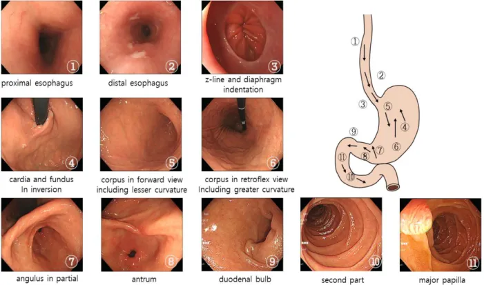
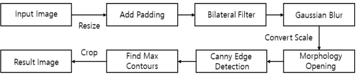
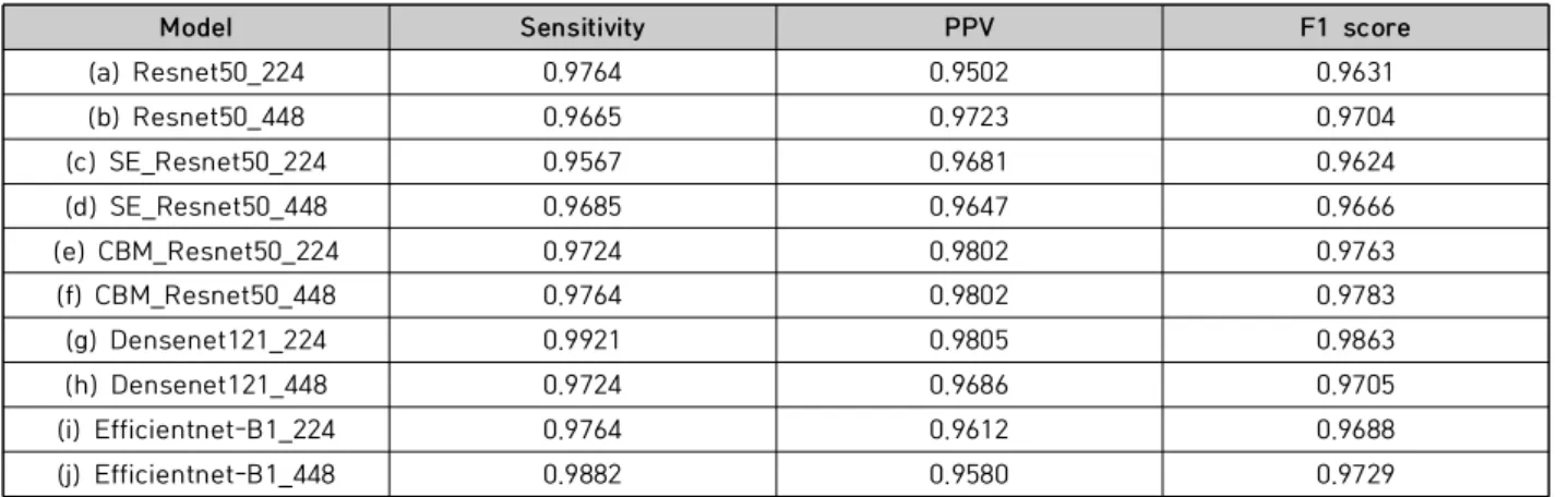
And countries including the US, the UK, and Japan, the World Endoscopy Organization (WEO) suggested guidelines about essential gastrointestinal parts to take pictures of so that all gastric lesions are observed. In this paper, we propose deep learning techniques for classification of anatomical sites, aiming for the system that informs practitioners whether they successfully did the gastroscopy without blind spots. The proposed model uses pre-processing modules and data augmentation techniques suitable for gastroscopy images. Not only does the experiment result with a maximum F1 score of 99.6%, but it also shows a error rate of less than 4% based on the actual data. Given the performance results, we found the model to be explainable with the potential to be utilized in the clinical area.
▸Key words: Deep Learning, Medical Image Analysis, EsophagoGastroDuodenoscopy, Stomach Anatomy Site Classification, Image Processing
[요 약]
위내시경 촬영은 조기에 위 병변을 진단하기 위해서 주로 사용한다. 하지만 위내시경을 했음에도 불구하고 위 내부를 자세히 관찰하지 못해서 10~20% 위 병변을 놓치는 경우가 생기는 것으로 보고 되고 있다. 미국, 영국, 일본 등의 일부 국가와 세계내시경협회(Wold Endoscopy Organization)에서는 위내시경 시에 맹점 없는 관찰을 위해서 반드시 촬영해야 할 부위에 대한 촬영지침을 제안한 바 있 다 . 이에 본 논문에서는 수련의가 내시경을 하는 데 있어 위 내부를 자동으로 맹점 없이 관찰하는데 필요한 딥러닝 기술인 해부학적 분류모델을 제안한다. 제안한 모델은 위내시경 이미지에 적합한 전 처리 모듈과 데이터 증강 기술들을 사용한다. 실험결과를 통해 최대 F1 점수 99.6% 분류 성능을 확 인하였다 . 또한, 실제 데이터를 통한 실험결과에서도 에러율이 4% 미만을 보였다. 이러한 성능을 바 탕으로 설명 가능한 모델임을 보여 임상에서의 사용 가능성을 확인하였다 .
▸주제어: 딥러닝, 의료영상분석, 상부위장관내시경, 해부학적 위치 분류, 영상처리
∙First Author: Jung-Whan Park, Co-author: Yoon Kim, Woo-Jin Kim, Corresponding Author: Seung-Joo Nam *Jung-Whan Park ([email protected]), Dept. of Computer Science and Engineering, Kangwon National University *Yoon Kim ([email protected]), Dept. of Computer Science and Engineering, Kangwon National University **Woo-Jin Kim ([email protected]), Dept. of Internal Medicine and Biomedical Informatics, Kangwon National
University
***Seung-Joo Nam ([email protected]), Dept. of Internal Medicine, Kangwon National University School of Medicine
∙Received: 2021. 01. 18, Revised: 2021. 02. 24, Accepted: 2021. 02. 24.
Copyright ⓒ 2021 The Korea Society of Computer and Information http://www.ksci.re.kr pISSN:1598-849X | eISSN:2383-9945

![Fig. 6. Grad CAM Layer를 가지고 있을 때, 학습한 모델을 변경 없이 히트 맵을 그릴 수 있게 하는 Grad-CAM[22]을 사용하였다](https://thumb-ap.123doks.com/thumbv2/123dokinfo/5310075.382434/8.892.96.786.154.553/layer를-가지고-있을-학습한-모델을-변경-없이-사용하였다.webp)
