59
DOI: 10.4046/trd.2011.71.1.59
ISSN: 1738-3536(Print)/2005-6184(Online) Tuberc Respir Dis 2011;71:59-61
CopyrightⒸ2011. The Korean Academy of Tuberculosis and Respiratory Diseases. All rights reserved.
Image of the Month
Obstructive Fibrinous Tracheal Pseudomenbrane Mimicking Tra- cheal Stents
Ju Sang Kim, M.D., Ji Hyun Yu, M.D., Yu Seung Kim, M.D., Il Kim, M.D., Joong Hyun Ahn, M.D.
Division of Pulmonology, Critical Care Medicine, Department of Internal Medicine, The Catholic University of Korea College of Medicine, Seoul, Korea
Obstructive Fibrinous Tracheal Pseudomenbrane (OFTP) is a rarely known but potentially fatal complication of endotracheal intubation. Sudden respiratory failure shortly after extubation is not infrequent in the ICU. However, these cases are commonly diagnosed as laryngospasm, retention of secretion or laryngeal edema. A 68-year-old woman presented with a 6-day history of progressive dyspnea. She had undergone invasive ventilator care for 24 hours. The patient was discharged from the hospital with improvement after having an extubation. However, after 3 days she revisited the emergency department with progressive dyspnea. The patient was diagnosed with OFTP from the results of chest CT and bronchoscopy. This is the first case studied in detail using CT images, pulmonary function test, and bronchoscopy.
Key Words: Trachea; Airway Obstruction; Fibrin; Stents
Address for correspondence: Joong Hyun Ahn, M.D.
Department of Internal Medicine, Incheon St. Mary's Hospital, The Catholic University of Korea College of Medicine, 665, Bupyeong 6-dong, Bupyeong-gu, Incheon 403-720, Korea
Phone: 82-32-280-5842, Fax: 82-32-280-5190 E-mail: [email protected]
Received: Apr. 27, 2011 Accepted: Jun. 28, 2011
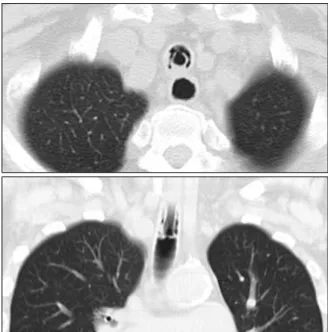
Figure 1. Chest axial and coronal CT images are showing tubular structure with a good patency. It looks like a tra- cheal stents at upper trachea. CT: computed tomo- graphy.
Case Report
A 68-year-old woman with ischemic heart disease presented with a 6-day history of progressive dyspnea.
Her symptoms had been progressively worsening for 6 days before the admission. On the first admission, her blood pressure was 130/70 mm Hg, and the pulse rate was 100/min. She was diagnosed with acute pulmonary edema, and she had undergone invasive ventilator care with a high volume-low pressure cuffed tube (Hi-LoTM; Mallinckrodt Medical, Athlone, Ireland) for 24 hours.
While incubating, the intra-cuff pressure was monitored, and it was stable to maintain below 25 cm H2O. She improved, and the tube was removed immediately. She was discharged from the hospital on the 3rd day after extubation. After 3 days from being discharged, she re-
visited the emergency department with progressive dyspnea. On physical examination, breathing sound was coarse and stridor was ausculated on anterior neck.
A chest CT scan was performed to search for an etiology
JS Kim et al: A comprehensive image of obstructive fibrinous tracheal pseudomembrane
60
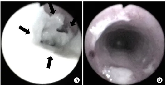
Figure 2. (A) Fiberoptic bronchoscopy showing a thick tubular, rubber-like, whitish pseudomembrane moulding covering 2∼3 cm of the tracheal wall at the level of the endotracheal cuff (the arrows indicate the pseudomembrane partially detached from trachea). (B) Fiberoptic bronchoscopy showing good patency after mechanical ablation with fiberoptic bronchoscopy.
Figure 4. Microscopic examination showing that the pseu- domembrane consists of fibrinous material with acute in- flammatory cell infiltration (H&E stain, ×100).
Figure 3. Flow-volume loops showed a fixed air- way obstruction before mechanical removal of OFPT (the arrows indicate flow-volume loops) (A) and complete recovery of a fixed airway obstruction af- ter mechanical removal of OFPT (B).
of tracheal stenosis. The chest CT showed tubular struc- ture mimicking stents at upper trachea (Figure 1).
Pulmonary function test showed fixed airway ob- struction (Figure 2). A bronchoscopy was performed under moderate sedation and showed a thick tubular, rubber-like, whitish pseudomembrane moulding, cover- ing 2∼3 cm of the tracheal wall at the level of the pre- viously placed endotracheal tube cuff (Figure 2). It was completely ablated without secondary development of tracheal stenosis using a fiberoptic bronchoscope and forceps alone. After the mechanical removal of OFTP using a fiberoptic bronchoscope and forceps alone, fol- lowing pulmonary function test showed no limitation of airway flow (Figure 3). A bronchoscopy was performed
Tuberculosis and Respiratory Diseases Vol. 71. No. 1, Jul. 2011
61 1 month later, but showed no residual lesion. The biop-
sy specimen showed fibrinous material with acute in- flammatory cells (Figure 4).
Discussion
Obstructive fibrinous tracheal pseudomenbrane (OFTP) is a potentially fatal complication of tracheal intubation, consisted of fibrinous material with acute inflammatory cell infiltration and respiratory failure1,2 with unknown causes. OFTP typically appears as sudden upper airway obstruction occurring shortly after extubation. The clin- ical feature may be various from subclinical airway ob- struction to acute respiratory failure. The delay from ex- tubation to initial symptoms varies from several hours to several days1,2. So, it could be misled to the other causes of upper airway obstruction related to intubation such as acute retention of secretion, laryngospasm, glot- tic edema, and post-intubation tracheal stenosis. OFTP could sometimes be unrecognizable.
One possible cause of pathogenesis is a tracheal is- chemic damage related to the cuff induced injury.
According to the previous reports, the duration of in- tubation varies from 3 hours to 16 days. The delay from extubation to initial symptoms is anywhere between 3 hours to 9 days, and the delay from initial symptoms to diagnosis can be anywhere between <1 hour to 4
days1. Fiberoptic bronchoscopy is the key for diagnosis.
The treatment choice is mechanical ablation with fiber- optic or rigid bronchoscopy1-4.
Unexplained respiratory failures with symptoms of upper airway obstruction, shortly after extubation, should lead to considering the diagnosis of OFTP.
Learning points of this case are as following: 1) consider OFTP if unexplained respiratory failures occur shortly after extubation; 2) confirm the diagnosis of OFTP and ablate OFTP with bronchoscopy if OFTP is suspected.
References
1. Deslée G, Brichet A, Lebuffe G, Copin MC, Ramon P, Marquette CH. Obstructive fibrinous tracheal pseudo- membrane. A potentially fatal complication of tracheal intubation. Am J Respir Crit Care Med 2000;162:1169- 71.
2. Lins M, Dobbeleir I, Germonpré P, Waelput W, Pauwels P, Jorens PG. Postextubation obstructive pseu- domembranes: a case series and review of a rare com- plication after endotracheal intubation. Lung 2011;189:
81-6.
3. Harbison J, Collins D, Lynch V, McNicholas WT. Acute stridor due to an upper tracheal membrane following endotracheal intubation. Eur Respir J 1999;14:1238.
4. Kang HH, Kim JW, Kang JY, Kim JS, Kim MS, Kim SS, et al. Obstructive fibrinous tracheal pseudomembrane after tracheal intubation: a case report. J Korean Med Sci 2010;25:1384-6.