© 2010 The Korean Academy of Medical Sciences.
This is an Open Access article distributed under the terms of the Creative Commons Attribution Non-Commercial License (http://creativecommons.org/licenses/by-nc/3.0) which permits unrestricted non-commercial use, distribution, and reproduction in any medium, provided the original work is properly cited.
pISSN 1011-8934 eISSN 1598-6357
Obstructive Fibrinous Tracheal Pseudomembrane After Tracheal Intubation: A Case Report
Obstructive fibrinous tracheal pseudomembrane is a rare, but potentially fatal complication associated with endotracheal intubation. It has been known that the formation of tracheal pseudomembrane is related with intracuff pressure during endotracheal intubation or infectious cause. But in the patient described in this case, pseudomembrane formation in the trachea was associated with subglottic epithelial trauma or caustic injuries to the trachea caused by aspirated gastric contents during intubation rather than tracheal ischemia due to high cuff pressure. We report a patient with obstructive fibrinous tracheal pseudomembrane after endotracheal intubation who presented with dyspnea and stridor and was treated successfully with mechanical removal using rigid bronchoscopy.
Key Words: Airway Obstruction; Intubation; Bronchoscopy Hyeon Hui Kang, Jin Woo Kim,
Ji Young Kang, Ju Sang Kim, Myung Sook Kim, Seung Su Kim, Yong Hyun Kim, Sang Haak Lee, and Hwa Sik Moon
Division of Pulmonary, Critical Care and Sleep Medicine, Department of Internal Medicine, The Catholic University of Korea, College of Medicine, Seoul, Korea
Received: 19 August 2009 Accepted: 21 October 2009 Address for Correspondence:
Sang Haak Lee, M.D.
Division of Pulmonary, Critical Care and Sleep Medicine, Department of Internal Medicine, St. Paul’s Hospital, The Catholic University of Korea, 620-56 Jeonnong-1-dong, Dongdaemoon-gu, Seoul 130-709, Korea
Tel: +82.2-961-4500, Fax: +82.2-968-7250 E-mail: [email protected].
DOI: 10.3346/jkms.2010.25.9.1384 • J Korean Med Sci 2010; 25: 1384-1386
CASE REPORT
Respiratory Diseases
INTRODUCTION
Obstructive fibrinous tracheal pseudomembrane (OFTP) is a rare condition associated with endotracheal intubation in which a pseudomembrane that encircles the tracheal wall is formed.
Little is known about the mechanisms involved in the develop- ment of this fibrinous pseudomembrane, but it requires early detection and urgent management, as it causes a life-threaten- ing tracheal obstruction. Here, we present a case of OFTP that developed 3 days after extubation in a patient who was intubat- ed for 3 days.
CASE REPORT
A previously healthy, nonsmoking 66-yr-old woman (height 160 cm, weight 58 kg) was admitted to the hospital because of altered mental status on November 8, 2007. According to her family, she was vomiting and becoming increasingly lethargic when found, and empty sleeping pill bottles were discovered beside her. On arrival to the hospital, the patient was somnolent and agitated, and unable to follow commands. Her blood pres- sure was 110/70 mmHg, pulse rate 80/min, body temperature 36°C, and respiration rate 24/min. Physical examination reveal- ed no evidence of trauma. The lungs were clear on auscultation.
The results of urinalysis and routine hematologic and blood
chemistry tests were normal. Brain computed tomography (CT) was normal. The arterial pH was 7.23, PaO2 35.3 mmHg, PaCO2
62.7 mmHg, and SaO2 55.8% while the patient was breathing room air.
After several unsuccessful and traumatic attempts, her trachea was intubated with a 7.5-mm cuffed tracheal tube (Hi-LoTM, Mallinckrodt Medical, Athlone, Ireland) to treat acute respira- tory distress. After mechanical ventilation, the patient’s condi- tion improved rapidly. Suctioning via the endotracheal tube re- vealed scanty amount of whitish sputum that showed no evi- dence of microorganisms. Seventy-two hours after intubation, the trachea was extubated. While intubated, the intra-cuff pres- sure was monitored and maintained below 15 cmH2O. Three days after extubation, she complained of dyspnea and inspira- tory wheezing. On examination, she was afebrile and tachypne- ic. Despite treatment with a short-acting bronchodilator and steroids, the symptoms became progressively worse. There were no signs suggestive of infection on a chest radiography, blood cultures, urinalysis and sputum cultures. But chest CT revealed an irregular luminal narrowing in the proximal trachea just be- low the vocal cords (Fig. 1).
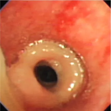
On flexible bronchoscopy under conscious sedation, a white, rubbery membrane encircling the trachea was seen just below the vocal cords, which resulted in narrowing of the tracheal lu- men by approximately 80% (Fig. 2). This pseudomembrane was
Kang HH, et al. • Obstructive Fibrinous Tracheal Pseudomembrane
http://jkms.org 1385
DOI: 10.3346/jkms.2010.25.9.1384
tubular in shape, 4 cm long, and adherent to the tracheal wall.
We tried to remove the pseudomembrane using bronchoscopic forceps, but the lesion was cut to pieces. Subsequently, rigid bronchoscopy was performed under general anesthesia and the pseudomembrane was removed entirely with mechanical forceps (Fig. 3). After the procedure, no findings suggestive of tracheomalacia or granulation tissue formation in the tracheal wall were detected. The patient’s symptoms improved immedi- ately after removing the pseudomembrane. The biopsy speci- men showed fibrinoid material with eosinophils and acute in- flammatory cells (Fig. 4). Tissue cultures grew no bacteria or fungus.
Bronchoscopy performed 3 months later showed no residual
lesion. The patient has remained asymptomatic for 17 months.
DISCUSSION
Tracheal complications associated with endotracheal intuba- tion include tracheal stenosis, ulcers, granuloma, and tracheo- malacia (1). Of these, tracheal stenosis is one of the most dire complications and requires appropriate evaluation and man- agement, such as endotracheal stenting or tracheal surgery.
Tracheal pseudomembranes are rare, but can occur follow- ing endotracheal intubation. Sigrist et al. (2) reported a rare, fa- tal case involving the development of a tubelike formation in the upper trachea after intubation. Harbison et al. (3) also described Fig. 2. Bronchoscopic image showing a thick, annular, rubberlike pseudomembrane encircling the tracheal wall.
Fig. 1. Chest computed tomography image at the level of the trachea just below the vocal cords showing luminal narrowing.
Fig. 3. Gross view of the pseudomembrane removed at rigid bronchoscopy. Fig. 4. Microscopic examination showing that the pseudomembrane consists of fib
rinous material with polymorphonulclear cell infiltration (H&E stain ×100).
Kang HH, et al. • Obstructive Fibrinous Tracheal Pseudomembrane
1386 http://jkms.org DOI: 10.3346/jkms.2010.25.9.1384
a patient who developed a tracheal fibrinoid membrane 3 days after extubation. Deslée et al. (4) reported a series of ten patients with tubular, rubberlike pseudomembranes molding the trachea after a short duration of intubation. In these patients, the mean duration of endotracheal intubation was 6.2±1.8 days, but in four cases it was less than 24 hr. Seven patients developed symp- toms of acute airway obstruction and these symptoms occurred shortly after extubation (59±27 hr). As in the reported cases, our patient was intubated for 72 hr and developed dyspnea and stri- dor 3 days after extubation.
Most reported cases of pseudomembrane formation in the trachea are associated with an infectious cause. Diphtheria is a well-known cause of pseudomembrane formation in the respi- ratory tract and other infectious etiologies have also been im- plicated, including fungi, bacteria, and viruses (5). Membranous laryngotracheobronchitis (membranous croup) may be consid- ered in the differential diagnosis of OFTP, but it is predominant- ly associated with signs of infection including high fever, a great- er degree of toxicity and presence of pathogenic bacteria in tra- cheal secretions or membrane (6).
The mechanism involved in the development of a pseudo- membrane following endotracheal intubation is not clear, al- though tracheal ischemia due to cuff pressure injury of the en- dotracheal tube has been suggested as the etiology (4). When the cuff pressure exceeds 30 mmHg, it can result in mucosal isch- emia, which causes ulceration and shedding of the tracheal epi- thelium. The pathological findings of tracheal pseudomembranes, such as superficial abrasions of the mucosa and desquamated necrotic tracheal epithelium, support this hypothesis (4).
However, we suggest that a pseudomembrane can be formed independently of cuff pressure. Indeed, the cuff pressure in our case was maintained below 15 cmH2O while she was intubated.
Considering the history of vomiting and several traumatic at- tempts to intubate the trachea, acid injury or traumatic injury to the tracheal mucosa, rather than ischemic injury, may have triggered the development of a pseudomembrane in our patient.
When we tried to intubate the trachea, the endotracheal tube might have been smeared with gastric acid in her oral cavity, which may eventually have damaged the tracheal mucosa in contact with the cuff, and trauma might have increased the dam- age to the tracheal mucosa. Several papers support our hypoth- esis that the formation of a pseudomembrane is associated with subglottic epithelial trauma or caustic injuries to the trachea
caused by aspirated gastric contents during intubation (7, 8).
If left untreated, OFTP may cause acute tracheal obstruction, which can be life-threatening. Once a diagnosis of OFTP is es- tablished, the lesion should be removed immediately. In many cases, the only successful treatment is excision of the lesion via rigid bronchoscopy (4). In our case, we tried to remove the pseu- domembrane with flexible forceps, but they were cumbersome and the procedure took too long. Subsequently, we performed rigid bronchoscopy and removed the pseudomembrane easily.
In summary, we described a case of OFTP that was removed via rigid bronchoscopy. OFTP is a very rare condition, but should be considered in every patient who develops symptoms of acute airway obstruction shortly after extubation. Tracheal ischemia has been considered as the main etiology in the development of a tracheal pseudomembrane, but we postulate that acidic or traumatic injury to the tracheal mucosa was a more likely cause in our case. Physicians should be alert to this fatal complication and bronchoscopy should be performed for the precise diag- nosis to allow early detection. Mechanical ablation using a rigid bronchoscope can cure this rare entity.
REFERENCES
1. Sandur S, Stoller JK. Pulmonary complications of mechanical ventila- tion. Clin Chest Med 1999; 20: 223–47.
2. Sigrist T, Dirnhofer R, Patscheider H. Rare complications following tra- cheotomy and intubation. Anaesthesist 1981; 30: 523–7.
3. Harbison J, Collins D, Lynch V, McNicholas WT. Acute stridor due to an upper tracheal membrane following endotracheal intubation. Eur Respir J 1999; 14: 1238.
4. Deslée G, Brichet A, Lebuffe G, Copin MC, Ramon P, Marquette CH.
Obstructive fibrinous tracheal pseudomembrane. A potentially fatal com- plication of tracheal intubation. Am J Respir Crit Care Med 2000; 162:
1169–71.
5. Salamone FN, Bobbitt DB, Myer CM, Rutter MJ, Greinwald JH Jr. Bacte- rial tracheitis reexamined: is there a less severe manifestation? Otolaryn- gol Head Neck Surg 2004; 131: 871–6.
6. Han BK, Dunbar JS, Striker TW. Membranous laryngotracheobronchitis (membranous croup). AJR Am J Roentgenol 1979; 133: 53–8.
7. Birch CW, Salkeld LJ. A rare tracheal lesion. Paediatr Anaesth 2005; 15:
73–6.
8. Leverment JN, Pearson FG. Tracheal damage associated with cuffed tra- cheostomy tubes: aspiration of gastric content as a cause of local damage in tracheotomised dogs. Anaesthesia 1977; 32: 603–13.