348
Pulmonary Atresia with Ventricular Septal Defect and Major Aorto-Pulmonary Collateral Arteries: Management Strategy at Our Hospital and the Results
Ji Seok Bang, MD1, Jae Suk Baek, MD1, Ling Zhu, MD1, Eun-Jung Bae, MD1, Chung Il Noh, MD1, Jung Yun Choi, MD1, Yong-Soo Yun, MD1,
Woong-Han Kim, MD2, Jeong Ryul Lee, MD2 and Yong-Jin Kim, MD2
1Department of Pediatrics and 2Pediatric Thoracic Surgery, College of Medicine, Seoul National University, Seoul, Korea
ABSTRACT
Background and Objectives:Based on our previous studies on pulmonary atresia with ventricular septal defect (VSD) and major aorto-pulmonary collateral arteries (MAPCAs), a management strategy for this disease was for- mulated at our hospital in 1999. To evaluate this strategy, it was applied to the new patients and their outcomes were measured. Subjects and Methods:The subjects were a cohort of newly diagnosed patients who were without any prior interventions and these patients were treated at our hospital from January 2000 to December 2003. The management strategy focused on promoting the growth of the pulmonary arterial confluence (PAC), if present, by performing a right ventricle-pulmonary artery conduit operation (RV-PA conduit). If the PAC was large or absent, then the management plan was discussed among the surgeons after performing an exhaustive work-up. Results:Seventeen patients were enrolled and their age at the time of diagnosis ranged from 8 days to 34 months with a median age of 3 months. The initial surgical method varied from one-stage total correction (4 cases) to a conduit operation with or without additional MAPCAs procedures (13 cases). All but three of the patients survived their initial surgery. There were two late deaths. In addition to the 3 survivors from one-stage total correction, 4 patients underwent a staged total corrective operation. The 5 remaining patients are still a subtotal correction state or a palliated state. Conclusion:When a large PAC is present, one-stage total correction is the best surgical option. However, when the PAC is small, then RV-PA conduit without any vascular procedures may be a good alternative. This staged approach makes a larger PAC, which enables surgeons to create a much easier vascular anastomosis later on. (Korean Circulation J 2007;37:348-352)
KEY WORDS:Pulmonary atresia;Ventricular septal defects.
Introduction
Pulmonary atresia with ventricular septal defect (VSD) and major aorto-pulmonary collateral arteries (MAPCAs) is still one of most difficult diseases to treat in pediatric cardiology.1-3) So far, various surgical techniques have been attempted, but the long-term prognosis at most medical centers has not been satisfactory.4-6) Staged oper- ations, as initially proposed by the McCartney group, have not proved to be satisfactory.7-9) Other reports have
suggested performing one-stage total correction,10-12) but this method has incurred high surgical mortality at our hospital.
In 1999 we devised a management strategy at our hos- pital to treat pulmonary atresia with VSD and MAPCAs, and this strategy was the result of a series of surgical attempts with using various techniques. The strategy focuses on promoting the growth of the pulmonary ar- terial confluence(PAC), if small, as early as possible.
The flow through the PAC is augmented by the right ventricle via a pulmonary artery conduit operation(RV- PA conduit), and later by balloon and surgical angio- plasty.13) Unifocalization is delayed unless absolutely necessary.14) If the PAC was absent or large, then the management plan was discussed among the surgeons after the patient underwent a complete work-up. This strategy was applied to patients who were consecutively
Received:May 14, 2007 Accepted:June 21, 2007
Correspondence:Jung Yun Choi, MD,Department of Pediatrics, Seoul National University Bundang Hospital, 300 Gumi-dong, Bundang-gu, Seongnam 463-707, Korea
Tel: 82-31-787-7281, Fax: 82-31-787-4054 E-mail: [email protected]
diagnosed with pulmonary atresia with VSD and MA PCAs since the year 2000. This study was undertaken to review our surgical results and the mid-term out- comes since our treatment policy changed in 2000. The first four cases were the subjects of a previous prelimi- nary report.15)
Subjects and Methods
The study subject consisted of all the consecutive cases seen at Seoul National University Children’s Hospital from January, 2000 to December, 2003. The criteria for inclusion were 1) pulmonary atresia with VSD and MAPCAs in the normal left ventriculo-aortic connection, and 2) those patients who had not had any prior inter- ventions. Those patients who had a patent ductus ar- teriosus were excluded, as were those patients with an abnormal ventriculo-arterial connection.
The hospital records, echocardiograms, catheterization data, angiocardiograms and surgical records were re- viewed to collect 1) the demographic findings, 2) the anatomic details of the disease, 3) the surgical methods and 4) the long-term outcomes. The size of the branch pulmonary artery was documented in the operation records. If these records were unavailable, then the angio- cardiograms and echocardigrams were used to measure the right and left branch pulmonary arteries. The size of the pulmonary artery confluence was(the diameter of the right pulmonary artery+the diameter of the left pulmonary artery)/2.
After performing palliative procedures, closure of the VSD was attempted when all or most of the PA branches were connected to the PAC and when the pressure of the right ventricle(RV) was expected to be normal or slightly elevated after the surgical closure of the VSD.
When the pressure of the RV was too high after the clo- sure of the VSD in the surgical field, then a fenestration was made in the patch, which is a subtotal correction.
The palliated patients were those who were determined to be unsuitable for VSD closure.
Statistical analysis was not performed because of the small number of cases.
Results
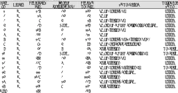
The demographic findings are shown in Table 1.
There were 17 cases, among which 10 were male and 7 were female. The age at the time of diagnosis ranged from 8 days to 34 months with a median of 3 months.
The median age at the first operation was 5 months, and the patients’ ages ranged from 17days to 37 month.
With regard to a PAC, 15 cases had a PAC and the other 2 cases did not have any PAC. When a PAC was present, the diameter ranged from 1.5mm to 8 mm.
There were various initial surgical approaches: one-stage total correction(4 cases), interposition of an RV-PA conduit(3 cases), conduit insertion with additional MAPCAs procedures(8 cases) and conduit interposition from the RV to the MAPCAs(2 cases). There were 3 initial surgical deaths.
Table 1. Demographic findings of the 17 patients Case
no Gender Age at diagn.
(day)
Size of PA confluence (mm)
Age at 1st.
Op. (day) 1st. Op. method Outcome of 1st. Op
01 M 0137 2.5 0155 RV-PA, unifocal. Rt Survived
02 M 0016 2.5 0024 RV-PA Survived
03 M 0008 4.0 00.17 RV-PA, ligation, Rt Survived
04 F 0029 None 0159 RV-neo-PA-MAPCA anastomosis, both side Survived
05 F 0107 5.0 0116 RV-PA, ligation, Rt Survived
06 F 0300 2.0 0311 RV-PA Survived
07 M 0128 3.5 0135 RV-PA, unifocal. Rt (1), ligation, Rt (2) Survived
08 M 0081 4.0 0099 RV-PA, pericardial defect repair Survived
09 M 0052 7.0 0056 Total correction Op. death
10 F 0029 None 0035 RV-neo PA-MAPCA anastomosis, Rt Op. death
11 M 0474 3.5 0476 RV-PA, ligation, Rt Survived
12 M 0228 6.0 0247 Total correction Survived
13 F 0039 3.0 0045 RV-PA, unifocal. Rt, ligation Lt Op. death
14 M 0111 2.0 0153 RV-PA, unifocal. both side Survived
15 F 1028 3.0 1114 Total correction Survived
16 M 0043 1.5 0229 RV-PA, unifocal. both side Survived
17 F 0150 8.0 0153 Total correction Survived
Mean 0174 0207
diagn.: diagnosis, PA: pulmonary artery, Op.: operation, RV-PA: conduit insertion between right ventricle and pulmonary artery, MAPCA:
major aorto-pulmonary collateral arteries, unifocal.: unifocalization, Rt: right, Lt: left, Ligation: MAPCA ligation, Size of the PA con- fluence: (diameter of the right pulmonary artery+the diameter of of left pulmonary artery)/2
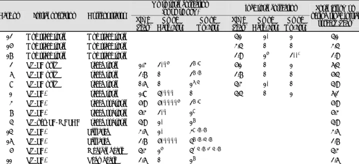
We followed up all the survivors of the initial sur- gery and the follow-up period varied from 2.6 years to 6.4 years with a mean of 4.6 years. Two patients died during follow up period, one of whom died of redo surgery and the other, who was in a palliated state, died of right heart failure at another hospital.
Table 2 shows the long-term outcomes according to the initial surgical methods. As a whole, 7 out of the 17 study patients underwent total correction. In addition to all three surgical survivors of one-stage total correction, 4 patients underwent staged total correction. While all 3 of the patients who had had a conduit operation without
any additional vascular procedure underwent total cor- rective operations, only 1 of the 8 patients who had had additional vascular procedures underwent total corrective operations. The additional vascular procedures seemed to have had an unfavorable effect.
Table 3 shows the long-term outcomes according to the initial PA morphology. In this study, the PA mor- phology was classified arbitrarily into 3 groups: no PAC, a PAC smaller than 4 mm and a PAC larger than 4 mm. Because of the small number of cases, any firm conclusions can not be drawn, but it looks as if the size of a PAC does not affect the long-term outcome.
Table 4. Follow up interventions by the initial surgical methods and by the final outcomes Up to total correction
(or up to now) Since total correction Case no Initial operation Current status
Time (year)
No of Cath. Int.
No of Op. Int.
Time (year)
No of Cath. Int.
No of Op. Int.
Total follow up period since initial
surgery (year)
12 One stage total One stage total 4.1 1* 1# 4.1
15 One stage total One stage total 3.4 0 0 3.4
17 One stage total One stage total 2.9 1§ 2**,# 2.9
02 RV-PA only Staged total 1.3 2*,*§ 2†,‡ 5.1 0 1# 6.4
06 RV-PA only Staged total 2.7 0 2†,† 2.7 0 0 5.4
08 RV-PA only Staged total 0.6 0 1¶≠ 4.3 1* 0 4.9
01 RV-PA+ Staged total 1.8 4*,*,*,* 0 4.4 0 1# 6.2
03 RV-PA+ Staged subtotal 5.9 5*,*,*,*,*§ 2†,‡ 5.9
07 RV-PA+ Staged subtotal 5.3 2*,* 1‡ 5.3
04 RV-neo-PA-MAPCA Staged subtotal 4.9 1* 1† 4.9
14 RV-PA+ Palliated 3.6 1* 3‡,†,† 3.6
16 RV-PA+ Palliated 2.7 5*,*,*,*,* 4†,†,≠,† 2.7
05 RV-PA+ Medical death 4.3 1§ 4∥≠,†,∥≠,† 4.3
11 RV-PA+ Redo death 2.6 0 1† 2.6
*: balloon angioplasty, †: reinsertion of conduit±angioplasty, ‡: surgical angioplasty, §: MAPCA coiling,∥: unifocalization,¶: MAPCA liga- tion, #: redo rastelli±angioplasty, **: residual VSD closure, ≠: diaphragm plication. Interpretation example: 5*,*,*,*,*§ means 5 times caths, 1st cath.: balloon angioplasty, 2nd cath.: balloon angioplasty, 3rd cath.: balloon angioplasty, 4th cath.: balloon angioplasty, 5th cath.:
balloon angioplasty and MAPCA coiling. Cath.: catheterization, Op.: operation, Int.: intervention, RV-PA: conduit insertion between right ventricle and pulmonary artery, MAPCA: major aorto-pulmonary collateral arteries, VSD: ventricular septal defect
Table 3. Final outcomes by the pulmonary artery morphology
PA status (n) Surgical death Medical death Palliated Subtotal correction Total correction (% of total correction)
Absent PA (2) 1 1 0 (00)
PA confluence <4 mm (9) 2 2 1 4 (44)
PA confluence >4 mm (6) 1 1 1 3 (50)
Total (17) 4 1 2 3 7 (41)
PA: pulmonary artery
Table 2. Final outcomes by the first surgical method
Method (n) Surgical death Medical death Palliated Subtotal correction Total correction (% of total correction)
One stage total correction (4) 1 3 (075)
RV-PA conduit (3) 3 (100)
RV-PA conduit+MAPCA ligation (3) 1* 1 1 0 (000)
RV-PA conduit+unifocalization (5) 1 2 1 1 (020)
RV-MAPCA+unifocalization (2) 1 1 0 (000)
Total (17) 4 1 2 3 7 (041)
*: redo death. RV: right ventricle, PA: pulmonary artery, MAPCA: major aorto-pulmonary collateral arteries
During the follow-up, most patients required a ca- theter or surgical interventions. The follow-up inter- ventions were tabulated according to the initial surgical methods and the final outcomes(Table 4). Three sur- vivors of one-stage total correction required balloon pulmonary angioplasty(1 case), redo Rastelli(2 cases) and surgical closure of the residual VSD(1 case). Four patients who had undergone staged total correction re- quired multiple catheters or surgical interventions before and after the total corrective operations. All those pati- ents who are still being palliated also required multiple catheters or surgical interventions.
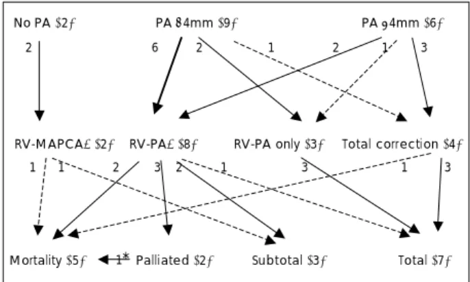
Fig. 1 shows the flow diagram of the patients. As ex- pected, this flow diagram is quite complicated partly because of the variable nature of the disease and partly because of the different surgical options that were used.
Those patients who had larger PAs were more likely to undergo total correction. Those patients who had smaller PAs were more likely to undergo RV-PA conduit with or without additional procedures such as MAPCAs unifocalization or ligation. Those patients who didn’t undergo additional vascular procedures seemed more likely to undergo a total corrective operation than those patients who underwent additional vascular procedures.
The most common reasons of not being able to undergo total correction were stenosis of multiple branches of the PA, anastomotic failure of the pulmonary segments, an insufficient number of unifocalized lung segments and pulmonary vascular obstructive changes.
All twelve surviving patients are being regularly ch- ecked at our outpatient clinic. All those who underwent total or subtotal correction are showing no symptoms and they are thought to be in the NYHA functional class 1. However, two patients who are still being pa- lliated have dyspnea or cyanosis.
Discussion
Pulmonary atresia with VSD and MAPCAs can vary
according to the PA anatomy and also according to the source of the pulmonary blood flow.16) Many surgical options have been devised and they have shown various success rates. The long-term prognosis of this disease is not very good and most failures are due to either an insufficient number of connected bronchopulmonary segments or to pulmonary vascular obstructive disease.
One-stage total correction at an early age can prevent the development of vascular obstructive disease, but not the development of pulmonary arterial branch ste- nosis after surgery. Moreover, one-stage total correction entails higher surgical mortality and this procedure is very difficult to perform in some cases. The staged app- roach also has many drawbacks. In short, there is no surgical option that is highly successful and at the same time, universally applicable. In fact, our study showed that this is indeed the case. Various surgical options have been used despite the high surgical mortality and the poor long-term morbidity rates. However, careful analysis of our material suggests there are better surgical options depending on the PA anatomy.
In our view, one-stage total correction seems feasible when there is a large confluent PA, and this procedure has a reasonable surgical mortality rate and a low long- term morbidity rate. For those patients with a small confluent PA, the RV-PA conduit seemed to be a good option that results in total correction. However, per- forming additional MAPCAs procedures, in association with a conduit operation, may jeopardize the long-term prognosis. The reason why additional procedures com- plicate the long-term prognosis is not clear, but addi- tional procedures are closely associated with the later development of stenosis in the pulmonary arterial tree.17)18) Simple ligation of MAPCAs is also associated with stenosis in the later stages. Moreover, this stenosis is very hard to balloon, even with performing surgical pulmonary angioplasty. Our result seems to suggest that procedures done on a small branch PA or MA PCAs are the least likely to succeed and the most likely to de- velop later stenosis, and so these procedures should be avoided. In contrast, simple anastomosis (RV-PA con- duit) shows better results. This conduit operation may enhance growth of the pulmonary artery19)20) and a larger PA, which in turn may improve the surgical result of anastomosis between the PA and MAPCAs. After the conduit procedure, close monitoring of the pulmonary arterial tree with echocardiography and catheterization is absolutely necessary and the liberal use of catheters or surgical interventions seems to be indicated. In summary, the surgical options for this complicated disease should be individualized and vigilant surveillance for any com- plication is absolutely necessary for total correction at a later date.
For a patient who already has increased pulmonary flow, the construction of a RV-PA conduit may further
No PA (2) PA <4mm (9) PA >4mm (6)
RV-MAPCA+(2) RV-PA+(8) RV-PA only (3) Total correction (4)
Mortality (5) Palliated (2) Subtotal (3) Total (7)
2 6 2 1 2 1 3
1 1 2 3 2 1 3 1 3
1*
Fig. 1. Flow diagram by the size of the pulmonary artery and the first surgery. *: Medical death. RV-MAPCAs+: RV-Neo PA-MAPCAs with other MAPCAs procedures. RV-PA+: RV-PA conduit op. with other MAPCAs procedures.
increase the pulmonary flow and worsen the patient’s heart failure. However, as discussed in a previous pa- per,15) the addition of another source of pulmonary flow, in theory, does not increase flow to the lungs. None- theless, meticulous assessment and careful decision mak- ing should be done to determine the best surgical option for a patient who has increased flow.
A major limitation of our study is that we were un- able to perform statistical analysis due to the small number of cases. Because this disease is so rare and it involves anatomy that can certainly vary, it is hardly likely that there are enough patients in a single insti- tution for conducting a meaningful statistical analysis.
Thus, designing a multi-institution study seems to be necessary to shed more light on this complex disease and its proper treatment.
REFERENCES
1) Ramsay JM, Macartney FJ, Haworth SG. Tetralogy of Fallot with major aortopulmonary collateral arteries. Br Heart J 1985;
53:167-72.
2) Faller K, Haworth SG, Taylor JF, Macartney FJ. Duplicate sour- ces of pulmonary blood supply in pulmonary atresia with ventri- cular septal defect. Br Heart J 1981;46:263-8.
3) Choi JY, Yun YS. Diagnosis and management of pulmonary ar- teries with major aorto-pulmonary collateral arteries. Korean Circ J 1989;19:473-82.
4) Millikian JS, Puga FJ, Danielson GK, Schaff HV, Julsrud PR, Mair DD. Staged surgical repair of pulmonary atresia, ventricu- lar septal defect, and hypoplastic, confluent pulmonary arteries.
J Thorac Cardiovasc Surg 1986;91:818-25.
5) Puga FJ, Leoni FE, Julsrud PR, Mair DD. Complete repair of pulmonary atresia, ventricular septal defect and severe periph- eral arborization abnormalies of the central pulmonary arteries:
experience with preliminary unifocalization procedures in 38 pa- tients. J Thorac Cardiovasc Surg 1989;98:1018-29.
6) Marelli AJ, Perloff JK, Child JS, Laks H. Pulmonary atresia with ventricular septal defect in adult. Circulation 1994;89:243-51.
7) Sullivan ID, Wren C, Stark J, de Leval MR, Macartney FJ, De- anfield JE. Surgical unifocalization in pulmonary atresia and ventricular septal defect: a realistic goal? Circulation 1988;78:
III5-III13.
8) Permut LC, Laks H. Surgical management of pulmonary atresia with ventricular septal defect and multiple aortopulmonary co-
llaterals. Adv Card Surg 1994;5:75-95.
9) Dinarevic S, Redington A, Rigby M, Shinebourne EA. Outcome of pulmonary atresia and ventricular septal defect during infancy.
Pediatr Cardiol 1995;16:276-82.
10) Reddy VM, Petrossian E, McElhinney DB, Moore P, Teitel DF, Hanley FL. One stage complete unifocalization in infants: when should the ventricular septal defect be closed? J Thorac Cardio- vasc Surg 1997;113:858-68.
11) Tchervenkov CI, Salasidis G, Cecere R, et al. One-stage midline unifocalization and complete repair in infancy versus multiple- stage uniforcalization followed by repair for complex heart dise- ase with major aortopulmonary collaterals. J Thorac Cardiovasc Surg 1997;114:727-37.
12) Carotti A, Di Donato RM, Squitieri C, Guccione P, Catena G.
Total repair of pulmonary atresia with ventricular septal defect and major aortopulmonary collaterals: an integrated apporach.
J Thorac Cardiovac Surg 1998;116:914-23.
13) Iyler KS, Mee RB. Staged repair of pulmonary atresia with ventricular septal defect and major systemic to pulmonary artery collaterals. Ann Thorac Surg 1991;51:65-72.
14) Sullivan ID, Stark J, deLeval M, Macartney FJ, Deanfield JE.
Surgical unifocalization in pulmonary atresia with ventricular septal defect an major aortopulmonary collateral arteries.
Circulation 1988;78(suppl III):III5-III13.
15) Choi JY, Yu JY, Zhu L, et al. Ventricular septal defect and pulmonary atresia with major aorto-pulmonary collateral artery:
diagnosis, management and review. J Korean Pediatr Cardiol Soc 2006;10:171-82.
16) Choi JY, Lee JK, Cha ES, Sul JH, Lee SK, Choe KO. Origins, distributions and characteristics of collateral circulation in pul- monary atresia with ventricular septal defect. Korean Circ J 1998;
28:1561-76.
17) Rome JR, Mayer JE, Castaneda AR, Lock JE. Tetralogy of Fallot with pulmonary atresia: rehabilitation of diminuitve pulmonary arteries. Circulation 1993;88:1691-8.
18) Haworth SG. Collateral arteries in pulmonary atresia with ven- tricular septal defect: a precarious blood supply. Br Heart J 1980;44:5-13.
19) Piehler JM, Danielson GK, McGoon DC, Wallace RB, Fulton RE, Mair DD. Management of pulmonary atresia with ventricu- lar septal defect and hypoplastic pulmonary arteries by right ventricular outflow construction. J Thorac Cardiovasc Surg 1980;
80:552-67.
20) Freedom RM, Pongiglioni G, Williams WG, Trusler GA, Rowe RD. Palliative right ventricular outflow reconstruction for patients with pulmonary atresia, ventricular septal defect, and hypoplastic pulmonary arteries. J Thorac Cardiovasc Surg 1983;86:24-36.