Veterinary Science
http://dx.doi.org/10.4142/jvs.2012.13.1.73
Received: 3 Mar. 2011, Revised: 3 Jan. 2012, Accepted: 16 Jan. 2012
Original Article
*Corresponding author: Tel: +886-4-2205-3366 (ext. 5208); Fax: +886-4-2207-8083; E-mail: [email protected]
†The first two authors contributed equally to this work.
ⓒ 2012 The Korean Society of Veterinary Science.
This is an Open Access article distributed under the terms of the Creative Commons Attribution Non-Commercial License (http://creativecommons.org/licenses/by-nc/3.0) which permits unrestricted non-commercial use, distribution, and reproduction in any medium, provided the original work is properly cited.
Development and characterization of a potential diagnostic monoclonal antibody against capsid protein VP1 of the chicken anemia virus
Yi-Yang Lien
1,†, Chi-Hung Huang
2,†, Fang-Chun Sun
3, Shyang-Chwen Sheu
4, Tsung-Chi Lu
2, Meng-Shiunn Lee
5, Shu-Chin Hsueh
1, Hsi-Jien Chen
6, Meng-Shiou Lee
7,*
Departments of
1Veterinary Medicine, and
4Food Science, National Pingtung University of Science and Technology, Pingtung 91201, Taiwan
2
Graduate School of Biotechnology, Hungkuang University, Taichung 43302, Taiwan
3
Department of Bioresources, Dayeh University, Chunghwa 51591, Taiwan
5
Department of Medical Research, Tungs’ Taichung MetroHarbor Hospital, Taichung 43503, Taiwan
6
Department of Safety, Health and Environmental Engineering, Mingchi University of Technology, Taipei 24301, Taiwan
7
School of Chinese Medicine Resources, China Medical University, Taichung 40402, Taiwan
Chicken anemia virus (CAV) is an important viral pathogen that causes anemia and severe immunodeficiency syndrome in chickens worldwide. In this study, a potential diagnostic monoclonal antibody against the CAV VP1 protein was developed which can precisely recognize the CAV antigen for diagnostic and virus recovery purposes.
The VP1 gene of CAV encoding the N-terminus-deleted VP1 protein, VP1Nd129, was cloned into an Escherichia (E.) coli expression vector. After isopropyl-β-D-thiogalactopy- ronoside induction, VP1Nd129 protein was shown to be successfully expressed in the E. coli. By performing an enzyme-linked immunoabsorbent assay using two coating antigens, purified VP1Nd129 and CAV-infected liver tissue lysate, E3 monoclonal antibody (mAb) was found to have higher reactivity against VP1 protein than the other positive clones according to the result of limiting dilution method from 64 clones. Using immunohistochemistry, the presence of the VP1-specific mAb, E3, was confirmed using CAV- infected liver and thymus tissues as positive-infected samples. Additionally, CAV particle purification was also performed using an immunoaffinity column containing E3 mAb. The monoclonal E3 mAb developed in this study will not only be very useful for detecting CAV infection and performing histopathology studies of infected chickens, but may also be used to purify CAV particles in the future.
Keywords: chicken anemia virus, immunoaffinity column, immunohistochemistry, monoclonal antibody, VP1
Introduction
Chicken anemia virus (CAV) is the sole member of the genus Gyrovirus of the Circoviridae family and causes severe anemia and chicken anemia disease, an immu- nosuppressive disorder [3,13,14]. Histopathological studies have shown that CAV infection leads to aplasia of the bone marrow [10,21]. This results in anemia and severe immunodeficiency syndrome due to the destruction of T lymphoid tissue [10,21].
The CAV genome consists of a circular single-stranded DNA genome of 2.3 kb encoding three viral proteins (VP):
VP1, VP2 and VP3 [3,13,14]. VP1 is the sole structural
protein of the CAV capsid. At a very late stage of the virus
life cycle, the assembled virus particles created by VP1
spread into various other tissues and organs of chickens
such as the thymus, spleen, and liver. Among these tissues
and organs, liver tissue has been reported to have the
highest accumulation of CAV virions [21]. Several
methods have been developed to conventionally detect
CAV infection such as serological tests for identifying
CAV antibodies. Recently, immunohistochemistry (IHC)
and immunofluorescence (IF) have been used as alternative
methods for detecting CAV antigen [1,11,12,17,18]. For
these methods to be successful, an excellent monoclonal
antibody is essential. Thus, antigen preparation is a critical
factor when producing these monoclonal antibodies. It has
been reported that VP2 and VP3 have been used as target antigens to generate monoclonal antibodies for immunological characterization or for developing diagnostic enzyme-linked immunoabsorbent assay (ELISA) kits [5,21]. However, VP1 has rarely been used as the antigen for generating antibodies or for producing diagnostic kits. This is because problems with VP1 expression have been reported in several host cell systems [5,8,15,18].
Research on VP1 antigen preparation has generally been unsuccessful because of a failure to find a good recombinant protein expression system. The highly enriched span of arginine residues at the N-terminus of VP1 has been proposed to be cytotoxic in an Escherichia (E.) coli expression system [15]. Thus, there is a need to overcome the difficulties of VP1 antigen preparation. If successful, this would allow the generation of a monoclonal antibody against VP1 capsids that may potentially be used diagnostically for the clinical detection of CAV infections.
Recently, our group has shown that the VP1Nd129 protein (amino acid residues 130∼450 of VP1), from which the first 129 amino acid residues of the VP1 N-terminus have been deleted, can be used to successfully express large amounts of the protein in prokaryotic cells [8].
In this study, the truncated recombinant VP1Nd129 protein was used as antigen in immune BALB/c mice to develop and produce a number of monoclonal antibodies for immunological applications. One of these monoclonal antibodies, E3, was selected for evaluating the ability of the monoclonal antibody to recognize VP1 in clinical samples infected with CAV including liver and thymus tissue. In addition, an immunoaffinity column containing E3 mAb as a ligand for virus particle purification also was investigated herein. The results of our study will be very useful for developing immunological tools to detect CAV, identifying CAV infection, or for performing CAV histopathology studies in chickens.
Materials and Methods
Virus strain and CAV-infected liver tissue
CAV (CIA89) was provided by Professor Yi Yang Lein of the National Pingtung University of Science and Technology (Taiwan). Two 1 day old specific pathogen free (SPF) hybrid white leghorn chickens purchased from the Animal Health Research Institute of the Council of Agriculture (Taiwan) were used to propagate the virus by passaging 20% liver homogenates (0.1 mL per bird). The phosphate buffered saline was used to dilute the homogenates containing virus particles. These animals were inoculated orally with CIA-89 containing titers of 10
7.5TCID
50. At 10-days post-infection, the chicken sera were collected and used to confirm the virus infection in terms of the per- formance of anti-CAV antibody using commercial CAV test kit (IDEXX, Netherlands). Then the chicken was
sacrificed. The individual livers of the sacrificed chickens were removed, collected, immersed in formaldehyde, and then stored at room temperature until required.
Plasmid construction and bacterial strain
The pGEX-6P-1-VP1 plasmid derived from pGEX-6P-1 plasmid (GE Healthcare, USA), which contains cDNA encoding the VP1 genes, was provided from Professor Yi-Yang Lien of National Pingtung University of Science and Technology (Taiwan) and initially used as the PCR template. To amplify the VP1 gene lacking the coding region for the first 129 amino acids from the full-length VP1 gene, two PCR primers, VP1-388FE and VP1-RHX, were designed and used as described in a previous study [8]. Using pGEX-6P-1-VP1 as the template, PCR reactions were performed at 95
oC for 5 min, 95
oC for 45 sec, 59
oC for 50 sec, and 72
oC for 1 min for 30 cycles. The last PCR cycle was carried out with a final elongation step of 10 min at 72
oC. The amplified DNA fragments were digested with XhoI and EcoRI (Takara, Japan), and then cloned into the prokaryotic expression vector pET28a (Merck, Germany). The constructed recombinant plasmid, pET28a-VP1Nd129, was used to transform One Shot Top10 cells (Invitrogen, USA) and BL-21 (DE3) competent E. coli for maintaining the recombinant plasmids and for protein expression, respectively. Transformants with the correct gene size were identified by PCR and screened using restriction enzyme digestion and sequencing. The above PCR was performed in a 25 μL reaction mixture containing 0.4 mM of dNTPs, 5 pmole each of VP1-388FE and VP1-RHX, 1U Pro-taq DNA polymerase (Protech, Taiwan) and 1× Pro-taq buffer (10 mM Tri-HCl, 50 mM KCl, 0.01% gelatin, 1.5 mM MgCl
2, 0.1% Triton X-100, pH 9.0). The PCR conditions was 95
oC for 5 min, followed by 35 cycles of 95
oC for 1 min, 57.7
oC for 1 min, and 72
oC for 1 min, and a final extension cycle at 72
oC for 10 min. Digestion was carried out at 37
oC for 1 h in a 50 μL reaction mixture containing 50 mM of potassium acetate, 20 mM Tris-acetate, 10 mM magnesium acetate, 1 mM DTT, 100 μg/mL BSA, 5U XhoI and 5U EcoRI. The resulting restriction digestion product was analyzed by 1% agarose electrophoresis and visualized using ethidium bromide staining and UV.
Expression and purification of VP1Nd129 protein in recombinant E. coli
Recombinant BL-21 (DE3) E. coli containing pET28a- VP1Nd129 were used for protein induction and expression.
The recombinant strains were grown overnight in Luria-
Bertani (LB) medium (BD-Difco, USA) in the presence of
kanamycin (50 μg/mL; Amresco, USA) at 37
oC. Next, 0.5
mL of the overnight culture was used to inoculate 50 mL
LB medium. The culture was grown at 37
oC for around 3
h by which time the optical density had reached 0.5. At
this point, 0.1 mM isopropyl-β-D-thiogalactopyronoside (IPTG) (Amresco, USA) was added to the culture to induce protein expression, which continued for 6 h. The presence of expressed VP1Nd129 protein was confirmed by 12.5%
SDS-PAGE followed by Western blotting using a monoclonal anti-His antibody (Invitrogen, USA), as described in previously work [8].
To purify the recombinant VP1Nd129 protein, a cell pellet was spun down using 5,000 × g, 15 min, at 4
oC from 50 mL of the culture supernatant and resuspended in denaturing binding buffer (20 mM NaH
2PO
4, 0.5 M NaCl, and 8 M urea, pH 7.8). The mixture was then sonicated on ice three times for 3 min with a 20% pulsed activity cycle (Sonics & Materials, USA), and then centrifuged for 10 min at 13,300 × g to remove cell debris. The resulting cell lysate was poured into an Enco-column (Bio-Rad, USA) with 2 mL of Ni
2+-NTA agarose and the resin was allowed to settle by gravity. The packed resin in column was washed by gravity with three volumes of denaturing binding buffer and then a similar volume of wash buffer (20 mM NaH
2PO
4, 0.5 M NaCl, and 8 M urea, pH 6.3).
Finally, the bound proteins were eluted with elution buffer (20 mM NaH
2PO
4, 0.5 M NaCl, and 8 M urea, pH 4). For each fraction, 2 mL of elute was collected. The fractions were monitored at OD
280using a U-2001 spectropho- tometer (Hitachi, Japan) with wash buffer as a blank.
Putative peaks corresponding to the recombinant CAV viral protein were identified and the eluate was collected for analysis. The total protein concentration of each fraction was determined using a Micro BCA kit (Pierce, USA) with bovine serum albumin as the reference protein.
The purity of the protein samples was analyzed using aliquots of the concentrated fraction; this was done by 12.5% SDS-PAGE and Coomassie brilliant blue staining.
Generation of a monoclonal antibody against CAV VP1 protein
SPF BALB/c mice (Charles River, MA) were immunized by subcutaneous injection of 20 μg purified VP1Nd129 protein emulsified with complete Freund’s adjuvant (Sigma-Aldrich, USA). After immunization, the BALB/c mice were sacrificed, the spleens were removed, and the 2
× 10
8splenocytes were fused with 2 × 10
7SP2/0 myeloma cells (ATCC CRL1581) by treatment with polyethylene glycol [6]. After 2 weeks of growth, the resultant hybridomas were examined the presence of antibodies against VP1Nd129 protein. Antibodies secreted from the various hybridomas were screened by ELISA. They were subcloned three times using the limiting dilution technique [20], and then ascitic fluid containing monoclonal antibodies was produced by introducing the cloned hybridomas into Pristane-primed mice (Charles River, USA). The immunoglobulin class of the hybridoma antibodies was determined by ELISA with a Mab kit
(Zymed Laboratories, USA) using rabbit antisera against mouse immunoglobulin (Ig)M, IgG1, IgG2a, IgG3, and IgA, and goat anti-rabbit IgG serum conjugated with horseradish peroxidase (HRP).
ELISA
To evaluate the specificity and reactivity of the mAb against CAV, an ELISA with the antibody was performed with purified VP1Nd129 protein or lysates from tissues infected with CAV as the antigen. After this, polyvinyl chloride 96-well plates were coated with recombinant VP1Nd129 or tissue lysates containing CAV. The antigen was buffered and diluted with carbonate-bicarbonate buffer (pH 9.6). Varying amounts of antigen from 1 to 500 ng were added to the 96-well strip plate and incubating overnight at 4
oC. After antigen coating, the plates were washed twice with phosphate-buffered saline (PBS), containing 0.05% Tween 20 (PBS-T), and then 0.2 mL of blocking reagent (PBS containing 5% skim milk) was added to each well. After 30 min of incubation at room temperature, the plates were washed twice with PBS. One hundred μL of the hybridoma supernatants were then added to the plate wells coated with CAV antigen or recombinant antigen and incubated at 37
oC for 1 h. The bound mAbs were detected using HRP-conjugated rabbit anti-mouse IgG (Jackson Immuno Research, USA) and the color was developed with o-phenylenediamine dihydrochloride (Sigma-Aldrich, USA).
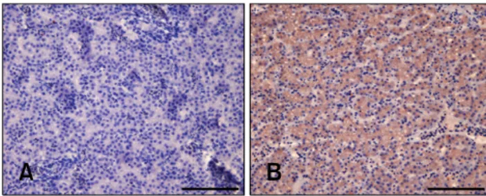
Histopathology and IHC
CAV-infected and uninfected liver and thymus tissues
(positive and negative controls, respectively) were all from
clinical cases of CAV infection confirmed by PCR using
specific primers as previously described [8]. The liver and
thymus samples were fixed using 30% neutral buffered
formaldehyde and embedded in paraffin. The paraffin-
embedded tissues were sectioned, mounted, and then
stained with hematoxylin and eosin. IHC staining was
carried out at room temperature. Tissue sections (5-μm
thick) of the paraffin-embedded tissues were washed with
phosphate saline buffer containing 0.3% hydrogen peroxide
to inactivate endogenous peroxidase. After washing three
times with phosphate saline buffer, antigen retrieval was
carried out by incubating the tissue sections with a 0.1% of
phosphate buffer diluted trypsin solution. The 250 fold
diluted monoclonal antibody E3 mAb, which is specific for
CAV VP1, was then applied as the primary antibody for 2
hours of the incubation. E3 mAb was recognized by
HRP-conjugated goat anti-mouse IgG secondary antibody
(Jackson ImmunoResearch, USA). 0.2 μg/mL of secondary
antibody solution was used for 2 h of the incubation. After
IHC staining, the sections were counterstained with
hematoxylin and examined under light microscopy.
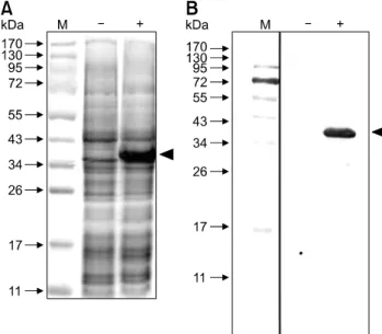
Fig. 1. Analysis of the VP1Nd129 protein expressed in recombinant Escherichia (E.) coli by SDS-PAGE (A) and Western blotting (B). The symbols ‘’ and ‘+’ represent pre- and post-induction with 1 mM of isopropyl-β-D-thiogalactopyronoside in E. coli, respectively. Anti-His tag monoclonal antibody was used for recognizing the VP1Nd129 protein. The arrowheads are the symbol for expressed VP1Nd129 protein.
Immunofluorescence
For IF microscopy, infected and un-infected MSB-1 cells on glass coverslips were fixed in 4% formaldehyde, washed with PBS, and permeabilized with 0.01% Triton X-100.
After washing with PBS, samples were incubated with blocking solution (0.5% bovine serum albumin in PBS) for 1 h, followed by 1 h of incubation with 2 μg/mL of E3 mAb solution. 0.2 μg/mL of fluorescein isothiocyanate- conjugated goat anti-mouse IgG (Jackson Immuno- Research, USA) was used to detect E3 mAb binding. Cell nuclei were counterstained with 4´,6-diamidino-2- phenylindole (Sigma-Aldrich, USA) and fluorescence images were captured using a laser scanning confocal microscope (Leica Microsystems, Germany).
Immunoaffinity column purification of CAV particles To create an immunoaffinity column, cytoplasmic extracts from chicken liver tissues infected with CAV were prepared by centrifugation at 13,300 × g for 30 min at 44
oC.
Next, 100 μL of cell extract were mixed with 50 μL of ascitic fluid and incubated overnight at 4°C. Then, 50 μL of protein A agarose beads (Sigma-Aldrich, USA) were added and incubated overnight at 44
oC with gentle rotation. The agarose beads were washed three times with buffer containing 50 mM Tris-HCl (pH 7.5), 500 mM NaCl, 0.2% NP-40, and 0.05% sodium deoxycholate.
Finally, viral protein and/or CAV virus particles were eluted from the protein A agarose beads by boiling with SDS-PAGE sample buffer (60 mM Tris-Cl pH 6.8, 2%
SDS, 10% glycerol, 5% β-mercaptoethanol, 0.01%
bromophenol blue) for 5 min. The extracted proteins were then subjected to SDS-PAGE followed by Western blotting using E3 mAb as primary antibody and HRP-conjugated goat anti-mouse IgG secondary antibody, respectively. In addition, CAV genomic DNA from the CAV virus particles was detected by PCR using CAV genome-specific primers.
The sequences of the CAV VP1 gene PCR primers were VP1F: 5´-ATGGCAAGACGAGCTCGCAGACCGAGA GG-3´ and VP1R: 5´-CTAACCATGGTGATGGTGATG GTGGGGCTGCGTCCCCCAGTA-3´. The sequences of the CAV VP3 primers were VP3F: 5´-CCGCTCGAGC AGTCTTATACACCTTCTTG-3´ and VP3R: 5´-GCGAA TTCATGAACGCTCTCCAAGAAGATAC-3´. The PCR conditions was 95
oC for 5 min, followed by 35 cycles of 95
oC for 1 min, 57.7
oC for 1 min, and 72
oC for 1 min, and a final extension cycle at 72
oC for 10 min.
Results
Expression, purification and characterization of CAV VP1Nd129 protein using a recombinant E.
coli system
To express the CAV VP1 as an antigen for immunization, the VP1Nd129 construct was created by PCR using VP1
cDNA as the template DNA. The VP1 cDNA partially overlapped partially the VP2 gene of the CAV genome, which had been previously cloned into the pGEX-6P-1 plasmid [4]. Using the primers VP1-388FE and VP1-RHX, the VP1Nd129 construct was amplified by PCR and cloned into the pET28 a vector at the EcoRI and XhoI sites, thereby creating a protein with an in-frame His-tag. This plasmid, pET28a-VP1Nd129, was used to transform E. coli BL-21 (DE3) cells. The E. coli were examined for protein expression after 4 h of induction with IPTG. VP1Nd129 protein was successfully expressed in
E.coli