features of intra-abdominal extra-hepatic metastases from gastrointestinal stro- mal tumors in patients who were treated with imatinib.
Materials and Methods: Eleven patients with intra-abdominal extra-hepatic metastases from gastrointestinal stromal tumors, who were treated with imatinib between May 2001 and December 2003, were included in this study. The clinical findings and CT scans were retrospectively reviewed. The metastatic lesions were assessed according to the location, size (greatest diameter), attenuation, and the enhancing pattern before and after imatinib treatment.
Results: Prior to the treatment, the sizes and attenuation values of the metastatic lesions ranged from 5 to 20 cm and from 63 to 131 H, respectively.
The metastatic lesions showed a heterogeneous enhancement pattern on the contrast-enhanced CT scans. After the treatment, the metastatic lesions became smaller in all 11 patients, and the corresponding attenuation value ranged from 15 to 51 H. The metastatic lesions became homogeneous and cystic in appear- ance on the follow-up CT scans, mimicking ascites.
Conclusion: Intra-abdominal extra-hepatic metastases of patients with gas- trointestinal stromal tumors treated with imatinib may appear as well-circum- scribed cystic lesions on contrast-enhanced CT. These metastases are likely to become smaller and resemble ascites, but may persist indefinitely on the follow- up CT.
astrointestinal stromal tumors, formerly classified as leiomyomas or leiomyosarcomas, constitute the most common form of mesenchymal tumor in the gastrointestinal tract, with the stomach being the most common site of origin (1). The diagnosis of these tumors became feasible through the application of CD117 immunohistochemistry, which allows these neoplasms to be distinguished from leiomyomas or leiomyosarcomas (2). Moreover, the recently developed KIT-tyrosine kinase inhibitor (STI 571, imatinib [Gleevec], Novartis, Basel, Switzerland) has dramatically improved the treatment of gastrointestinal stromal tumors (3).
The radiologic findings of gastrointestinal stromal tumors were recently described in the radiologic literature (1, 4 8), and are ostensibly similar to those of the previously described leiomyomas or leiomyosarcomas (9). The characteristic features of this disease are its recurrence at the primary site and the presence of metastases, primarily in the liver and the peritoneum (4). Recently, it was reported that the hepatic
metastases in patients treated with imatinib resembled cystic lesions (10 12). In line with these observations, our findings indicate that when gastrointestinal stromal Seung Hong Choi, MD1
Heon Han, MD2 Sam Soo Kim, MD2 Sang Hyun Lee, MD3 Joon Koo Han, MD1 Byung Ihn Choi, MD1
Index terms :
Gastrointestinal stromal tumor Neoplasms, metastases Abdomen, CT
Korean J Radiol 2004;5:157-163 Received October 19, 2003; accepted after revision February 9, 2004.
1Department of Radiology, Seoul National University College of Medicine, Institute of Radiation Medicine, SNUMRC, and Clinical Research Institute, Seoul National University Hospital; 2Department of Radiology, Kangwon National University College of Medicine; 3Radiation Medicine Branch, National Cancer Center
This study was supported in part by the 2003 BK21 Project for Medicine, Dentistry and Pharmacy.
Address reprint requests to:
Jeong Min Lee, MD, Department of Radiology, Seoul National University Hospital, 28 Yongon-dong, Chongno-gu, Seoul, 110-744, Korea.
Tel. (822) 760-2584 Fax. (822) 743-6385
e-mail: [email protected]
G
Table 1. Summary of the 11 Patients with Intraabdominal Extrahepatic Metastasis from Gastrointestinal Stromal Tumor Treated with Imatinib PatientAge/Site ofSite ofClinicalInitial Findings of Metastasis1st F/ULast F/U under Imatinib therapyComment NumberSexPrimaryMetastaticSymptomMetastatic IntervalHounsfieldAttenuationSizeIntervalHounsfieldAttenuationSize IntervalHounsfieldAttenuation TumorLesionsLesionbetweenMeasurementsPatternafterbetweenMeasurementsPatternafterbetweenMeasurementsPattern Size (cm)Operation andImatinibInitialImatinibInitial Detection ofTherapyDetectionTherapyDetection Metastasis(cm)and 1st F/U(cm)and Last F/U (months)(weeks)(months) 0159/MStomachLocalAbdominal150773Hetero030836Homo< 122Homo recurpain 0246/MStomachPeritonealRoutine051272Hetero030825Homo< 110HomoAggravated seeding,F/U11 months liverlater 0365/MStomachPeritonealAbdominal200988Hetero150439HeteroF/U loss seeding,pain liver 0426/MStomachLocalRoutine 0925106Hetero031025Homo20518Homo recurF/U 0568/MStomachPeritoneal Routine151281Hetero080828Homo51320Homo seedingF/U 0642/MRectumPeritonealRoutine053867Hetero030839Homo11017Homo seeding,F/U liver 0752/MMesenteryPeritonealRoutine0728131Hetero050451Hetero1.50822Homo seedingF/U 0858/MMesenteryPeritonealAbdominal101579Hetero041033Hetero40421Homo seedingpain 0941/MSmallPeritoneal Routine 133574Hetero100415Homo30615Homo bowelseedingF/U 1058/MSmallPeritonealAbdominal051863Hetero030844Homo< 114Homo bowelseedingpain 1134/MSmallPeritonealRoutine101682Hetero050835Homo31128HomoAggravated bowelseeding,F/U2 months liverlater
mean age was 50 years, ranging from 34 to 68 years, and all were men. The mean time between the initial diagnosis of the gastrointestinal stromal tumor and the diagnosis of metastases was 19 months (range, 7 38 months). The primary sites were the stomach (n=5), the small bowel (n=3), the rectum (n=1), and the mesentery (n=2). In all patients, the primary tumors were removed by curative surgery. The presence of metastatic lesions was confirmed by biopsy (n=5) or radiologic studies and clinical follow-up (n=6). The criterion for the diagnosis of metastasis was the presence of new lesions detected in the follow-up CT scan, which were not detected in the initial CT scan and which
All patients underwent CT scans prior to the administra- tion of imatinib and 4 10 weeks after the initiation of the imatinib therapy. Ten patients underwent two or more follow-up CT scans 4 22 months after the initiation of the treatment with imatinib. The CT scan data were available on a picture archiving and communications system (PACS;
Marotech, Seoul, Korea) for all patients. The CT scans were performed using a Somatom Plus-4 (Siemens Medical Systems, Erlangen, Germany), a HiSpeed Advantage scanner (General Electric Medical Systems, Milwaukee, WI), or a MX8000 four-detector row CT scanner (Philips Medical Systems, Cleveland, OH). Each patient received
A B
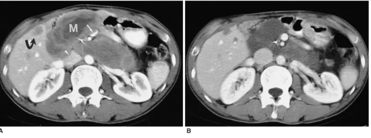
Fig. 1. A 34-year-old man with peritoneal seeding and liver metastases after resection of gastrointestinal stromal tumor of the duodenum.
A. CT scan before treatment shows multiple heterogeneous masses (82 H) (M) compressing inferior vena cava (arrowhead) and superior mesenteric vein (arrow). Metastatic lesion in liver (curved arrow).
B. CT scan obtained after 8 weeks of treatment with imatinib shows peritoneal and hepatic metastases that have decreased in size and density (35 H). Note the decompressed inferior vena cava and superior mesenteric vein and shrunk metastatic lesions in the liver.
C. CT scan obtained 2 months after stopping imatinib therapy shows increased size and density of peritoneal and hepatic metastases. After resumption of imatinib therapy, the metastatic lesions showed cystic changes again (now shown).
C
120 mL of a nonionic contrast material (Iopromide, Ultravist 370; Schering Korea, Seoul, Korea) through an 18-gauge angiographic catheter inserted into a forearm vein. The contrast material was injected at a rate of 2.5 mL/sec using an automatic injector. In the case of the single-detector scanner, a helical CT scan was performed with the following parameters: 5 7 mm collimation, 1:1 table pitch, and 5 7 mm reconstruction intervals. In the case of the MX8000 scanner, the parameters were 2.5 mm detector collimation, 20 mm/sec table speed, 5 mm slice thickness, and a 5 mm reconstruction interval. The delay between the contrast material administration and scanning was 55 70 seconds.
Two radiologists reviewed all of the CT scans retrospec- tively, and the final interpretations were reached by consensus. All images were reviewed on a 2,000 2,000 PACS monitor. The presence of the metastatic lesion and its size before and after the imatinib treatment were compared. The metastatic lesions were assessed according to their location, size (greatest diameter), attenuation, and
enhancing pattern. If multiple metastatic lesions were detected, the largest lesion was recorded. For the objective analysis, the CT attenuation value was measured in a circular region of interest with a diameter of 10 mm. The CT attenuation value was measured three times by a single radiologist and the mean value was recorded. In the case of a heterogeneous mass, the CT attenuation value was measured in the solid portion of the tumor. The CT attenu- ation value before and after the imatinib treatment was compared using the paired t-test. Statistical analyses were performed using a computer software package (SPSS, version 10.0; SPSS, Chicago, Ill). A p value of less than .05 was considered to indicate a statistically significant differ- ence.
RESULTS
The clinical and radiologic findings are summarized in Table 1. One patient (patient 3) was lost to follow-up after 1 month of imatinib therapy. Two patients (patients 2 and
A B
Fig. 2. A 68-year-old man with peritoneal seeding after resection of gastrointestinal stromal tumor of the stomach.
A. CT scan before treatment shows 15 13 cm heterogeneous metastatic lesion (81 H) (G) in left subphrenic space. Note small metastatic nodule (arrow) in right subphrenic space and ascites (15 H).
B. CT scan obtained after 8 weeks of treatment with imatinib shows metastatic lesion (G) that has decreased in size to 8 6 cm and is cyst-like in appearance lesion (28 H) around spleen (S).
Note the disappearance of the ascites.
C. CT scan obtained after 13 months of treatment with imatinib shows 5 3.5 cm cystic lesion (20 H).
C
geneous enhancement pattern on the contrast-enhanced CT scans.
After the treatment, the mean size of the metastatic lesions was 5.8 3.6, ranging from 3 to 15 cm, on the first follow-up CT scan, showing a reduction in size for all 11 patients. On the first follow-up CT scan, the attenuation of the metastatic lesions was homogeneous in eight patients (Figs. 1 and 2), and heterogeneous in three patients (Fig.
3). In cases of peritoneal seeding, the metastatic lesions developed a cystic appearance, mimicking ascites (Figs. 2 and 3). In reviewing the original CT reports, it was found that the cystic change of the tumor was described as ascites or fluid collection in three patients.
Prior to the treatment, the mean attenuation value of the metastatic lesions was 83 20 H, ranging from 63 to 131 H. On the first follow-up CT scan, the mean attenuation value was 34 13 H, ranging from 15 to 51 H. This differ- ence in the mean CT attenuation value was statistically significant (p < 0.01).
On the subsequent CT scans, the metastatic lesions became smaller, homogeneous and cystic during imatinib therapy. However, they did not disappear completely and were always detected throughout the study in all patients.
metastatic lesions increasing in size and attenuation, and showing a heterogeneous enhancement pattern on the CT scans (Fig. 1C). These two patients resumed imatinib therapy, and their metastatic lesions subsequently became smaller and homogeneous on the follow-up CT scans.
DISCUSSION
Conventional chemotherapeutic agents are rarely effective against gastrointestinal stromal tumors. The new chemotherapeutic agent, imatinib, has been applied and the results are extremely encouraging. The rationale behind imatinib treatment for gastrointestinal stromal tumors lies in the fact that the KIT (encodes the human homolog of the proto-oncogene c-kit) gene mutation has been detected frequently in gastrointestinal stromal tumors. This mutation induces the constitutive activation of the tyrosine kinase receptor, causing the proliferation of tumor cells (2). Imatinib is highly effective in bringing about a reduction in KIT tyrosine kinase activity.
Gastrointestinal stromal tumors frequently spread to the liver and the peritoneum (4). On the CT scan of the portal venous phase, the metastases within the liver are usually
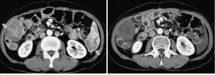
Fig. 3. A 52-year-old man with peritoneal seeding after resection of gastrointestinal stromal tumor of the mesentery.
A. CT scan before treatment shows multiple peritoneal implants (arrows) in both paracolic gutters.
B. CT scan obtained after 4 weeks of treatment with imatinib shows that the metastatic lesions in the right paracolic gutter have some solid components, while that in the left paracolic gutter resembles ascites.
A B
heterogeneous and peripherally enhanced, similar to primary tumors (4). The low attenuation in the center of these metastatic lesions often indicates the presence of necrosis in the center of the solid mass. The peripheral enhanced portion represents viable solid tumor. Peritoneal metastasis shows a CT appearance similar to that of metastasis in the liver.
In the peritoneum, metastatic lesions treated with imatinib may appear as ascites or fluid collection. In reviewing the original CT reports, we found that the cystic change of the tumor was described as ascites or fluid collection in three patients. Although long-term follow-up is needed, metastatic lesions in the peritoneum gradually decrease in size, although they may persist for months or years, which is not the case for ascites. The density of the metastases decreased to 15 51 H on the first CT scan after the treatment and then to 15 28 H on the follow-up CT scan, which is close to that of ascites. Metastases can be distinguished from ascites by reviewing the change in the attenuation value and the previous CT scan. Ideally, the scans should be interpreted by a radiologist who is familiar with scans of peritoneal metastases from gastrointestinal stromal tumors following imatinib treatment, in order to avoid the underestimation of the extent of the tumors.
The mechanism that induces the cystic change after imatinib treatment is not clear. In several reported cases, histological examination of the tumors treated with imatinib showed areas with extensive necrosis, hyalinized areas with sparse, scattered tumor cells containing small, condensed nuclei and areas with viable tumor cells (10 12).
The optimal duration of the treatment is not yet known (13). It is not clear whether viable tumor cells with malignant potential persist within the cystic lesions and, consequently, the continuous maintenance of imatinib treatment is required. In this study, two patients whose metastatic lesions became small and cystic after imatinib therapy, experienced aggravation of metastasis after termination of the imatinib treatment.
Traditionally, the response to cancer treatment in solid tumors is evaluated by subsequent clinical or radiological assessments, and is defined as a significant decrease in the measurable tumor dimensions. A reduction in the viable tumor cell fraction, however, does not always result in a volume reduction, since tumor tissue can be replaced by necrotic or fibrotic tissue, and morphological images are often unable to differentiate between these different tissue types. In recent years, metabolic imaging with positron emission tomography (PET) has become increasingly important in cancer management. Although the perfor- mances of PET and CT are comparable in terms of the
process of staging before the initiation of imatinib therapy, PET can evaluate the tumor response as early as 1 week after the start of treatment, preceding the CT response by several weeks (14). Treatment-induced changes resulting in tumor cell death or growth arrest should therefore result in a subsequent reduction in glucose uptake, making this technique a sensitive and early marker for response evaluation.
There are several limitations to this study. First, this was a retrospective review of cases collected over a number of years for which CT scans were performed irregularly, depending on the condition of the patients. Second, unenhanced images were not obtained in all patients, and it is unclear whether or not any subtle enhancement changes are present in the metastatic lesions. Third, we did not provide any pathologic correlation in any of the cases.
Pathologic correlation with the radiologic findings for metastatic lesions is helpful for clinicians as well as for radiologists.
In conclusion, after treatment with imatinib, responsive intra-abdominal extra-hepatic metastases of gastrointesti- nal stromal tumors appear as well-defined cystic lesions on contrast-enhanced CT. These metastases become smaller and resemble ascites, but may be detected for a long time on the follow-up CT scans.
References
1. Levy AD, Remotti HE, Thompson WM, Sobin LH, Miettinen M.
Gastrointestinal stromal tumors: radiologic features with pathologic correlation. RadioGraphics 2003;23:283-304 2. Hirota S, Isozaki K, Moriyama Y, et al. Gain-of-function
mutations of c-kit in human gastrointestinal stromal tumors.
Science 1998;279:577-580
3. Demetri GD, von Mehren M, Blanke CD, et al. Efficacy and safety of imatinib mesylate in advanced gastrointestinal stromal tumors. N Engl J Med 2002;347:472-480
4. Burkill GJ, Badran M, Al-Muderis O, et al. Malignant gastroin- testinal stromal tumor: distribution, imaging features, and pattern of metastatic spread. Radiology 2003;226:527-532 5. Levy AD, Remotti HE, Thompson WM, Sobin LH, Miettinen M.
Anorectal gastrointestinal stromal tumors: CT and MR imaging features with clinical and pathologic correlation. AJR Am J Roentgenol 2003;180:1607-1612
6. Nishida T, Kumano S, Sugiura T, et al. Multidetector CT of high-risk patients with occult gastrointestinal stromal tumors.
AJR Am J Roentgenol 2003;180:185-189
7. Kim H-C, Lee JM, Kim SH, et al. Primary gastrointestinal stromal tumors in the omentum and mesentery: CT findings and pathologic correlations. AJR Am J Roentgenol 2004;182:1463- 1467
8. Kim H-C, Lee JM, Son KR, et al. Gastrointestinal stromal tumors of the duodenum: CT and barium study findings. AJR Am J Roentgenol 2004;183:415-419
9. Megibow AJ, Balthazar EJ, Hulnick DH, Naidich DP, Bosniak MA. CT evaluation of gastrointestinal leiomyomas and leiomyosarcomas. AJR Am J Roentgenol 1985;144:727-731