107 https://doi.org/10.22889/KJP.2020.51.2.107
대장 상피세포에서 p-Hydroxycinnamic Acid의 항염증 효과와 염증성 장질환에 대한 치료 효과
이현수1·이승호2·최혁재2·정길생1*
1
계명대학교 약학대학,
2영남대학교 약학대학
Anti-inflammatory Effect of p-Hydroxycinnamic Acid on HT-29 Intestinal Cells and Its Therapeutic Effect of Immune Bowel Disease
Hyun-Su Lee1, Seung-Ho Lee2, Hyukjae Choi2, and Gil-Saeng Jeong1*
1
College of Pharmacy, Keimyung University, Daegu 42601, Korea
2
College of Pharmacy, Yeungnam University, Gyeongsan 38541, Korea
Abstract Inflammatory bowel disease (IBD) is a chronic inflammatory disorder on the large intestine that has been considered as an incurable not only in Western society but also in Eastern Asia in recent years. Despite enormous efforts to develop novel therapeutics for this disease, strategy using bioactive compounds from natural product is still considered as important. p- hydroxycinnamic acid (HCA) is an intermediate substance found in several plants and has been known to possess anti-inflam- mation but little evidence is reported whether HCA has an inhibitory effect on intestinal inflammation. In the present study, we observed HCA does not show cytotoxic and apoptotic in HT-29 cells. Quantitative PCR analysis revealed that HCA effec- tively blocks the activity of HT-29 cells stimulated with TNF- treatment. HCA inhibits translocation of p65 and MAPK path- ways in activated HT-29 cells by TNF- treatment. Besides, oral administration of HCA attenuates manifestation of DSS- induced inflammatory disease in vivo. Histological analysis exhibited that oral administration of HCA recovers IBD symptoms.
The expression of pro-inflammatory cytokines were reduced by oral administration of HCA on intestinal tissues. Therefore, these results suggest that HCA has a potent anti-inflammatory effect on intestinal cells as well as show a therapeutic potential for treating IBD in vivo.
Keywords p-Hydroxycinnamic acid, HT-29, TNF-, MAPK, Immune bowel disease, Anti-inflammation
염증성 장질환은 만성 염증 질환으로써 주로 대장에서 발 병하는 병이다. 염증성 장질환은 크게 병인에 따라 크게 2 개로 분류되는데 크론병(Crohn’s disease, CD)과 궤양성 대 장염(Ulcerative colitis, UC)으로 구분된다.
1)CD는 소화관 전체에 걸쳐서 발생한다고 알려져 있지만 UC는 주로 대장 과 직장의 가장 안쪽에서 발생한다고 알려져 있다.
2)임상적 으로 IBD의 주요 증상은 심한 설사, 통증, 피로, 체중감소 및 림프절의 확대와 관련이 있다고 보고 되었다.
3)또한, 계 속되는 염증으로 인한 IBD는 대장암의 위험을 증가시킨다 는 보고와 관련해 다양한 면역관련 매개인자들이 CD와 UC 의 진행에 중요한 역할을 한다는 보고가 있다.
4)종양괴사인 자(tumor necrosis factor-; TNF-), 인터루킨(interleukin-
1β; IL-1β, interleukin-6; IL-6, interleukin-8; IL-8), 그리고 nitric oxide(NO)등이 염증성 장질환에 관여한다고 알려져 있다.
5,6)이러한 면역관련 매개인자들의 과도한 발현은 일반 적인 염증성 장질환의 증상을 촉진하기 때문에 IBD 치료에 는 염증 요인의 분비에 대한 효과적인 조절이 필수이다.
p-hydroxycinnamic acid(HCA)는 phenolic acid의 한 종류 로써 다른 여러가지 phenolic compounds의 전구체로써 역 할을 하는 화합물로써 tyrosine으로부터 생합성 되어 진다 고 알려져 있다. Turnip tops, corn bran, oregano를 포함한 여러 식물에서 발견되는 HCA는 anti-inflammatory, anti- oxidant, anti-mutagenic, anti-platelet, anti-ulcer 그리고 anti- cancer를 포함한 여러 가지 활성을 가지고 있다고 보고되었 다.
7-9)특히 HCA는 염증반응에서 중요한 세포 중 하나인 T 세포의 활성을 효과적으로 억제 함으로써 마우스를 이용한 아토피 피부염 모델에서 탁월한 치료 효과를 나타냈다는
*교신저자(E-mail):[email protected] (Tel): +82-53-580-6649
연구 결과가 발표되었다.
10,11)이러한 탁월한 항염 효과에도 불구하고 HCA가 대장세포의 염증을 억제하고 염증성 장질 환에서 완화효과를 나타낸다는 보고는 없기 때문에 본 연 구에서는 HCA가 어떠한 메카니즘으로 대장세포의 염증매 개 인자의 발현을 조절하는지 그리고 마우스를 이용한 염 증성 장질환 모델에서 HCA의 치료 효과를 확인하였다.
재료 및 방법
세포 배양 및 MTT 분석 HT-29 대장 상피 세포는 American Type Culture Collection(ATCC, Rockville, MD, USA)에서 구입하였으며, 10% FBS, 100 units/ml 페니실린 및 100 mg/ml streptomycin이 첨가된 RPMI 배지로 5%
CO
2환경에서 37
oC로 배양 하였다.
실험 동물(마우스) 생후 8주 된 암컷 BALB/c 마우스는 Samtako에서 구입하여 특정 병원균이 없는(SPF) 조건에서 사육 되었다. 모든 실험은 계명대학교 약학대학 동물보호 및 사용위원회(승인번호: KM2020-004)에 의해 승인되었다.
시약 및 기기 본 실험에서 p-hydroxycinnamic acid는 영 남대학교 이승호 교수와 최혁재 교수에 의해 계지(桂枝)에 서 분리되어 사용되었다. Recombinant Human TNF-는 PeproTech EC Ltd.(London, UK)사의 제품을 사용하였으며, ECL western blotting detection reagents와 PVDF membranes, BCA protein assay kit는 Thermo Scientific (Rockford, IL, USA)에서 구입하였다. RPMI 1640 media와 fetal bovine serum(FBS)은 Welgene Inc.(Gyeongsan, Korea)사의 제품을 사용하였다. 2′, 7′-dichlorofluorescin diacetate와 NE-PER kit는 Thermo Scientific(Rockford, IL USA)사의 제품을 사용하였으며, 3-(4,5-Dimethylthiazol-2- yl)-2,5 diphenyltetrazoliumbromide(MTT)시약은 Amresco Inc.(OH, USA)사의 제품을 사용하였다. Mesalazine(MSZ) 는 Sigma-Aldrich(St. Louis, MO, USA)에서 구매하였고 dextran sulfate sodium(DSS)는 MP biomedicals(Irvine, CA, USA)에서 구매하였다. 또한 phospho-JNK, JNK, phospho-ERK, ERK, phospho-p38, p-38, p65, PARP, phospho-IκB, IκB, β-actin의 1차 항체와 각각의 2차 항 체는 Cell Signaling Technology Inc.(Beverly, MA, USA) 사의 제품을 사용하였다. Western blot 단백질 분석을 위해 LAS 4000(GE Healthcare Life Science, Tokyo, Japan) 감 광 검출기를 사용하였으며, Infinite m200 pro 흡광도 측정 기(Tecan, Männedorf, Switzerland), 냉장 초고속 원심분리 기(Labogene, Seoul, Korea), BX-51 편광현미경(Olympus, Tokyo, Japan), 세포 이미징 시스템 IncuCyte(Essenbioscience, Ann hrbor, MI)를 사용하였다.
세포독성 확인 HT-29 세포를 96 well plate에 5×10
3cells/ml의 농도로 배양한 후 IncuCyte imaging을 위하여
AnnexinV 염색 시약과 caspase3/7 염색시약을 동시에 넣어 주고 HCA를 5, 10, 20, 40 μM로 24 hr 동안 처리하였다.
처리 후 IncuCyte imaging system을 통해 AnnexinV와 caspase3/7의 발현양을 각각 확인하였고 세포 confluency를 IncuCyte software를 통해 확인하였다. 세포 생존율을 구하 기 위해 MT T 분석을 진행하였다.
Quantitative PCR 분석을 통한 유전자 발현 확인 염 증 인자 발현을 확인 하기 위해 quantitative PCR 분석을 수 행하였다. 세포 및 조직을 얻은 후 T RIZOL에 녹였다. Total RNA를 얻은 후 cDNA 합성을 진행하였고 표기된 유전자 들의 발현을 확인하기 위해 primer와 함께 quantitative PCR 을 이용해 분석했다. 사용된 primer의 sequence는 T able I에 정리되어 있다.
세포질 및 핵 단백질 추출 p65의 세포핵 내로의 전좌 (translocation)를 확인하기 위한 세포질 및 핵의 단백질 추 출을 위해 Thermo Scientific.(Rockford, IL USA)사의 NE- PER kit 제품을 사용하여 분리하였다. HT-29 세포를 1×10
5cells/ml의 밀도로 60 mm 세포 배양 접시에 배양 후 40 μM 의 HCA를 1 hr 처리한 뒤 10 ng/ml의 T NF-를 1 hr 처리 하였다. 그 후 얻어진 세포를 kit 제조사의 지시에 따라 사 용, 분리하여 세포질 단백질과 세포핵 단백질을 각각 추출 하였다.
Western Blot Analysis HT-29 세포를 RIPA 버퍼에 용 해시키고 13,000 rpm에서 20 min 원심 분리 하였다. 농도정 량을 통해 동일한 농도의 세포 용해액(lysate)을 만든 후 sodium dodecyl sulfate polyacrylamide gel electrophoresis (SDS-PAGE)로 분리 하였다. 전기 영동 후 단백질을 PVDF 막을 사용하여 옮겼다. 5% Skim milk를 함유 한 T BS-T로 blocking 한 후, Membrane에 각각의 1차 항체를 밤새 처리 한 다음 PBS-T로 5분마다 3회 세척하고, 2차 항체를 사용 하여 상온에서 1.5 hr 반응시킨 후, PBS-T로 5분마다 3회 세척하였다. 그 후 ECL 용액을 처리하고 LAS 4000(GE Healthcare Life Science, Tokyo, Japan)에서 감광시켜 단백 질 발현을 확인하였다.
염증성 장질환 모델의 유도 DSS를 이용해 염증성 장질
환 모델을 마우스에서 유도하였다. 마우스를 임의적으로 5
마리씩 각 그룹으로 나눈 뒤 10일 동안 3% DSS가 함유된
식용수를 자유투여 함으로써 IBD를 유도하였다. Control 그
Fig. 1. The chemical structure of p-hydroxycinnamic acid.
룹은 DSS가 함유 되지 않은 식용수를 투여한 그룹, DSS 그 룹은 3% DSS가 함유된 식용수를 투여한 그룹, DSS+
HCA_20 그룹은 3% DSS가 함유된 식용수를 투여와 동시 에 매일 1회 20 mg/kg의 HCA를 구강 투여한 그룹, DSS+HCA_50 그룹은 3% DSS가 함유된 식용수를 투여와 동시에 매일 1회 50 mg/kg의 HCA를 구강 투여한 그룹, DSS+MSZ 그룹은 3% DSS가 함유된 식용수를 투여와 동 시에 매일 1회 70 mg/kg의 meslalzine을 구강 투여한 그룹 으로 나누어 실험하였다. 매일 구강투여가 끝난 후 몸무게 측정과 대변 상태 확인을 진행하여 질병 유도의 추이를 확 인하였다. 실험 종료 시 마우스 마취 후 개복 하여 대장 조 직을 적출하였고 실험을 진행하였다.
질병 활동성 지표 계산(Disease Activity Index) DSS 를 이용한 IBD 유도 중 질병 활동 지수 점수를 평가하여 대장염의 임상 진행을 평가하였다. 질병 활동 지수는 초기 체중, 대변의 일관성 및 출혈과 비교하여 체중 감량의 총점 인데 다음과 같이 정의된다: 체중감소: 0(무감소), 1(1-5%), 2(5-10%), 3(10-20%), 4(>20%); 대변 일관성: 0(정상), 2(진 대변), 4(설사); 출혈: 0(정상), 1(출혈), 2(출혈 및 시각적 피 응고), 4(출혈 및 항문 주위의 혈액 자국).
12)조직 검사 대장을 약 1 cm
3의 크기로 잘라 5 ml의 4%
paraformaldehyde에 넣어 하룻동안 고정시킨다. 고정된 대
장을 조직은 물을 제거하기 위해 에탄올에 수 차례 담그었 다 빼기를 반복한다. 에탄올을 제거 후 xylene을 이용해 조 직을 세척한 후 녹여진 paraffin wax에 넣고 굳힌다. Paraffin wax에 의해 침투된 조직을 임베딩 한 뒤 단면 두께 5 μm 로 잘라 슬라이드를 준비한다. 조직학적 분석을 위해 hematoxylin과 eosin(H&E)으로 염색하였다.
통계분석 모든 실험은 평균과 표준오차로 표시하였으 며, SPSS(Version 22.0, IBM, Armonk, NY, USA)를 사용 하여 one-way analysis of variance(ANOVA) test를 실시한 뒤, 유의수준 p-value<0.05에서 검증하였다.
결과 및 고찰
이전 많은 연구에서 HCA는 세포독성을 일으키지 않는 것으로 확인되었다.
10)HT-29 대장 상피세포에서 HCA의 세 포독성을 확인하기 위해 HCA를 농도 의존적으로 처리한 후 AnnexinV, caspase3/7의 발현을 IncuCyte imaging system과 MTT 분석을 통해 확인하였다(Fig. 2). 세포독성 실험을 통 해 HCA가 80 μM의 농도까지 세포 독성에 영향을 주지 않 음을 확인하였다. HT-29 대장 상피세포에서 T NF-에 의한 자극은 여러 가지 염증과 관련된 염증 매개 인자들의 분비 를 촉진하는데 IL-8, T NF-, IL-1β와 같은 사이토카인을 Table I. Primer sequences used for detection of expressions of pro-inflammatory cytokines by qPCR analysis
Gene name Direction Sequence(5'-3')
human il8 Forward GTG CAG TTT TGC CAA GGA GT
Reverse T T A T GA AT T CT C AGC CCT CT T CAA AAA human tnfa Forward CCT ACC AGA CCA AGG T CA AC
Reverse AGG GGG TAA TAA AGG GAT TG human il1b Forward GGA TAT GGA GCA ACA AGT GG Reverse ATG TAC CAG TTG GGG AAC TG human gapdh Forward CGG AGT CAA CGG AT T T GG T CG T AT
Reverse AGC CT T CT C CAT GGT GGT GAA GAC mouse il8 Forward ATG GCT GCT CAA GGC TGG TC
Reverse AGG CTT TTC ATG CTC AAC ACT AT mouse il1b Forward ATA ACC TGC TGG TGT GTG AC
Reverse AGG TGC TGA TGT ACC AGT TG
mouse tnfa Forward GGC AGG TCT ACT TTG GAG TCA TTG C Reverse ACA TTC GAG GCT CCA GTG AAT TCG G mouse il6 Forward CCG GAG AGG AGA CTT CAC AG
Reverse GGA AAT TGG GGT AGG AAG GA mouse inos Forward AAA GCC ACG AGG CTC TGA CA Reverse GTG AGA GGC AAA GGA GGA GA mouse gapdh Forward GCA CAG TCA AGG CCG AGA AT
Reverse GCC TTC TCC ATG GTG GTG AA
Fig. 2. HCA does not show cytotoxic and apoptotic in HT-29 cells. (A-D) HT-29 cells(5×10
3) were seeded in 96-well plate and treated with 5 to 80 μM of HCA for 24 hr. Before incubation, AnnexinV and caspase3/7 staining reagents were added to HT-29 cells for IncuCyte cell imaging analysis. For (A-C), microscopic images including DIC (A), AnnexinV (B) and caspase3/7 (C) were obtained by IncuCyte imaging system and cell confluency, integrated intensities of AnnexinV and caspase3/7 were calculated in IncuCyte software. Normalized values with control were presented in bottom panel. For (D), cell viability was determined by MTT assay. Normalized value was presented in bar graph.
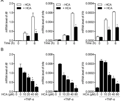
Fig. 3. HCA effectively blocks TNF- induced activation of HT-29 cells. (A) HT-29 cells were pre-treated with HCA(40 μM) for
1 hr and stimulated with TNF-(10 ng/ml) for 3 and 6 hr. After harvested, cells were lysed and genes expressions indicated were
determined by quantitative PCR. (B) HT-29 cells were pre-treated with the indicated concentrations of HCA for 1 hr and stimulated
with TNF-(10 ng/ml) for 6 hr. After harvested, cells were lysed and genes expressions indicated were determined by quantitative
PCR. Mean values of three experiment ±SEM were presented in bar graph. *p<0.05 compared to the group treated with TNF-.
Fig. 4. HCA inhibits translocation of p65 and MAPK pathways in HT-29 cells stimulated with TNF-. (A) HT-29 cells were pre- treated with HCA(40 μM) for 1 hr and stimulated with TNF-(10 ng/ml) for 30 min. After harvested, nucleic extract was isolated from cytosolic extract to measure the translocation of p65. The level of indicated proteins were detected by western blot analysis.
Protein levels were normalized with PARP for nucleic proteins and β–actin for cytosolic proteins. (B) HT-29 cells were pre-treated with the indicated concentration of HCA for 1 hr and stimulated with TNF-(10 ng/ml) for 30 min. After harvested, cells were lysed for western blot analysis to assess the phosphorylated level of the indicated proteins. The phosphorylated levels were nor- malized with total proteins. Mean values of three experiment ±SEM were presented in bar graph. *p<0.05 compared to the group treated with TNF-.
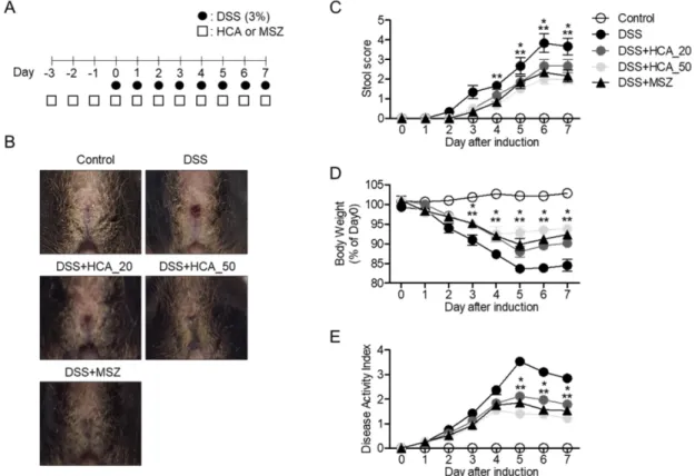
Fig. 5. Oral administration of HCA attenuates manifestation of DSS-induced IBD in vivo. (A) The experimental scheme of animal
experiment. ●: 3% DSS in drinking water, □: oral administration of HCA or MSZ. (B) The pictures of mice anus at day 7. (C) The
progress of stool score from each group. (D) The progress of body weight from each group. Body weight was normalized with
weight at day 0. (E) The progress of disease activity index from each group. Mean values of five mice ± SEM were presented in a
line graph. *p<0.05 compared to the group treated with DSS.
포함한다고 알려져 있다.
13)따라서 T NF-에 의해 염증 반 응이 유도되었을 때 HCA가 염증 사이토카인의 발현을 억 제하는지 확인하기 위해 HCA를 시간 의존적, 농도 의존적 으로 처리한 후 il8, tnfa 그리고 il1b의 mRNA 수준을 확인 하였다. Fig. 3을 통해서 HCA가 염증 사이토카인의 발현을 감소 시킴으로써 효과적으로 HT-29 대장 상피세포 활성을 억제하는 것을 확인할 수 있다. HT-29 대장 상피세포는 TNF-에 의한 자극이 있을 때 NF-κB 전사인자를 통해서 염증성 사이토카인의 발현을 유도한다고 알려져 있다.
14)HCA의 염증인자 발현 억제 효과가 NF-κB 전사인자를 통 해서 일어나는지 확인하기 위해 p65의 핵 내로의 전좌 (translocation)를 확인하였다(Fig. 4A). 뿐만 아니라 HCA는 IκB의 분해(degradation)와 IκB의 인산화를 효과적으로 억 제함을 확인하였다. NF-κB 전사인자의 하위 신호경로인 MAPK는 염증성 사이토카인의 발현에 굉장히 중요한 신호 경로로 알려져 있기 때문에
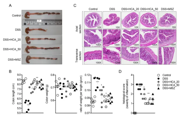
15), EKR, p38, JNK의 인산화 수 준을 western blot으로 확인하였다. 농도 의존적으로 HCA 를 처리한 후 T NF- 자극을 주었을 때 MAPK의 인산화 Fig. 6. Oral administration of HCA histologically attenuates IBD manifestations. (A) Large intestines were removed at day 7 post- induction of IBD and images were obtained from each group. (B) The length and weight were measured after removal of large intestine. The ratio of weight per length was determined and presented in dot graph. (C) Removed large intestines were treated for histological analysis and sliced tissues were prepared with 5 μm thickness for H&E staining. Large intestines were cut in axial (top panel) and transverse section(bottom). (D) Histological scores were measured from H&E stained slides from each group. Mean val- ues of five mice±SEM were presented in a scattered dot graph. *p<0.05 compared to the group treated with DSS.
Fig. 7. Oral administration of HCA decreases the expression of pro-inflammatory cytokines in IBD induced mice. (A) Large intes-
tines were removed at day 7 post-induction of IBD and mRNA levels of pro-inflammatory cytokines were observed by quantitative
PCR analysis. Mean values of five mice±SEM were presented in a bar graph. *p<0.05 compared to the group treated with DSS.
수준이 농도 의존적으로 감소함을 확인하였다(Fig. 4B). 이 러한 세포수준의 연구결과를 토대로 HCA의 구강투여가 실 제로 DSS로 유도된 염증성 장질환에서 완화 또는 치료 효 과를 나타내는지 동물실험을 수행함으로써 확인했다.
Mesalazine은 실제 IBD 환자들에게 처방되는 약으로써 이 실험에서는 양성 대조군으로 사용되었다.
16)HCA를 일주일 동안 투여한 결과 DSS에 의해 유도되는 염증성 장질환의 증상이 완화가 됨을 농도 의존적으로 확인하였다(Fig. 5B).
대변의 변형 정도나 몸무게, 질병 활동성 지표 모두 유의하 게 염증성 장질환의 증상이 완화됨을 나타내었다(Fig. 5C, 5D와 5E). 실험 종류 후 대장을 적출해서 비교했을 때 HCA 구강투여 한 그룹에서 염증으로 인한 대장의 길이 감소가 농도 의존적으로 완화되었음을 확인하였고 증가된 대장의 길이 대비 무게가 상대적으로 줄어들었음을 검증하였다(Fig.
6A와 6B). 조직학적 변화를 확인하기 위해 H&E 염색을 수 행하였는데 HCA를 구강 투여한 그룹에서 DSS가 일으킨 염증에 의한 대장 세포의 손상 정도가 상대적으로 줄어들 었음을 확인하였다(Fig. 6C와 6D). 마지막으로 HCA를 구 강 투여한 그룹에서 DSS에 의해 유도되는 염증 인자들의 발현에는 어떤 변화가 있는지 살펴보기 위해 대장 조직에 서 염증 사이토카인의 유전자 발현을 확인하였다. il8을 비 롯하여 il1b, tnfa, il6 그리고 inos의 유전자 발현이 HCA를 구강 투여한 그룹에서 농도 의존적으로 현저히 감소되어 있 음을 확인하였다(Fig. 7A). 본 연구 결과는 HCA가 대장 상 피세포에서 T NF- 자극에 의해 유도된 염증 반응에서 염 증 사이토카인의 발현을 NF-κB 전사인자의 활성 억제와 MAPK 신호전달을 억제함으로써 항염증 작용이 있다는 것 을 나타내었다. 또한 이를 토대로 DSS로 유도된 염증성 장 질환에서 여러 증상들이 유의성 있게 완화됨을 확인하였다.
이러한 결과는 HCA가 염증성 장질환 치료제로서 충분한 가능성을 가지고 있음을 시사한다.
결 론
본 연구는 p-hydroxycinnamic acid가 대장 상피세포와 염 증성 장질환에 미치는 보호 효과에 대해 평가하였다. HT- 29 세포에서 독성을 나타내지 않았던 HCA는 T NF-에 의 해 유도된 염증인자들의 유전자 발현을 농도의존적으로 억 제 하였고 이런 억제 효과는 NF-κB와 MAPK 신호 전달의 감소를 통해 일어난 다는 것을 확인하였다. DSS를 이용한 IBD 모델 마우스에서 HCA의 구강투여가 IBD의 증상 완화 에 효과가 있음을 검증하였고 조직학적으로 염증 정도가 완 화됨을 확인하였다. 이러한 염증완화는 구강 투여된 HCA 가 염증인자들의 유전자 발현을 억제함으로써 일어난 것을 검증하였다.
사 사
본 논문은 2020년도 정부(교육부)의 재원으로 한국연구재 단의 지원을 받아 수행된 기초연구사업임(과제번호: 2016 R1A6A1A03011325).
인용문헌