Introduction
The elderly population has increased disproportionately worldwide. It has been predicted that those over 65 years will constitute 20% of the total population in 2030
1. The propor- tion of old people who suffer from asthma has increased more than expected previously. In a study of old persons over the age of 60 in Switzerland, the prevalence of physician-reported asthma in old people was found to be around 7%
2. In South Korea, a report using national database showed that the prevalence of asthma in the elderly (>65 years old) was 7% in 2013, which was remarkably higher than in other age groups of asthma
3. However, elderly asthma is frequently misdiag- nosed/underdiagnosed and undertreated due to factors such as frailty, co-morbidities, low perception of their symptoms, poor compliance, and side effects associated with medication
Expression of Muscarinic Receptors and the Effect of Tiotropium Bromide in Aged Mouse Model of Chronic Asthma
Ji Young Kang, M.D., Ph.D. , In Kyoung Kim, Ph.D., Jung Hur, Ph.D., Seok Chan Kim, M.D., Ph.D., Sook Young Lee, M.D., Ph.D., Soon Seog Kwon, M.D., Ph.D. and Young Kyoon Kim, M.D., Ph.D.
Department of Internal Medicine, College of Medicine, The Catholic University of Korea, Seoul, Korea
Background: Efficacy and safety of tiotropium bromide, a muscarinic receptor antagonist, in treatment of asthma have been reported. However, its effect on airway remodeling in chronic asthma of the elderly has not been clearly verified.
The objective of this study was to investigate the effect of tiotropium and expression of muscarinic receptors as its related mechanism in an aged mouse model of chronic asthma with airway remodeling.
Methods: BALB/c female mice age 6 weeks, 9 and 15 months were sensitized and challenged with ovalbumin (OVA) for three months. Tiotropium bromide was administered during the challenge period. Airway hyperresponsiveness (AHR) and pulmonary inflammation were measured. Parameters of airway remodeling, and expression levels of M
2and M
3receptors were examined.
Results: Total cell with eosinophils, increased in the OVA groups by age, was decreased significantly after treatment with tiotropium bromide, particularly in the age group of 15 months. AHR and levels of interleukin (IL)-4, IL-5, and IL-13 were decreased, after tiotropium administration. In old aged group of 9- and 15-months-treated groups, hydroxyproline contents and levels of α-smooth muscle actin were attenuated. Tiotropium enhanced the expression of M
2but decreased expression of M
3in all aged groups of OVA.
Conclusion: Tiotropium bromide had anti-inflammatory and anti-remodeling effects in an aged mouse model of chronic asthma. Its effects seemed to be partly mediated by modulating expression M
3and M
2muscarinic receptors.
Tiotropium may be a beneficial treatment option for the elderly with airway remodeling of chronic asthma.
Keywords: Receptors, Muscarinic; Ageing; Asthma; Tiotropium Bromide; Airway Remodeling
Address for correspondence: Young Kyoon Kim, M.D., Ph.D.
Department of Internal Medicine, Seoul St. Mary’s Hospital, College of Medicine, The Catholic University of Korea, 222 Banpo-daero, Seocho- gu, Seoul 06591, Korea
Phone: 82-2-2258-6060, Fax: 82-2-596-2158 E-mail: [email protected]
Received: Jul. 5, 2018 Revised: Sep. 29, 2018 Accepted: Oct. 4, 2018
cc It is identical to the Creative Commons Attribution Non-Commercial License (http://creativecommons.org/licenses/by-nc/4.0/).
Copyright © 2019
The Korean Academy of Tuberculosis and Respiratory Diseases.
and polypharmacy. Older patients diagnosed with asthma exhibits unique features, including severe symptoms, uncon- trolled status to standard therapy, and higher mortality
4, differ- ent from younger asthma patients.
During the aging process, changes in the respiratory tract are largely divided into two categories. The first category is im- munosenescence which refers to gradual and various altera- tions in immune system with age. Its manifestations include imbalances in lymphocyte subset, thymus involution with decreased production of new T cells, defects in apoptosis, mi- tochondrial function, and malfunction of immune regulatory cells
5. These series of age-related changes will increase the susceptibility to infections and develop a status of subclinical, sustained inflammation. The second category is physiologic changes in the lung with aging, including the following: (1) decreased strength of respiratory muscles, (2) decreased lung recoil, and (3) increased stiffness of the chest wall
6. Conse- quently, the elderly takes a breath at higher lung volumes than the young, requiring an additional load on their respiratory muscles. These aforementioned changes associated with ag- ing can affect the development and pathogenesis of asthma in older people.
Acetylcholine is not only a parasympathetic neurotrans- mitter, but also an autocrine or paracrine hormone in the respiratory tract
7. It is also released from non-neuronal cells, including inflammatory, epithelial, and smooth muscle cells.
Therefore, in addition to its traditional role in smooth muscle contraction and mucus secretion, it plays proinflammatory, proliferative, and pro-fibrotic roles via interaction with musca- rinic receptors. In clinical studies
8-10, the addition of tiotropium bromide to inhaled corticosteroids (ICS) or ICS plus a long- acting β-agonist (LABA) can also delay initial exacerbation in both in adults and adolescents who have poorly controlled severe asthma. In the 2016 Global Initiatives of Asthma (GINA) report, tiotropium bromide was recommended at steps 4 and 5 as an add-on therapy to the existing treatment for at-risk symptomatic adults with poorly controlled symptoms and forced expiratory volume per 1 second less than 60%
11.
In a previous study
12, we found a distinct change in the pathogenesis of asthma according to the aging process, in- cluding the expression of muscarinic receptors in an acute asthma model. Therefore, the objective of the present study was to investigate the effect of related tiotropium bromide on airway remodeling and the expression of muscarinic recep- tors in an aged mice model of chronic asthma with airway remodeling.
Materials and Methods
1. Sensitization and antigen challenge protocol
Three different age groups (6 weeks, 9 and 15 months) of
female BALB/c mice (Orient, Seongnam, Korea) were used in this study. To obtain mice with age of 9 and 15 months, six-month-old mice were purchased and raised to the cor- responding age in our laboratory. Each age group was di- vided into three subgroups: control, ovalbumin (OVA), and OVA+tiotropium (5 to 8 mice per group). Based on a previous protocol to make a chronic asthma model
13, mice were sen- sitized by subcutaneous injection with 25 µg of OVA (grade V, Sigma-Aldrich, St. Louis, MO, USA) adsorbed to 1 mg of aluminum hydroxide (Aldrich, Milwaukee, WI, USA) in 200 µL of phosphate-buffered saline (PBS) on days 0, 7, 14, and 21.
OVA challenge [20 ng/50 µL in PBS] under isoflurane (Vedco, St. Joseph, MO, USA) anesthesia was done intranasally start- ing on day 31 twice per week for 9 weeks. Control groups were treated similarly with PBS. Mice were sacrificed at 24 hours after the final OVA challenge. The Animal Subjects Committee of the Catholic University of Korea approved all animal experi- mental protocols in this study.
2. Administration of tiotropium bromide
Tiotropium bromide was generously donated by Boeh- ringer Ingelheim Co. Ltd. After the sensitization phase, 0.1 mM tiotropium bromide in 50 µL of PBS was administered from day 38 via intranasal nebulization at 30 minutes before the OVA challenge for 5 days a week for a total of 2 months, refer- ring to the previous protocols
13,14. Control and OVA groups were treated similarly with PBS.
3. Measurement of airway hyper-responsiveness Airway hyper-responsiveness (AHR) was assessed using a flexiVent system (SCIREQ, Montreal, QC, Canada) at 24 hours after the final OVA inhalation based on changes in airway re- sistance (Rrs; cm H
2O/mL/sec) to aerosolized methacholine (Sigma) as described previously
15. Under anesthesia, trache- ostomized mice were mechanically ventilated at 150 breaths/
min and tidal volume of 10 mL/kg with a positive end-expira- tory pressure of 3 cm H
2O. Mice inhaled saline at baseline and sequentially increasing amounts of methacholine (6.25, 12.5, 25, and 50 mg/mL) via aerosol nebulizer for about 4 minutes at each concentration. Rrs was continuously recorded. Data were shown as peak Rrs value for each methacholine concen- tration.
4. Measurement of pulmonary inflammation
To evaluate pulmonary inflammation, total and differen-
tial cell counts were determined in bronchoalveolar lavage
(BAL) fluid followed by lung histology. Based on our previous
protocols
12,13, after AHR measurement, mice were thoracoto-
mized under anesthesia. BAL fluid was obtained in the right
lung through instillation with sterile PBS. After a centrifuge
at 353 ×g for 10 minutes at 4°C, supernatants were stored at –70°C. Total and differential cell types such as macrophages, eosinophils, lymphocytes, and neutrophils in the BAL fluid were counted using a hemocytometer. To perform histologic analysis, lungs were processed with 4% paraformaldehyde with overnight incubation and were then cut to sections with thickness of 5 to 6 µm. These sections of the left lung were stained with hematoxylin and eosin (H&E).
5. Assay of cytokines by enzyme-linked immunosorbent assay
Concentrations of type 2 cytokines such as interleukin (IL)- 4, IL-5, and IL-13 in supernatants of BAL fluid were measured using enzyme-linked immunosorbent assay (R&D Systems, Minneapolis, MN, USA; BioSource International, Camarillo, CA, USA) according to manufacturers’ recommendations.
6. Measurement of airway remodeling
To evaluate airway remodeling in a chronic asthma model, three crucial indicators (goblet cell hyperplasia, subepithelial collagen deposition, and smooth muscle hypertrophy) were examined. To determine goblet cell hyperplasia, left lung epithelium was stained with periodic acid Schiff (PAS) and quantified with a modified five-point scoring system (grades 0 to 4) of Padrid et al.
16. To investigate MUC5AC gene expres- sion involved in mucus hypersecretion in the respiratory tract, total RNA was isolated from lung homogenates with TRIzol (Invitrogen, Grand Island, NY, USA) and reverse-transcribed.
Real-time polymerase chain reaction (PCR) was carried out with a CFX96 Real-Time PCR Detection System (Bio-Rad Lab- oratories, Hercules, CA, USA) using specific primers and an iQ SYBR gene expression assay (Bio-Rad Laboratories) accord- ing to manufacturer’s instructions. To evaluate subepithelial fibrosis, total collagen content in the lung was quantified by measuring hydroxyproline content with spectrophotometry.
Collagen V was stained immunohistochemically. In collagen V assay, lung samples were incubated with a primary mono- clonal antibody against collagen V (Abcam, Cambridge, UK) overnight at 4°C and then incubated with a biotinylated sec- ondary antibody. The immunoreactivity in the lung tissue was detected using peroxidase reagent and 3-amino-9-ethylcarba- zole chromogen. In α-smooth muscle actin (α-SMA) assay, an immunohistology kit (IMMH-2; Sigma-Aldrich) was used to let α-SMA react with a primary antibody. Immunostained area of the lung was quantified using an image analysis system (BX50;
Olympus, Tokyo, Japan). It was expressed as per micrometer length of the basement membrane with 150–200-µm-sized internal diameter, referring to our previous protocols
12,13.
7. Evaluation for M
2and M
3muscarinic receptors subtypes by western blot
In the lung tissue, protein was quantified using the Bradford assay followed by electrophoresis using 12% sodium dodecyl sulfate‒polyacrylamide gels. Following the same method described in the previous studies
12,13, after protein were trans- ferred to nitrocellulose membrane followed by a blocking process, samples were incubated with the primary antibodies such as polyclonal rabbit anti- mAChR M
2and mAChR M
3(Santa Cruz Biotechnology, Santa Cruz, CA, USA) and then incubated with corresponding secondary anti-rabbit IgG antibodies. Proteins were detected with chemiluminescence system (PRO-MG; DaeSung Company, Korea) and quantified with Image J software (http://imagej.nih.gov/ij)
8. Statistical analyses
A comparison between groups was assessed using a t-test or Mann-Whitney U test. Data involving more than two groups were analyzed by one-way ANOVA pairwise and nonparamet- ric Kruskal-Wallis test followed by post-hoc Dunn’s multiple comparison test. All values are expressed as mean±standard error of the mean. A p-value less than 0.05 was considered sig- nificant. The SPSS version 12.0 (SPSS Inc., Chicago, IL, USA) was used for all statistical analyses.
Results
1. Effect of tiotropium bromide on AHR in an aged mouse model of chronic asthma
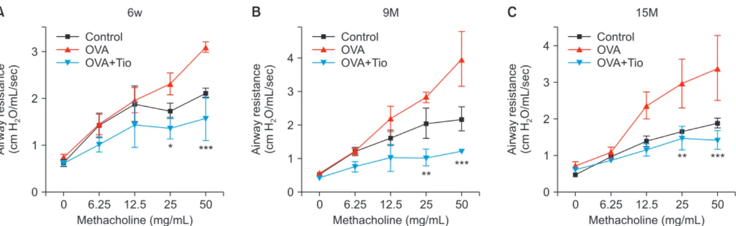
Among OVA groups, the aged mice (9 and 15 months) showed higher Rrs than the younger (6 weeks) during inha- lation of methacholine at concentration of 12.5 mg/mL of concentration. In OVA groups at 6 weeks, 9 months, and 15 months, Rrs values were 1.95±0.60, 2.19±0.76, and 2.36±0.64, respectively under 12.5 mg/mL of methacholine, showing no significant differences among groups (p>0.05). Under 25.0 mg/mL of methacholine, these values were 2.30±0.51, 2.82±0.31, and 2.96±1.31, respectively, showing significant dif- ferences between 6-week and 15-month groups (p<0.05). Un- der 50 mg/mL of methacholine, these values were 3.08±0.25, 3.97±1.62, and 3.38±1.77, respectively (p<0.01 between 6-week and 9-month groups). After tiotropium bromide administra- tion, the resistance was decreased significantly in all the three groups irrespective of age (Figure 1).
2. Effect of tiotropium bromide on pulmonary inflam-
mation in an aged mouse model of chronic asthma
In the evaluation of cell counts in BAL fluid, numbers of
total cells and eosinophils were increased remarkably in OVA groups with increasing age, whereas they showed little differ- ences among control groups. OVA+tiotropium groups showed significant reduction in the total cell number and in eosino- phils compared to OVA groups at same age, which was partic- ularly pronounced in aged mice (Figure 2A). H&E staining of lung tissue also showed an increased infiltration of peribron- chial inflammatory cells in all OVA groups, which appeared more intense in the aged groups than in the younger group.
Tiotropium bromide administration attenuated pulmonary inflammation in all corresponding OVA age groups (Figure 2B). However, few differences were detected in inflammation among control groups at different ages.
3. Effect of tiotropium bromide on type 2 cytokines in an aged mouse model of chronic asthma
With respect to type 2 cytokines, the three OVA age groups showed increased levels of IL-4, IL-5, and IL-13 than their cor- responding control groups. In OVA groups, IL-4 and IL-13 lev- els were the lowest in the 9-month-old group while IL-5 level showed a decreased tendency by increasing age. However, no age-related consecutive pattern of changes was observed (Fig- ure 3). Levels of these cytokines were significantly decreased after administration of tiotropium bromide irrespective of age.
4. Effects of tiotropium bromide on airway remodeling in an aged mouse model of chronic asthma
Pivotal structural alterations of airway remodeling in chronic asthma were analyzed in lung tissues, including gob- let cell hyperplasia, peribronchial fibrosis, and smooth muscle hypertrophy. Regarding goblet cell hyperplasia, compared with the same-age control groups, all OVA groups showed considerable elevation both in PAS-positive area and in the
score regardless of age. However, after treatment with tiotro- pium bromide, these groups showed significant decline in the score, especially with remarkable changes observed for the 15-month-old group (Figure 4A). The expression of MUC5AC gene in the lung showed a decreased tendency with age in all groups (control, OVA, and OVA+tiotropium groups). Treat- ment with tiotropium bromide decreased the expression of MUC-5AC in the OVA group at a corresponding age, although the decrease was not statistically significant (Figure 4B).
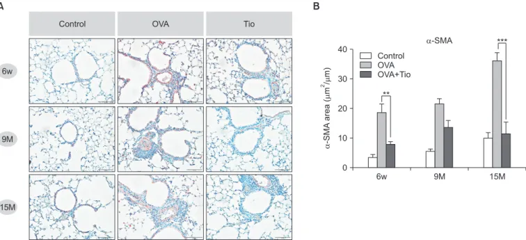
As for peribronchial fibrosis, in the three aged OVA groups, immunohistochemically stained area for collagen V was decreased by tiotropium bromide (Figure 5A). The increase in the hydroxyproline level was higher in older OVA groups than that in the younger OVA group compared to control groups with the same age. Tiotropium bromide decreased the content of hydroxyproline in all OVA groups, especially in the 9-month-old group (Figure 5B). For smooth muscle hyper- plasia, immunostained area for peribronchial α-SMA among OVA groups showed a gradual increase with age. Similar age- related patterns of this change were also observed in control groups, although less distinctly. Administration of tiotropium bromide to OVA mice significantly reduced the area of α-SMA compared to the corresponding area in OVA group with the same age. The most significant change was observed in the 15-month-old age group (Figure 6).
5. Effect of tiotropium bromide on expression of M
2and M
3muscarinic receptors in an aged mouse model of chronic asthma
In control groups, M
3expression showed an age-related in- crement. Mice in the 15-month-old group exhibited the high- est M
3but the lowest M
2level. However, in OVA groups, no consecutive trend in the expression of M
2or M
3was observed with aging. The 9-month-old OVA group had a unique pattern
A B C
Airwayresistance (cmHO/mL/sec)2
3
2
1
0 0
Methacholine (mg/mL) 6.25 12.5 25 50
* *** Airwayresistance (cmHO/mL/sec)2
4
3
2
1
0 0
Methacholine (mg/mL) 6.25 12.5 25 50
** ***
Airwayresistance (cmHO/mL/sec)2
4
3
2
1
0 Control
OVA OVA+Tio
Control OVA OVA+Tio
Control OVA OVA+Tio
0
Methacholine (mg/mL) 6.25 12.5 25 50
***
**
6w 9M 15M
Figure 1. Effect of tiotropium bromide on airway hyperresponsiveness in a mouse model of chronic asthma with aging: 6-week-old group (A),
9-month-old group (B), and 15-month-old group (C). 6w: 6 weeks old; 9M: 9 months old; 15M: 15 months old; OVA: ovalbumin; Tio: tiotro-
pium bromide. *p<0.05, **p<0.01, ***p<0.001 compared with the OVA group.
characterized by the lowest expression of M
3and the highest expression of M
2. After treatment with tiotropium bromide, all OVA groups showed an increase in M
2expression and a
decrease in M
3expression. A clear suppression of M
3receptor was shown in the 15-month-old-treated group, although the change was not statistically significant (Figure 7).
6w 80
70 60 50 40 30 20
4 Totalcellcounts(x10/mL) 10 0
9M 15M
Control OVA OVA+Tio
***
***
6w 35
30 25 20 15 10 5
Eosinophils(x10/mL)4
0
9M 15M
Control OVA OVA+Tio
*
***
6w 7.5
5.0
2.5 Neutrophilsx104 (/mL)
0.0
9M 15M
Control OVA OVA+Tio
*
6w 1.2
0.9
0.6
4 Lymphocytesx10(/mL) 0.3
0.0
9M 15M
Control OVA OVA+Tio
A
6w
9M
15M
B
Control OVA OVA+Tio
Total cell counts Eosinophils
Neutrophils Lymphocytes
Figure 2. Effects of tiotropium bromide on pulmonary inflamma- tion in a mouse model of chronic asthma with aging. (A) Inflamma- tory cell counts in bronchoalveolar lavage fluids. (B) Hematoxylin and eosin – stained section of lungs (×200). Values represent mean±standard error of the mean (n=5–8 per group). 6w: 6 weeks old; 9M: 9 months old; 15M: 15 months old; OVA: ovalbumin; Tio:
tiotropium bromide. *p<0.05 and ***p<0.001 compared with the
OVA group.
IL-4(pg/mL) 75
50
25
0 6w Control OVA OVA+Tio
9M 15M
*
***
IL-13(pg/mL)
250
200
150
100
50
0 6w Control OVA OVA+Tio
9M 15M
*
*
IL-5(pg/mL)
60 50 40 30 20 10 0
6w Control OVA OVA+Tio
9M 15M
*** *
*** *
IL-4 IL-5 IL-13
A B C
Figure 3. Effects of tiotropium bromide on levels of type 2 cytokines (A, interleukin [IL]-4; B, IL-5; and C, IL-13) in bronchoalveolar lavage fluid in a mouse model of chronic asthma with aging. Values are presented as mean±standard error of the mean (n=5–8 per group). 6w: 6 weeks old; 9M: 9 months old; 15M: 15 months old; OVA: ovalbumin; Tio: tiotropium bromide. *p<0.05 and ***p<0.001 compared with the OVA group.
PAS point scoring
Gobletcellhyperplasia
4
3
2
1
0
6w 9M 15M
RelativemRNAexpression
2.0
1.5
1.0
0.5
0.0
6w 9M 15M
*** ***
***
MUC5AC
Control OVA OVA+Tio
Control OVA OVA+Tio 6w
9M
15M
Control OVA Tio
B A
Figure 4. Effect of tiotropium bromide on goblet cell hyperplasia in airway remodeling in a mouse model of chronic asthma with aging. (A) Representative photomicrographs of periodic acid Schiff (PAS)‒stained lung sections (×200) and quantification of the PAS-stained area by point scoring as described in Materials and Methods. (B) Expression of mRNA of MUC5AC gene by real-time polymerase chain reaction. 6 w:
6 weeks old; 9M: 9 months old; 15M: 15 months old; OVA: ovalbumin; Tio: tiotropium bromide. ***p<0.001 compared with the OVA group.
6w
9M
15M
Control OVA Tio
B A
Hydroxyproline(M)
1,500
1,000
500
0
6w 9M 15M
**
Control OVA OVA+Tio
Hydroxyproline
Figure 5. Effect of tiotropium bromide on collagen deposition in airway remodeling in a mouse model of chronic asthma with aging. (A) Im- munohistochemical staining for collagen V in lung tissues (×200). (B) Measurement of hydroxyproline content to quantify collagen expres- sion in lung tissues. 6 w: 6 weeks old; 9M: 9 months old; 15M: 15 months old; OVA: ovalbumin; Tio: tiotropium bromide. **p<0.01 compared with the OVA group.
6w
9M
15M
Control OVA Tio
B A
-SMAarea(m/m)2
40
30
20
10
0
6w 9M 15M
**
Control OVA OVA+Tio
-SMA ***
Figure 6. Effect of tiotropium bromide on smooth muscle hyperplasia in airway remodeling in a mouse model of chronic asthma with aging.
(A) Representative photomicrographs of lung sections immunostained α-smooth muscle actin (α-SMA) (×200). (B) Image analysis of areas of immunostaining per micron length of basement membrane in bronchioles. 6 w: 6 weeks old; 9M: 9 months old; 15M: 15 months old; OVA:
ovalbumin; Tio: tiotropium bromide. **p<0.01 and ***p<0.001 compared with the OVA group.
Discussion
The current study demonstrated that tiotropium bromide, a muscarinic receptor antagonist, affected airway remodel- ing as well as airway inflammation and AHR in OVA-induced chronic asthma model not only in 6-week-old mice group, but also in aged, 15-month-old mice group. Furthermore, tiotro- pium treatment increased the expression of M
2receptor but decreased the expression of M
3receptor in all aged groups, especially with distinct changes of M
3in the oldest OVA group.
The cholinergic system is known to modulate type 2 im- mune response such as allergic asthma via nicotinic or mus- carinic receptors
17. In particular, the muscarinic receptors show Th2 polarization by dendritic cells, Th2 differentiation from naive T cells, and activation of mast cells, accentuating allergic response in asthma. Among five muscarinic recep- tors (M
1–M
5) in the respiratory tract, M
3subtype is expressed on almost all cell types in the airway. It plays a pivotal role in airway inflammation and remodeling
18. Kistemaker et al.
19have reported that only M
3receptor knock-out mice (not M
1or M
2receptors knock-out mice) do not induce any change of airway remodeling, including goblet cell metaplasia, smooth muscle thickening, and collagen deposition after allergen exposure. In this context, tiotropium bromide, a long-acting muscarinic receptor antagonist with a kinetic selectivity for M
3receptors and long half-life of dissociation, is thought to
exhibit a strong benefiit in terms of its effect and usage. In a few animal and in vitro experiments
13,14,19-21, tiotropium bro- mide has improved airway inflammation or remodeling in an asthma model. However, all these previous studies were con- ducted with young aged mice only.
We first investigated how airway remodeling in a chronic asthma model was altered by age and whether tiotropium reg- ulated its pathogenesis in aged mice. Collagen deposition and smooth muscle hypertrophy in airway remodeling of chronic asthma were aggravated more in aged OVA mice (9 and 15 months old) than those in younger OVA mice (6 weeks old).
However, differences in these parameters of airway remodel- ing according to age were less obvious in the control group than those in the OVA group. After treatment with tiotropium bromide, all OVA groups irrespective of age showed an anti- remodeling effect. In particular, smooth muscle hypertrophy was improved siginficantly in 15-month-old OVA mice than that in 6-week-old OVA mice. This result is consistent with a previous study with a Guinea pig model of ongoing asthma by Gosens et al.
14. In clinical settings, besides the well-known bronchodilating effect of tiotropium bromide, few clinical studies have addressed its anti-inflammatory or anti-remod- eling effects of tiotropium in asthma
22,23. Hoshino et al.
23have assessed FeNO, an inflammatory marker, and airway wall thickness measured by computed tomography, a remodel- ing marker, in 53 symptomatic asthmatic patients with ICS/
M(foldchage)2
1.5
1.0
0.5
0.0
6w 9M 15M
M(foldchage)3
3
2
1
0
6w 9M 15M
Control OVA OVA+Tio
Control OVA OVA+Tio
B A
M2
M3
-Actin OVA Tio (0.1 M)
M2
M3
6w 9M 15M

![Figure 3. Effects of tiotropium bromide on levels of type 2 cytokines (A, interleukin [IL]-4; B, IL-5; and C, IL-13) in bronchoalveolar lavage fluid in a mouse model of chronic asthma with aging](https://thumb-ap.123doks.com/thumbv2/123dokinfo/4861167.285977/6.892.63.824.119.370/figure-effects-tiotropium-bromide-cytokines-interleukin-bronchoalveolar-chronic.webp)