Expression and by Clinico-pathologic Correlation Analysis in Non-small Cell Lung Cancer
1Division of Allergy, Respiratory and Critical Care Medicine, Department of Internal Medicine, 2Department of Pathology, Chung-Ang University College of Medicine, Seoul, Korea
Jong Wook Shin, M.D.1, Jae Ho Choi, M.D.2, In Won Park, M.D.1, Jae Hyung Yoo, M.D.2
비소세포폐암에서 COX-2, MMP-9와 돌연변이형p53의 발현이 생존에 대한 예후 분석
신종욱1, 최재호2, 박인원1, 유재형2
1중앙대학교 의과대학 내과학교실, 2병리학교실
Background: In pathogenesis and prognosis of lung cancer, significance of enormous types of genetic expression were very compounding and undetermined. We performed this study to search association between clinical characteristics and expression of COX-2, MMP-9 and p53 in non-small cell lung cancer.
Methods: Ninety-one patients with adenocarcinoma or squamous cell carcinoma were enrolled. We had searched clinical data retrospectively and performed immunohistochemical staining for COX-2, MMP-9 and p53. We had analyzed significance of these three genes in clinical features and prognosis for survival.
Results: 1) In squamous cell carcinoma, male was predominant and was significantly correlated with smoking. 2) Major prognostic determinants for overall survival were curative resection. 3) Expression of COX-2 was more frequent in adenocarcinoma than in squamous cell carcinoma. 4) Negative staining of COX-2, MMP-9 and p53 was more frequent in squamous cell carcinoma than adenocarcinoma. 5) Survival duration was longer in the group with positive expression of p53 and negative for COX-2 and MMP-9 (median duration of survival = 165.6 weeks) than groups with the other expressional patterns. 6) Significant correlation was found between expression of MMP-9 and COX-2. In squamous cell carcinoma, expression of MMP-9, COX-2 and mutant p53 were mutually correlated. 7) COX-2 expression was significant prognostic factor for survival in resected cancer group. In unresected inoperable non-small cell lung cancer group, MMP-9 was statistically significant prognostic factor for overall survival.
Conclusion: COX-2 and MMP-9 might have some roles for progression or prognosis in some selected patients with non-small cell lung cancer. COX-2 and MMP-9 may have some roles for disease progression or prognosis in selected patients with NSCLC.
(Tuberc Respir Dis 2007; 63: 31-41)Key Words: COX-2, MMP-9, P53, Non-small cell lung cancer.
Address for correspondence: Jae Hyung Yoo, M.D.
Department of Pathology, Chung-Ang University College of Medicine, 221 Huk-seok-dong, Dongjak-gu, Seoul 156-756, Korea
Phone: 82-2-820-5657, Fax: 82-2-813-5387 E-mail: [email protected]
Received: May. 3. 2007 Accepted: May. 29. 2007
Introduction
Lung cancer is the leading type of cancer and the leading cause of mortality from cancer -i.e., 17.8% of all cancer deaths worldwide
1. A variety of oncobiologic studies have been focusing on onco- genes and tumor suppressor genes and enormous
genes related to cell cycle control and apoptosis. So
far, the mechanism of development and progression
of non-small cell lung cancer (NSCLC) was
explained firstly as multistep, multifocal and yet
clonal processes by chromosomal instabilities
2-6,
allelic losses and microsatellite instabilities
7-12.
Another hypothesis for carcinogenesis is the field
cancerization by which tobacco smoke and its 60
carcinogens cause the whole bronchial epitleial cells
to be exposed to increased risk of developing lung
cancer
13-15. In recent data, many other genes related
to inflammation, extracellular matrix also be con-
cerned in tumorigenensis. More defined knowledge
of lung carcinogenesis must be ascertained and
exploited to enhance survival rates of patients suffering from lung cancer.
In this study, we evaluated expression of Cyclooxygenase-2(COX-2), Matrix Metalloprote- inase-9(MMP-9), p53 in NSCLC and analyzed prognostic significance.
COX-2 is induced by inflammatory stimuli.
COX-2 over-expression has been found in many solid tumors-hepatocellular carcinoma
16, squamous cell carcinoma of esophagus
17, squamous cell car- cinoma of skin
18, colorectal adenocarcinoma
19-21.
There is evidence that cyclooxygenase-2 (COX-2) is overexpressed in lung cancer and promotes tumor proliferation, invasion, angiogenesis, and resistance to apoptosis
22.
Wild type p53 inhibits G1 to S transition through induction of p21 and facilitating apoptosis and DNA repair through BAX induction and DNA repair enzymes
23-25. The mechanism of activation in this protein comes from modification of the protein rather than genetic transcriptional or translational upregulation
26-29. Mutant type p53 expression has been consistently associated shorter survival but some studies have failed to fine such results
30,31.
Recently, several matrix metalloproteinases have shown promise as prognosticators in non-small cell lung cancer. Excessive or inappropriate expression of matrix metalloproteinase may contribute to the pathogenesis of tissue destructive processes in a wide variety of diseases including lung diseases.
MMP-9 is known to have some important role for metastasis in various cancers.
It is necessary to determine the clinical significance of these three genes in NSCLC. Genetic mechanisms for cancer development were very complicated and main schemata were concentrated on tumor suppressor genes and oncogenes and genes related cell cycle and apoptosis.
Co-expression of carcinogenic genes was more
meaningful than a single as biological predicators on survival rate of NSCLC, it is worth performing further studies.
COX-2 is related to tumor proliferation and progression, MMP-9 is important in interaction between tumor cells and adjacent interstitium and metastasis. Some studies had reported data about prognostic implication and correlation among p53, MMP-9 and COX-2 but these results are incon- clusive. Some immunohistochemical findings question a relationship between COX-2 and p53 as a marker of malignancy in colorectal cancer tissues
32. An immunohistochemical study of human colon car- cinomas designed to look for coexpression of MMP and COX-2 revealed that 80% of the specimens were overexpressed both COX-2 and matrilysin in the neoplastic epithelium
33.
In NSCLC, prognosis in overall survival can be affected by expression of COX-2, MMP-9 and p53.
We intended to determine whether expression of COX-2, MMP-9 and mutant p53 could contribute to prognosis in overall survival in patients with NSCLC.
Subjects and Methods 1. Subjects and Materials
We retrospectively searched clinical data based
on clinical charts and radiologic films of 91 patients
with non-small cell lung cancer diagnosed and
treated in Chung-Ang University hospital from
1990 to 2004. Among clinical data, we could not
definitely estimate clinical performance status. We
performed clinical or pathologic staging of cancers
based on the TNM criteria of American Joint
Committee for Cancer 2001. Eligibility criteria were
that were thatthe study patients should be those
with pathologically confirmed non-small cell lung
Number of cases, n(%) (range)
Male / Female 74 (81.3) / 17 (18.7) Age 64 years (19 - 84 years) Smoker (- / +/ unknown) 22 (24.2) /46 (50.5) /
23 (25.3) Squamous cell carcinoma 57 (62.6)
Adenocarcinoma 34 (37.4)
Stage
IA 6 (6.6)
IB 25 (27.5)
IIA 2 (2.2)
IIB 1 (1.1)
IIIA 22 (24.2)
IIIB 14 (15.4)
IV 21 (23.1)
Curative resection of lung 38 (41.8)
Chemotherapy 25 (27.5)
Radiotherapy 12 (13.3)
Survived / Deceased 26 (28.6) / 65 (71.4) Median duration of survival
(weeks) 62.7 (2.7 - 709.4)
Table 1. Clinical characteristics of study subjects cancer as squamous cell carcinoma and adeno-
carcinoma. Two histologic types could be enrolled due to eligible statistical analysis. Exclusion criteria were other histologic types of NSCLC, which was lesser numbers for statistical analysis and patients who could not be followed up.
2. Immunohistochemistry
With 4 - 5 ㎛-thick paraffin-embedded tissues of pulmonary carcinoma, we performed deparaffini- zation and soaking them into 90%, 75% and 50%
alcohol solution for 2 minutes. Next, those tissues were treated with 0.3% hydrogen peroxide- methanol for 10 minutes and washed with distilled water and sequentially with 50 mM Tris-buffered soultion(TBS, pH 7.5). Then those were treated with goat serum for 30 minutes. After removal of remnant solution, those were incubated with primary human antibodies for COX-2(1:50, Zymed, South San Francisco, CA, USA), MMP-9(1:50, Zymed,South San Francisco, CA, USA), p53 (1:50, Zymed, South San Francisco, CA, USA) for 2 hours in room temperature. Subsequently, these were washed three times with TBS. As a next procedure, secondary antibody labeled with biotin (Streptavidin biotin kit, Zymed, South San Francisco, CA, USA) was applied to them for 20 minutes and then stained with conventional Avidin-Biotin Complex method.
For development, 3-amino-9-ethylcarbazole(AEC) was used. For contrast staining, Mayer's hemato- xylin was used. And staining results were reported with light microscopy.
As for COX-2 and MMP-9 expression, positive result was defined if tumor cells with intracy- toplasmic or membranous staining were more than 10 percent in overall field. Mutant p53 expression was defined as positive if tumor cells with intranuclear staining were more than 10% among
total carcinoma cells in the whole field under light microscopy.
3. Statistical analysis
Statistical analyses were performed with the use of statistical software package, SPSS (version 11.5).
Between two groups, differences of frequency were compared with chi-square test. For comparing the mean between two groups, unpaired T test was used. Correlation analysis was done using Pearson's correlation analysis. Survival analysis was perfor- med with Kaplan-Meier method and Log rank test.
To define significant prognostic factors for overall
survival, Cox's proportional hazards model was
applied. In all statistical test in this study,
ADC*
(n = 34)
SCC†
(n = 57) p Value Male / Female 21 / 13 53 / 4 0.000 Age 63.3 years 62.14 years 0.622 Nonsmoker/Smoker 15 / 11 7 / 35 0.001 Stage I / II 10 / 3 21 / 0 Stage III / IV 9 / 12 27 / 9 0.534
Distant metastasis 121 9 0.058
Resectable 12 26 0.207
Chemotherapy(+) 11 14 0.285
Radiotherapy(+) 5 7 0.501
COX-2(+) 29 (85.3%) 27 (47.4%) 0.000 P53(+) 14 (41.2%) 32 (56.1%) 0.122 MMP-9(+) 18 (52.9%) 21 (36.8%) 0.100 median survival
(weeks)
54.4 (4.6 - 709.4)
67.0
(2.7 - 486.7) 0.658
*adenocarcinoma, †squamous cell carcinoma.
Table 2. Clinical characteristics and immunohis- tochemical staining between adenocarcinoma and squamous cell carcinoma in NSCLC.
significance was determined if p value was less than 0.05.
Results
1. Clinical characteristics of study subjects with NSCLC (Table 1, 2)
In 91 cases, proportion of male patients was 81.3%. In squamous cell carcinoma in the lungs, proportion of male was predominant than that of female(p=0.000). Proportion of smokers was also significantly higher than that in adenocarcinoma (p=0.001). Smoking history was higher in men than women (p=0.000) (Table 2).
Thirty four cases (37.4%) had early stage (stage I, II) of NSCLC, so cases with NSCLC in advanced stage(stage III, IV) were double to early ones.
Curative resection of the lung was performed in 41.8%. In the end of study, 71.4% of patients were dece ased and the others were censored. Median duration of survival in all the enrolled patients was 62.7 weeks.
2. Clinical characteristics and immunohisto- chemical staining positivity between adeno- carcinoma and squamous cell carcinoma in the lungs (Table 2)
Expressional rates of COX-2, MMP-9 and p53 were sequentially 61.5%, 42.9% and 50.5%. In staging, distant metastasis, therapeutic modalities, there was no significant differences between two histologic types of NSCLC. Interestingly, expression of COX-2 gene was higher in tissues of adeno- carcinoma than squamous cell carcinoma(p=0.000)..
In expression of p53 and MMP-9, there was no statistically significant difference between two carcinoma groups.
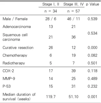
3. Expression of COX-2, MMP-9, p53 in NSCLC according to early (stage I, II) and advanced stage (stage III, IV) (Table 3)
Baseline clinical characteristics were similar
between early and advanced carcinoma. Rate of
curative resection for patients with early cancer
was 76.5% and significantly higher than for patients
with advanced stage(p=0.000). Accordingly, duration
of overall survival was longer in early stage than in
advanced stage(p=0.001). Expression of three genes
between early versus advanced NSCLC was
similar. But, in stage IV NSCLC, two cases with
grade III positive staining for COX-2 and 1 case
grade II positive for MMP-9, which were the
strongest staining among total cases.
Stage I, II Stage III, IV p Value
n = 34 n = 57
Male / Female 28 / 6 46 / 11 0.539
Adenocarcinoma 13 21
0.534 Squamous cell
carcinoma 21 36
Curative resection 26 12 0.000
Chemotherapy 6 19 0.082
Radiotherapy 5 7 0.501
COX-2 17 39 0.118
MMP-9 14 25 0.489
P-53 15 31 0.232
Median duration of
survival (weeks) 119.7 51.10 0.001
Table 3. Baseline clinical characteristics and immunohistochemical expression of COX-2, MMP-9, p53 in NSCLC according to early (stage I, II) and advanced stage (stage III, IV)
COX-2 MMP-9 P53 Total
number
Stage I, IIEarly
cancer
Advanced cancerStage
III, IV
ADC* SCC†
median survival (weeks)
+ + + 19 6 13 8 11 57.7
+ + - 12 4 8 8 4 30.0
+ - + 12 3 9 4 8 47.7
- + + 6 2 4 1 5 22.1
sSubtotal (%) 53.8% 44.1% 59.7% 61.8% 49.1%
+ - - 13 4 9 9 4 67.0
- + - 2 2 0 1 1 71.7
- - + 9 4 5 1 8 165.6
Subtotal (%) 26.4% 29.4% 24.6% 32.4% 22.8%
- - - 18 9 9 2‡ 16‡ 46.3
Subtotal (%) 19.8% 26.5% 15.7% 5.8% 28.1%
91 34 57 34 57
*adenocarcinoma, †squamous cell carcinoma, ‡p=0.025.
Table 4. Stage, histologic diagnosis and median duration of survival according to synchronous or single expression of COX-2, MMP-9 and P53 in NSCLC
4. Stage, histologic diagnosis and median dura- tion of survival according to synchronous or single expression of COX-2, MMP-9 and p53 in NSCLC (Table 4)
Between early stage of carcinoma and advanced diseases, there was no significant difference in expression of COX-2, MMP-9 and p53. In 28.1% of squamous cell carcinoma and 5.8% of adeno- carcinoma showed no expression of COX-2, MMP-9 and p53, statistical difference of which was significant(p=0.025, Table 4).
Median duration of overall survival was similar
among these groups. Notably, survival duration was
longer in the group with positive expression of p53
and negative for COX-2 and MMP-9(median
duration of survival = 165.6 weeks).
A. All study patients
COX-2
p value
(-) (+)
MMP-9 (-) 27 28
0.006
(+) 9 33
P53 (-) 21 26
0.147
(+) 15 35
B. Squamous cell carcinoma
COX-2
p value
(-) (+)
MMP-9 (-) 24 12
0.007
(+) 6 15
P53
(-) 17 8
0.061
(+) 13 19
Table 5. Correlation among COX-2, MMP-9 and p53 expression in non-small-cell lung cancer
A. NSCLC with curative resection (Univariate Analysis), n= 38
Factor RR
95.0% CI for RR
p Value Lower Upper
COX-2 0.591 0.383 0.913 0.018
B. NSCLC without curative resection (Univariate Analysis), n = 53
Factor RR
95.0% CI for RR
p Value Lower Upper
MMP-9 2.371 1.044 5.388 0.039
P53 2.395 0.959 5.985 0.062
Age 1.040 0.976 1.108 0.225
Albumin 0.707 0.372 1.343 0.290 C. Univariate regression analysis in all of study patients, n = 91
Factor RR 95% CI for RR
p Value Lower Upper
Curative
Resection 0.305 0.173 0.536 0.000
Age 1.033 1.002 1.066 0.035
Stage 2.234 1.332 3.746 0.002
Curative
Resection 0.300 0.175 0.514 0.000 Albumin 0.525 0.343 0.805 0.003
FEV1 0.999 0.999 1.000 0.002
COX-2 0.930 0.718 1.205 0.582
MMP-9 1.183 0.768 1.823 0.445
P53 0.883 0.690 1.131 0.325
D. Multivariate regression analysis in all of study patients, n = 91
Factor RR 95.0% CI for RR
p Value Lower Upper
Curative Resection
0.4823.0
85 0.2622.3460.8886.17
2 0.0190.000
Age 1.006 0.977 1.035 0.693
Stage 0.603 0.884 2.908 0.120
Albumin 0.615 0.374 1.012 0.056 NSCLC: Non-Small Cell Lung Cancer; RR: Relative Risk;
CI: Confidence Interval.
Table 6. Univariate and multivariate regression analysis for prognostic factors for overall survival using Cox's proportional hazards model
5. Correlation among COX-2, MMP-9 and p53 expression in NSCLC
MMP-9 was significantly associated with expression of COX-2(p=0.006, Table 5A). Seventy five percent of patients with negative stain for COX-2 were also negative in MMP-9. Among patients with positive stain for MMP-9, 78.6%
patients were positive stain for COX-2. In squamous cell carcinoma, COX-2 expression was correlated significantly with expression of MMP-9 (p=0.007), and also seemed to be correlated with mutant type p53 (p=0.061) (Table 5B).
6. Prognostic factors for survival in resectable cancer group with (A) or without (B) curative resection (A) and unresectable NSCLC group (B) using Cox's proportional hazards model (Table 6)
With the use of Cox's proportional hazards
model, COX-2 expression was significant prog-
nostic factor for survival in resectable cancer group
(A) (B) (C)
(D) (E) (F)
Figure 1.
Overexpression of COX-2 (A, D), MMP-9 (B, E) and p53 (C, F) in Adenocarcinoma (A, B, C) and Squamous cell carcinoma (D, E, F) of the lungs with immunohistochemical staining (x100).(relative risk = 0.591; 95% confidence interval 0.383 - 0.913; pp=0.018: Table 6A). In unresectable NSCLC group with curative resection, MMP-9 was statistically significant prognostic factor for overall survival (relative risk = 2.371; 95% confidence interval 1.044 - 5.388; pp=0.018: Table 6B).
Considering all carcinoma cases, prognostic factor for overall survival curative resection factor was only significant statistically (relative risk = 0.305;
95% confidence interval 0.173 - 0.536; p=0.000:
Table 6C).
As the results of univariate analysis for all the study patients, significant predictors for survival were tumor stage, patient’s age, curative resection, albumin, FEV1 (Table 6C) but not COX-2, MMP-9 and p53.
Curative resection was the only statistically significant prognostic factor for overall survival according to multivariate regressional analysis (relative risk = 0.482; 95% confidence interval 0.262
- 0.888; p=0.019: Table 6D).
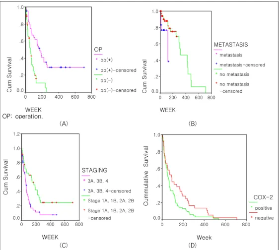
7. Survival analysis in overall group and in subgroup according to clinical factors and genetic expression.
In cases with curative resection, survival duration was significantly prolonged than those without surgery (median duration of survival in resected group versus that in inoperable group: 170.3 weeks vs. 57.7 weeks, p=0.0000, Figure 2A).
Median duration of survival in metastasis was
45.4 weeks, which was significantly shorter than
93.3 weeks in cases without metastasis (p=0.0009,
Figure 2B). In cases of advanced carcinoma, median
survival duration was 62.7 weeks, which was
significantly shortened comparing 170.3 weeks in
early carcinoma group (p=0.0028, Figure 2C). As
prognostication results based on the expression of
COX-2, MMP-9 and p53 among total study
WEEK
800 600 400 200 0
Cum Survival
1.0
.8
.6
.4
.2
0.0
OP op(+) op(+)-censored op(-) op(-)-censored
WEEK
800 600 400 200 0
Cum Survival
1.0
.8
.6
.4
.2
0.0
METASTASIS metastasis metastasis-censored no metastasis no metastasis -censored
OP: operation.
(A) (B)
WEEK 800 600 400 200 0
Cum Survival
1.2
1.0
.8
.6
.4
.2 0.0
STAGING 3A, 3B, 4 3A, 3B, 4-censored Stage 1A, 1B, 2A, 2B Stage 1A, 1B, 2A, 2B -censored
Week
800 600 400 200 0
Cummulative Survival
1.0
.8
.6
.4
.2
0.0
COX-2 positive negative
(C) (D)
Figure 2.
A. Survival analysis of all study patients according to curative resection. Median survival duratrion in resected cases was 170.3 weeks (95% CI ; 109.1 - 231.5 weeks), which was more prolonged from that in inoperable cases [57.7 weeks (95% CI ; 39.6 - 75.8 weeks)]( p=0.0000;Log rank test )B. Survival analysis of all study patients according to metastasis. Median survival duration in 71 cases without metastasis was 93.3 weeks (95% CI; 55.1 - 131.5 weeks) was superior to that in 20 cases with metastasis [45.4 weeks (95% CI; 15.9 - 74.9 weeks)] (p=0.0009; Log rank test).
C. According to stage, median survival duration of 34 cases with early stage (stage I,II) was 170.3 weeks (95%
CI; 107.2 -233.4 weeks), which was significantly longer than survival duration of 57 cases with advanced lung cancers [62.7 weeks (95% CI; 45.0 - 80.5 weeks)] ( p=0.0028; Log rank test).
D. Survival curves according to the expression of COX-2, MMP-9 and p53. According to COX-2 expression, positive group seemed to have shorter survival duration (88.0 weeks) than that of negative group (57.7 weeks)(p=0.08). There were no significant differences between positive and negative expression between positive versus negative group based on the expression of MMP-9 and p53.