Original Article
원고 접수일 2012년 10월 17일, 원고 수정일 2012년 11월 8일, 게재 확정일 2012년 11월 27일
책임저자 이의석
(152-703) 서울시 구로구 구로동로 148, 고려대학교구로병원 구강악안면외과 Tel: 02-2626-3268, Fax: 0303-0482-7575, E-mail: [email protected]
RECEIVED October 17, 2012, REVISED November 8, 2012, ACCEPTED November 27, 2012
Correspondence to Eui Seok Lee
Department of Oral and Maxillofacial Surgery, Korea University Guro Hospital
148, Gurodong-ro, Guro-gu, Seoul 152-703, Korea
Tel: 82-2-2626-3268, Fax: 82-303-0482-7575, E-mail: [email protected]
CC This is an open access article distributed under the terms of the Creative Commons Attribution Non-Commercial License (http://creativecommons.org/licenses/
by-nc/3.0) which permits unrestricted non-commercial use, distribution, and reproduction in any medium, provided the original work is properly cited.
가토 두개골 결손부에 이식된 저골(猪骨)과 혈소판 풍부 섬유소의 골형성 효과
박정익ㆍ전성배ㆍ송영일ㆍ도형식ㆍ이진용ㆍ장현석ㆍ권종진ㆍ임재석ㆍ이의석
고려대학교 의과대학 구강악안면외과학교실
Abstract
The Effect of Porcine Cancellous Bone for Bone Regeneration and Application of Platelet-rich Fibrin in Rabbit Calvarial Defect
Jeong Ik Park, Seong Bae Jeon, Young Il Song, Hyung Sik Do, Jin Yong Lee, Hyun Seok Jang, Jong Jin Kwon, Jae Suk Rim, Eui Seok Lee
Department of Oral and Maxillofacial Surgery, Korea University College of Medicine
Purpose: The purpose of this study was to evaluate the effectiveness of the platelet-rich fibrin (PRF) used in combination with the porcine cancellous bone as a scaffold, in promoting bone regeneration in the bone defects ofthe rabbit calvaria.
Methods: Ten rabbits were used in the study. Three round-shaped defects (diameter 8.0 mm) were created in the rabbit calvaria and were filled with nothing (control group), porcine cancellousbone (Experimental Group 1, porcine bone) and PRF-mixed porcine cancellous bone (Experimental Group 2). TS-GBB is a xenogenic bone-substitute product comprised of a high heat-treated mineralized porcine cancellous bone. Animals were sacrificed at 6 weeks and 12 weeks for the histological and radiographic evaluations.
Results: In the micro computed tomography and histological results, the experimental groups 1 and 2 showed more bone formation, remodeling, and calcification than the control group. The new bone formation ratio showed theGroup 2 to be larger than Group 1 at6 and 12 weeks. However, there was no significant difference between the experimental groups 1 and 2 in the new bone formation area, at the 6 and 12 weeks (
P
>0.05).Conclusion: The PRF-mixed group showed more bone formation than the porcine cancellousbonegroup (TS-GBB), butthere was a no significant difference. The PRF may not lead to enhanced bone healing when grafted with the porcine cancellous bone.
Key words: Xenograft, Bone regeneration
서 론
구강악안면영역의 골 결손부에 대한 골이식술은 나날이 발전되 어 오고 있다. 특히 임플란트 수술 시 치조골 결손부에 대한 다양한 골이식술이 개발되었다. 골이식술의 예후를 증진시키기 위해 다양한 골형성유도 인자를 함유한 혈액제제에 대한 연구가 진행되고 있다. 혈소판 풍부 섬유소(platelet rich fibrin, PRF)는 혈소판 풍부혈장(platelet rich Plasma, PRP)과 달리 제조가 간편 하며 제조과정 시 이물이 들어가지 않아 골이식술에 적용될 가치 가 높다. 혈소판 풍부 섬유소는 Transforming Growth Factor (TGF-β1), Transforming Growth Factor (TGF-β1), Vascular Endothelial Growth Factor (VEGF), Platelet-Derived Growth Factor (PDGF) 등 다양한 성장인자를 함유하여[1] 염증을 조절하 는 능력이 있으며, 치유를 촉진시켜 조직의 생존을 높이기 위한 2세대 혈소판 농축 물질로 Choukroun 등[2]에 의해 2000년 소개 되었다. PRF는 혈소판과 다양한 성장인자가 내부에 농축되어 있다[3,4]. PRF는 fibrin과 구조가 비슷하며 좀 더 효과적인 세포 의 이주 및 재생, 혈관형성을 유도한다. 이러한 기전에 의하여 골이식술에서 PRF의 사용은 골치유를 촉진시킨다고 보고되고 있다[2,5].
골이식에는 자가골, 동종골, 이종골, 합성골 등이 사용되어 왔으며, 이 중 골형성능(osteogenetic potential), 골유도능(osteoinduction), 골전도능(osteoconduction)이 있는 자가골이 가장 이상적인 것 으로 알려져 있다. 그러나 자가골 이식은 노인 환자나 어린 환자에 서는 충분한 양을 얻기 어려우며, 공여부의 동통, 골 채취를 위한 부가적인 수술 및 수술 시간의 지연, 수술 시 실혈량의 증가 등의 문제점이 있다[6]. 이러한 이유로 인하여 이종골, 동종골, 합성골 등을 골이식술에 사용하고 있다. 이종골, 동종골, 합성골은 자가 골에 비해 상대적으로 골전도능은 있으나, 골유도능 혹은 골형성 능력이 높지 않기 때문에 이에 대한 다양한 연구들이 진행되고 있다. 이 중 돼지 해면골로 이루어진 이종골인 저골(猪骨, por- cine cancellous bone)은 인체와 유사하게 수산화인회석 (hydroxyapatite)으로 대부분 구성되어 있다. 저골의 칼슘과 인 의 비율은 인체와 유사하다는 연구결과가 있다[7]. Włodarski 등[8]은 저골은 이식 시 면역반응을 일으키지 않는다고 보고하였 으며, Nannmark와 Sennerby[9]는 저골은 골전도능을 통해 신생 골을 형성한다고 보고하였다. Orsini 등[10]은 저골이 상악동 거상 술에서 신생골 형성을 방해하지 않고 생체적합성이 있다고 보고하 였다. 그 외 많은 임상연구들에서도 상악동 거상술에서 저골이 유용한 결과를 나타냄을 보고하였다[11,12]. 현재 가장 많이 사용 되고 있는 우골의 경우 생체적합성이나 신생골 형성에 있어서 조직학적으로 임상적으로 적합하다는 연구 결과들이 보고되었다 [9,13,14]. 하지만 이식된 우골의 1년이 지나도 흡수가 잘 진행되 지 않는다는 연구 결과들도 보고되었다[15,16]. 저골의 경우
Nannmark와 Sennerby[9]의 연구에서 가토 상악골에 이식하여 조직학적으로 분석한 결과 4주에서 흡수소견이 시작되는 것을 보고하였다. 임상적인 연구에서도 18명의 환자의 상악동에 이식 한 저골을 5개월째에 조직학적 검사를 시행한 결과 흡수소견이 관찰되었음을 보고하였다[13]. 이에 저골이 이종골로서 흡수가 1년 이내에 시작되면서 신생골로 대치될 수 있다는 보고를 토대로 저골이 골이식술에 사용 가능할 것으로 생각하였다.
따라서 저골로 제조한 골이식재에 혈소판 풍부섬유소를 적용한 골이식술에 대한 연구가 필요하였다. 본 연구는 가토의 두개골 결손부에 저골과 PRF를 이식하여 PRF가 골 결손부의 골재생에 어떠한 영향을 미치는지 알아보고자 시행하였다.
연구방법
1. 실험동물 및 실험재료
본 실험에서는 2.5∼3.5 kg 이상인 New Zealand white rab- bit 10마리를 실험동물로 사용하였다. 실험동물은 실험 전 실온에 서 고형사료와 물을 이용하여 2주 이상 실험실에서 사육하였다.
저골(high heat-treated mineralized porcine cancellous bone, TS-GBB, TAESAN SOLUTIONS Ltd., Seoul, Korea)을 골이식 재로 사용하였고, PRF는 실험 전 가토의 귀의 중심동맥으로부터 혈액 5 mL를 채취하였다[17,18]. 그 후, 원심분리기로 3,000 rpm에 서 10분간 처리한 뒤에 3층으로 분리된 혈액에서 젤 형태로 굳어진 중간층인 혈소판 농축 섬유소를 채취하여 사용하였다. 본 연구는 고려대학교 동물실험윤리위원회의 승인(KIACUC-20110803-2)을 받고 진행되었다.
2. 동물실험
수술 전 실험동물의 대퇴부 근육에 tiletamine과 zolazepam 의 1:1 혼합액 50 mg/kg (Zoletil, Yuhan Corp., Seoul, Korea) 과 xylazine hydrochloride 10 mg/kg (Rompum, Bayer Korea, Seoul, Korea)을 근주를 하여 전신마취를 시행하였다.
가토의 수술부위인 두부에 제모를 시행한 뒤 iodine solution으 로 소독을 하였다. 1:100,000 epinephrine을 함유한 2% lido- caine을 두부에 주사하였다. 두개부의 정중앙에 5 cm 정도 길이 의 절개를 한 뒤 골막하 박리를 측방으로 시행하여 거상하였다.
노출된 두개골 양측 및 중앙부에 8 mm 직경의 trephine bur를
사용하여 3개의 8 mm 직경의 골 결손부를 형성하였다. 형성된
좌측 골 결손부에는 저골만 이식하고, 우측 골 결손부에는 저골과
PRF를 이식하여 실험군으로 설정하였다. PRF는 소독된 거즈로
눌러서 편평한 막의 형태로 변형한 후 가루 형태로 잘라서 기존의
저골과 혼합하였다. 중앙에 위치한 골 결손부는 이식을 시행하지
않고 대조군으로 설정하였다. 적절한 지혈을 시행하고 4-0 Vicryl
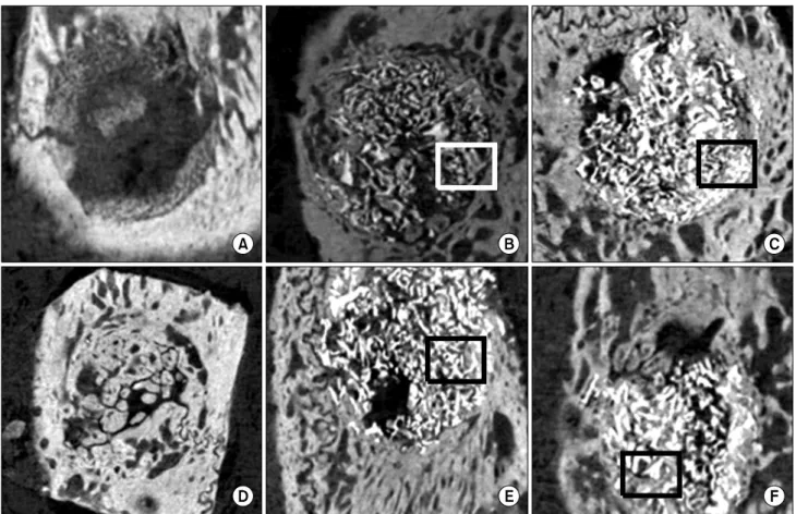
Fig. 1. Representative micro computed tomography images of the bone graft groups. New bone formation was excellent in the experiment
1 group (B, E) and experiment 2 group (C, F) rather than in the control group (A, D) at 6, 12 weeks. However, there is no significant difference between the experiment 1 and experiment 2 groups at 6 and 12 weeks. (A) 6 weeks pure bone defect, (B) 6 weeks, porcine cancellous bone graft, (C) 6 weeks porcine cancellous bone+PRF graft, (D) 12 weeks pure bone defect, (E) 12 weeks porcine cancellous bone graft, (F) 12 weeks porcine cancellous bone+PRF graft. PRF, platelet-rich fibrin.을 사용하여 골막과 피부를 봉합하였다. 모든 실험동물들은 술 후 감염 방지를 위해 5 mg/kg gentamycin (Kukje Pharm, Seongnam, Korea)을 근주하였다. 이후 실험동물을 5마리씩 나 누어 각각 6주와 12주 후에 희생을 시행하였다. 얻어진 두개골 조직은 10% formaldehyde solution에 48시간 고정시킨 후 mi- cro-computerized tomography를 촬영한 뒤 조직표본을 제작 하였다.
3. 방사선학적 분석
10% formaldehyde solution에 48시간 고정시킨 골조직을 Skyscan1172 high-resolution micro CT (Skyscan N,V., Konitch, Belgium)를 이용하여 micro CT 영성을 얻었다. 얻어진 data는 CTAn을 이용하여 8 mm의 두개골 결손부 전체를 시편의 tissue volume에 대한 bone volume의 비율로 계산하였다.
4. 조직학적 분석
실험 6주, 12주 후에 채취된 시편은 각각 10% formaldehyde solution에 48시간 고정시킨다. 그 후 formic acid로 탈회한
뒤에 파라핀 포매를 하였다. 형성된 골 결손부보다 넓은 영역을 Serial cross-section을 하여 조직절편을 제작하였다. H&E stain 을 시행하여 골과 조직의 형태를 관찰하였다.
5. 통계학적 분석
각 군 간의 신생골 부피차이를 비교하기 위하여 SPSS 12.0 (SPSS Inc., Chicago, IL, USA)을 이용하여 Wilcoxon sign- ed-rank test를 시행하였다.
결 과
1. 육안적 소견
육안적으로 실험부위를 관찰한 결과 감염, 염증, 수술부위의
이개 등의 현상은 모든 군에서 보이지 않았다. 수술 후 6주 소견에
서 이식재 사이로 신생골이 형성되는 양상을 보였으며 12주가
지나도 저골은 모두 흡수되지 않은 소견을 볼 수 있었다.
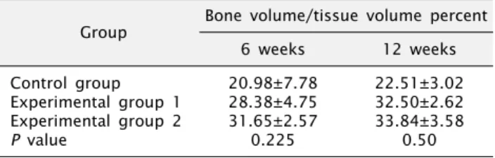
Fig. 2. Average ratio of the bone volume/tissue bone volume per
each group. Control: pure bone defect, Experimental 1: porcine cancellous bone, Experimental 2: porcine cancellous bone+platelet rich fibrin.Table 1. Area of new bone formation (bone volume/tissue volume)
Group Bone volume/tissue volume percent6 weeks 12 weeks
Control group Experimental group 1 Experimental group 2
P value
20.98±7.78 28.38±4.75 31.65±2.57
0.225
22.51±3.02 32.50±2.62 33.84±3.58
0.50 Values are presented as mean±standard deviation. Statistically significant P <0.05, Wilcoxon signed-rank test. Control: pure bone defect, Experimental 1: porcine cancellous bone, Experimental 2:
porcine cancellous bone+platelet rich fibrin.
2. 방사선학적 분석결과
Micro CT 소견에서 모든 실험 동물에서 술 후 6주 및 12주까지 저골은 모두 흡수되지 않은 상태를 보였다. 저골 사이로 신생골이 형성되는 양상을 확인할 수 있었다.
Micro CT 영상에서 저골은 두개골과 신생골에 비하여 밝은 색으로 나타나서 구분이 되었다. 두개골과 신생골은 비슷한 정도 의 밝기를 나타내었다.
6주에 희생한 이식재를 넣지 않은 대조군은 신생골이 거의 형성되지 않는 모습을 보였으며, 저골만 이식한 실험군에서는 이식재 사이로 신생골 형성 소견을 나타내었다. 저골과 PRF를 같이 이식한 실험군에서도 이식재 사이로 신생골 형성 소견을 보였다.
12주에 희생한 이식재를 넣지 않은 대조군은 6주보다 상대적으 로 신생골의 형성이 성숙된 소견을 보였다. 저골만 이식한 실험군 에서는 이식재 사이로 신생골 형성 소견이 6주보다 증가한 소견을 보였다. 저골과 PRF를 같이 이식한 실험군에서도 이식재 사이로 신생골 형성 소견이 증가한 소견을 보였다(Fig. 1).
각 군에서 형성된 신생골의 형성 정도(bone volume/tissue volume percent, BV/TV, %)를 분석 프로그램(CTAn, Skyscan N,V.)을 통해 분석하였다(Table 1). 신생골 형성률(BV/TV, %)은 6주 대조군, 저골만 이식한 군, 저골+PRF군에서 각각 20.98±7.78%, 28.38±4.45%, 31.65±2.57%로 나타났다. 이 중 저골과 저골 +PRF를 이식한 군의 신생골 형성률을 통계학적으로 비교한 결과 유의성이 없는 것으로 나타났다. 12주 대조군, 저골만 이식한 군, 저골+PRF군에서 신생골 형성률은 각각 22.51±3.02%, 32.50±2.62%, 33.84±3.58%로 나타났다. 12주에서 저골군과 저골+PRF군의 신생골 형성률을 통계학적으로 비교한 결과 유의 성이 없는 것으로 나타났다. 6주와 12주 모두에서 신생골 형성률 은 저골+PRF, 저골, 골 결손부 순으로 높게 나타났다(Fig. 2).
3. 조직학적 분석 결과
실험부위는 가토의 두개골에 형성한 골 결손부로 좌, 우측으로 두개골이 있으며 상하방으로는 두개골막 및 뇌막으로 경계를 이루
고 있다. 모든 실험군에서 저골이 있던 자리로 추정되는 부위가 시간이 지날수록 작아지는 양상을 보였으며, 시간이 경과함에 따라 신생골, 신생혈관 등이 다양한 정도의 성숙도를 보였다.
6주 소견에서는 신생골이 형성되는 것이 시작된 소견을 보인 다. 실험군에서 교직골(woven bone)이 저골 이식재 사이로 형 성되는 것을 관찰할 수 있다. 두 가지 실험군 모두 골형성 활동이 교직골을 주변으로 일어나는 것을 관찰하였다. 12주 소견에서는 형성된 신생골들이 층판구조를 형성하는 소견을 볼 수 있다. 실험 군의 신생골 내부에 혈관이 형성된 것도 확인할 수 있다. 저골이 있었던 자리로 추정되는 빈 공간도 주변의 파골세포로 인해 줄어 든 것을 확인할 수 있었다(Fig. 3).
고 찰
PRF는 2000년도에 Choukroun[2]에 의해 처음 소개된 이후 활발히 연구되고 있는 물질로 PRP에 이어 2세대 혈소판 응축물질 (fibrin concentrate)로 Dohan 등[19]에 의해 정립되었다. PRF 는 PRP의 제조 시 혈소판 활성(platelet activation)과 섬유소 중합(fibrin polymerization)을 위해 bovine thrombin과 cal- cium chloride 등 항응고제를 사용하지 않고 단순하고 저렴한 방법으로 제조가 가능한 장점이 있다[20]. 2009년 Dohan Ehrenfest 등[1]은 PRF가 방출하는 인자들을 7일 동안 분석한 결과 TGFβ-1, PDGF, VEGF 등 주요한 성장인자들을 다량 방출함을 보고하였 다. 이러한 특성으로 인해 PRF가 조직의 치유가 효과적으로 일어 날 수 있도록 도와준다[21]. 또한 채취한 PRF를 거즈로 눌러 PRF gel 내부의 액을 제거하면 질긴 성질을 가진 막을 만들 수 있으며, 골이식술 시 차단막으로 활용될 수 있다고 하였다[22,23].
또한 제조과정에서 추가적으로 들어가는 다른 물질이 없고 환자
자신의 혈액을 이용하므로 면역학적으로 안전하여 이물반응에
Fig. 3. Histological results. (A) 6 weeks porcine cancellous bone (×40), (B) 6 weeks porcine cancellous bone+PRF (×40), (C) 12 weeks
porcine cancellous bone (×40), (D) 12 weeks porcine cancellous bone+PRF (×40). (A∼D) H&E staining. PRF, platelet-rich fibrin.대한 우려가 없다. 제조과정도 PRP에 비해 비교적 간단하여, 추가 첨가물이 없기에 제조비가 저렴하다. Diss 등[24]은 상악동 거상술 시 PRF를 사용하여 평균 3.2 mm의 상악동 내부의 골이 형성됨을 보고하였다. Zhang 등[25]은 PRF와 우골(Bio-oss)을 함께 사용한 상악동 거상술에서 우골만 사용한 대조군에 비해 1.4배 많은 신생골이 형성되었음을 보고하였다. 가토의 상악동에 서 삼칼슘 인산염(tricalcium phosphate)만 이식한 대조군과 tricalcium phosphate와 PRF를 혼합하여 이식한 실험군의 골재 생 효과를 연구한 논문에서 tricalcium phosphate와 PRF를 혼합 하여 이식한 군이 더 높은 신생골 형성을 보였으며 통계학적으로 유의한 차이가 있음을 보고하였다[26]. 이와 같이 골결손 부위에 PRF를 적용한 연구에서 PRF는 신생골 형성을 증가시키는 결과를 나타내었다. 또한 silk를 scaffold로 사용하여 가토 두개골에 이식 한 연구 결과, Micro CT에서 PRF만을 이식한 대조군 보다 silk와 PRF를 혼합한 실험군의 4주 소견에서 무기질 밀도(tissue miner- al density)가 통계적으로 유의하게 높았다[27].
그러나 본 연구의 6주와 12주 소견상, 저골과 PRF를 함께 적용한 실험군 2의 결과가 대조군에 비해 모두 높게 나타났으나, 통계적으로 유의성이 없었다( P >0.05). 이는 저골의 골재생 효 과가 상당히 좋게 나타난 것으로 이전의 연구에서의 사용했던 silk나 tricalcium phosphate에 비하여 PRF의 효과가 상대적으
로 발현되지 못한 것으로 생각한다. Gürbüzer 등[28]은 발치와에 PRF를 이식한 결과 이식하지 않은 발치와와 4주 후 골재생능을 비교 시 유의할만한 차이가 없었다고 보고하였다. PRF에 함유된 성장인자와 염증조절인자들이 골세포의 증식을 증가시키지 않으 며, 이식편의 혈관신생을 돕는다는 연구내용과 여러 성장인자가 혼합되어 있을 때는 상승작용을 나타내기도 하지만 길항작용을 할 수 있다는 연구가 PRF가 골재생에 큰 기여를 하지 못한다는 가능성의 근거가 될 수도 있다[23,29,30].
Orsini 등[10]은 10명의 환자를 대상으로 저골을 이용한 상악동 거상술을 시행한 결과 본 연구와 비교하여 조금 높은 정도 (36±2.8%)의 신생골을 형성하였다고 보고하였다. 본 연구와 비슷한 형태의 돼지 두개골 결손부에 우골을 이식한 실험에서 6주에서 신생골 형성이 36.5%로 나온 결과를 보였다[31]. 이러한 연구들과 비교하여 볼 때 저골과 우골을 이식하여 얻는 신생골 형성이 큰 차이가 없음을 알 수 있었다.
본 실험에서는 저골이 골전도기능을 가진 이종골 이식재로서 신생골을 형성 역할을 충분히 수행하였기 때문에 PRF를 추가하여 얻어지는 골형성 인자의 효과를 뚜렷한 차이를 발견하기 어려웠다.
Choukroun 등[23]의 연구에 의하면 PRF를 생체막으로서 기
능을 한다는 보고가 있다. PRF의 적용은 이식재 공간을 차단하는
막으로서의 기능을 하여 이식재와 PRF를 혼합하여 넣은 경우
저골만 이식된 경우보다 골이식재를 함유하고 적용하기 쉽게 하는 역할을 한다고 할 수 있다. 저골을 사용한 연구 결과들을 토대로 볼 때[9,13,14], 본 실험에서 저골 단독사용도 이종골 골이식재로 충분히 골전도 효과가 있었다. 또한 선행 연구와 비교하여 본 연구에서 사용한 국내에서 제조한 저골 이식재도 신생골 형성 역할을 충분히 수행하였다.
PRF를 첨가한 실험군에서 조금의 신생골 형성이 증가된 결과 를 살펴볼 때, PRF는 다양한 골형성 인자를 함유하여 신생골 형성을 증가시키는데 도움을 준 것으로 생각할 수 있으나 통계적 인 유의성을 나타내지 못하였지만, 저골이 골전도 효과를 가진 이종골 이식재로 적용할 수 있음을 확인할 수 있었다. 따라서 충분한 골형성 효과가 있는 이식재에 PRF를 단순히 적용하는 방법보다는 차단막이나 다양한 운반체로서의 역할을 수행하는 것에 대한 연구가 필요할 것으로 생각한다. 이와 더불어 PRF는 다른 골이식 재료와 혼합 및 적용방법이 용이하므로 이러한 PRF 적용방법에 따른 PRF의 골형성 효과를 확인하는 추가적인 연구가 필요할 것이라 생각한다.
결 론
본 연구에서 저골만 이식한 실험군보다 저골과 PRF를 혼합 이식한 실험군에서 신생골 형성이 높게 나타났으나 통계적으로 유의할만한 차이가 없었다(>0.05). 저골 이식 시에 PRF를 사용 했을 때 신생골 형성에 있어서 큰 증가 효과를 나타내지 못하였으 나, 저골 단독 사용만으로 충분한 골형성 효과를 보였으므로, 저골의 골이식재로의 사용 가능성을 확인하였다.
References