Korean J Intern Med 2015;30:675-683 http://dx.doi.org/10.3904/kjim.2015.30.5.675
1Department of Internal Medicine, Kosin University College of Medicine, Busan; 2Department of Internal Medicine, College of Medicine, Seoul St. Mary’s Hospital, The Catholic University of Korea, Seoul;
3Department of Hematology- Oncology, Chonnam National University Hwasun Hospital, Hwasun; 4Department of Internal Medicine, Samsung Medical Center, Sungkyunkwan University School of Medicine, Seoul; 5Department of Oncology, Asan Medical Center, University of Ulsan College of Medicine, Seoul; 6Department of Internal Medicine, National Cancer Center, Goyang; 7Department of Internal Medicine, Inje University Busan Paik Hospital, Busan;
8Department of Internal Medicine, Pusan National University Hospital, Busan; 9Department of Internal Medicine, Dong-A University College of Medicine, Busan; 10Department of Internal Medicine, Korea University College of Medicine, Seoul;
11Department of Hematology and Oncology, Ulsan University Hospital, Ulsan; 12Department of Internal Medicine, Keimyung University School of Medicine, Daegu;
13Department of Internal Medicine, Ewha Womans University School of Medicine, Seoul, Korea
Received : September 5, 2014 Revised : October 13, 2014 Accepted : October 15, 2014
Correspondence to Ho Sup Lee, M.D.
Division of Hematology and Oncology, Department of Internal Medicine, Kosin University Gospel Hospital, 262 Gamcheon-ro, Seo-gu, Busan 49267, Korea Tel: +82-51-990-6363
Fax: +82-51-990-5820
E-mail: [email protected]
Background/Aims: The purpose of this study was to determine the correlations between inflammatory factors—including absolute lymphocyte count, lactate de- hydrogenase, β2-microglobulin, albumin, C-reactive protein, and ferritin—and the prognosis for survival in patients with multiple myeloma (MM) treated with induction chemotherapy containing thalidomide and who underwent autologous stem cell transplantation (ASCT).
Methods: Data from patients at 13 university hospitals in South Korea were col- lected retrospectively between December 2005 and May 2013.
Results: The median age of the 232 patients was 57 years (range, 33 to 77) and the male to female ratio was 1.09:1. In the multivariate analysis, fewer than two com- bined abnormal inflammatory factors was the only independent prognostic fac- tor for superior progression-free survival (relative risk [RR], 0.618; 95% confidence interval [CI], 0.409 to 0.933; p = 0.022), and platelet count > 100 × 109/L and fewer than two combined abnormal inflammatory factors were independent prognostic factors for superior overall survival (RR, 4.739; 95% CI, 1.897 to 11.839; p = 0.001 and RR, 0.263; 95% CI, 0.113 to 0.612; p = 0.002, respectively).
Conclusions: Patients with two or more than two combined inflammatory factors who were treated with thalidomide induction chemotherapy and who underwent ASCT showed significantly shorter survival compared to those with fewer than two combined inflammatory factors. These results could be helpful for predict- ing prognosis in patients with MM.
Keywords: Multiple myeloma; Thalidomide; Prognosis; Inflammation
The prognostic impact of inflammatory factors in patients with multiple myeloma treated with thalidomide in Korea
Cheolsu Kim1, Ho Sup Lee1, Chang-Ki Min2, Je Jung Lee3, Kihyun Kim4, Dok Hyun Yoon5,
Hyeon Seok Eom6, Hyewon Lee6, Won Sik Lee7, Ho-Jin Shin8, Ji Hyun Lee9, Yong Park10, Jae-Cheol Jo11, Young Rok Do12, and Yeung-Chul Mun13
INTRODUCTION
Many advances have been made in treatments for mul- tiple myeloma (MM), as novel agents, such as thalido- mide, bortezomib, and lenalidomide, have been devel- oped [1-3]. In particular, thalidomide was the first novel agent introduced that improved the overall response rate (ORR) in transplant eligible or ineligible patients with MM [4-6]. It was first confirmed in 1999 that tha- lidomide was active in patients with relapsed and/or refractory MM. Since then, thalidomide has become an important part of MM treatment as initial therapy for previously untreated patients, as maintenance therapy following definitive treatment, and as salvage therapy [3-9].
Many prognostic factors have been investigated to- gether with the development of MM treatments. The prognostic parameters correlated with survival in pa- tients with MM are serum β2-microglobulin (β2MG), albumin, absolute lymphocyte count (ALC), C-reactive protein (CRP), lactate dehydrogenase (LDH), serum ferritin, bone marrow plasma cell percentage, serum creatinine, hemoglobin, platelet count, age, Eastern Co- operative Oncology Group performance status, and se- rum free light chains or their ratio [10-16]. Prognostic scoring systems have also been developed, such as the Durie-Salmon Staging System (D-S stage) and the Inter- national Staging System (ISS) [17,18]. However, the sig- nificance of the D-S stage or the ISS for predicting prog- nosis decreased after the development and introduction of novel agents. Therefore, a new prognostic model or factors are needed for a more precise prediction of prog- nosis. Cytogenetic abnormalities, which are confirmed by conventional chromosomal studies or fluorescence in situ hybridization (FISH), have emerged as important prognostic markers [11,19-21]. However, FISH is used less frequently to predict prognosis in clinical practice because of the high cost and time expenditure involved.
We identified useful prognostic clinical or laboratory factors that are easily tested in patients with newly di- agnosed MM.
Serological inflammatory markers, such as ALC, LDH, β2MG, albumin, CRP, and ferritin, have been correlat- ed with the prognosis of patients with MM [10,13,15,16].
However, few studies of the prognostic impact of com- bined inflammatory status in patients with MM have
been conducted.
Therefore, the purpose of this study was to determine the correlation between the frequency of combined in- flammatory factors—including ALC, LDH, β2MG, and albumin—and the prognosis for survival in patients with MM treated with induction chemotherapy con- taining thalidomide who also underwent autologous stem cell transplantation (ASCT).
METHODS
Patients and treatment
Data from patients at 13 university hospitals in South Korea were collected retrospectively between December 2005 and May 2013. All patients were treated with thalid- omide-containing chemotherapy and then underwent ASCT. Patients who were treated with an induction che- motherapy regimen without thalidomide or those who underwent tandem ASCT were excluded. The thalido- mide combination induction chemotherapy consisted of thalidomide (fixed oral dose of 50 to 100 mg on days 1 to 28) plus intravenous (IV) dexamethasone (20 mg/m2 or oral on days 1 to 4 and days 15 to 18) every 4 weeks (TD), cyclophosphamide (fixed oral dose of 150 mg on days 1 to 4 every 4 weeks) plus TD (CTD), doxorubicin (9 mg/m2 IV rapid infusion on days 1 to 4) plus TD (TAD) every 4 weeks, and thalidomide plus vincristine 4 mg/
m2 IV infusion on days 1 to 4, 9 mg/m2 doxorubicin IV rapid infusion on days 1 to 4, and 20 mg/m2 dexametha- sone IV or oral on days 1 to 4, 9 to 12, and 17 to 20 (VAD) every 4 weeks. Only four patients were treated with this thalidomide plus VAD regimen. Low-dose aspirin was used to prevent thrombosis during induction chemo- therapy. A total of 221 patients who achieved PR or better underwent ASCT with high-dose melphalan (100 to 200 mg/m2).
Analysis
Progression-free survival (PFS) and overall survival (OS) were estimated using clinical parameters in all patients, including age, sex, hemoglobin, platelet count, ALC, β2MG, serum albumin, LDH, cytogenetic risk, ISS score, response before ASCT, and inflammatory score. A con- ventional chromosomal study was performed in 204 pa- tients and FISH including t(11;14), t(4;14), 13q deletion, 17p
deletion, t(14;16), tri 1q, and t(14;20) were also performed in more than 100 patients. The cytogenetic risk, which was determined by conventional cytogenetics or FISH, was divided into standard, intermediate, and high risk.
Standard risk included normal cytogenetics and hyper- diploidy, t(11;14), and t(6;14). Intermediate risk included t(4;14), 13q deletion, and hypodiploidy; and high risk in- cluded 17p deletion, t(14;16), and t(14;20) [19,21-23]. Each inflammatory factor of ALC level < 1.0 × 10⁹/L, β2MG level > 3.5 mg/L, serum albumin < 3.5 g/dL, and LDH lev- el above normal was defined as a combined abnormal inflammatory factor, and the sum of these abnormal
factors was analyzed. Responses before and after ASCT were assessed according to the International Myeloma Working Group uniform response criteria [24].
Statistical analysis
We investigated independent prognostic factors associ- ated with survival using the listed clinical and laboratory parameters. PFS was defined as the duration from the start date of induction chemotherapy to the date of dis- ease progression, relapse, or death from any cause after ASCT. OS was calculated from the date of diagnosis to the date of death from any cause or the final follow-up Table 1. Clinical and laboratory characteristics (n = 232)
Characteristic Value
Age, yr 57 (33–77)
Gender
Male 122 (52.6)
Female 111 (47.4)
Follow-up duration, mon 24 (7–107) Multiple myeloma subtype
IgG, κ, or λ 124 (53.4)
IgA, κ, or λ 35 (15.1)
Light chain disease 62 (26.7)
Othersa 11 (4.7)
Serum monoclonal protein, g/dL 2.4 (0.0–9.8)
Hemoglobin, g/dL 10.1 (4.2–15.4)
Platelet count, × 109/L 196.5 (37–454) Absolute lymphocyte count, × 109/L 1.730 (0.025–8.100) Serum calcium, mg/dL 9.1 (1.3–15.4) Serum total protein, g/dL 8.2 (2.9–15.2)
Serum albumin, g/dL 3.65 (1–5.6)
Serum β2-microglobulin, mg/L 3.4 (1.16–32) Lactate dehydrogenase, IU/L 310 (80–1,940) Cytogenetic risk
Standard 176 (75.9)
Intermediate 36 (15.5)
High 20 (8.6)
International Staging System
I 78 (33.6)
II 75 (32.3)
III 69 (29.7)
Unknown 10 (4.3)
Abnormal inflammatory factors
0 64 (27.6)
1 75 (32.3)
2 67 (28.9)
3 18 (7.8)
4 5 (2.2)
Treatment regimen
TD 129
CTD 96
TAD 3
TVAD 4
Response before ASCT
CR or sCR 81 (34.9)
VGPR 61 (26.3)
PR 79 (34.1)
< PR 7 (3.1)
Unknown 4 (1.7)
Table 1. Continue
Characteristic Value
Values are presented as median (range), number (%), or number.
TD, thalidomide plus dexamethasone; CTD, cyclophosphamide plus thalidomide and dexamethasone; TAD, thalidomide plus doxorubicin and dexamethasone; TVAD, thalidomide plus VAD (vincristine, doxorubicin and dexamethasone); ASCT, autologous stem cell transplantation; CR, complete response; sCR, stringent complete response; VGPR, very good partial response; PR, partial response.
aIgD κ, IgD λ or non-secretory.
date. Survival probabilities were calculated according to the Kaplan-Meier method and compared using the log-rank test. The Cox proportional hazards regression model was used for the multivariate analysis of inde- pendent prognostic factors of survival. Information about the baseline medical status and treatment modal- ities was collected from the medical records. Approval for this study was obtained from each Institutional Re- view Board.
RESULTS
Clinical and laboratory characteristics
The median age of the 232 patients was 57 years (range, 33 to 77), and the male to female ratio was 1.09:1. The medi- an serum monoclonal protein level was 2.4 g/dL (range, 0.0 to 9.8), the median hemoglobin level was 10.1 g/dL (range, 4.2 to 15.4), and the median platelet count was 196.5 × 10⁹/L (range, 37.0 to 454.0). Standard cytogenetic risk was found in 173 patients (74.6%), intermediate risk in 39 (16.8%), and high risk in 20 (8.6%). The frequencies of the combined abnormal inflammatory factors of 0, 1, 2, 3, and 4 were found in 64 (27.6%), 75 (32.3%), 67 (28.9%), 18 (7.8%), and five (2.2%) patients, respectively. Other clinical and laboratory characteristics are summarized in Table 1.
Treatment outcomes after induction chemotherapy and ASCT
The response rates achieved before and after ASCT were as follows: complete response (CR) or stringent CR in 81 (34.9%) and 142 (60.2%) patients, very good partial re- sponse (PR) in 61 (26.3%) and 47 (20.3%), PR in 79 (34.1%) and 32 (13.8%), and < PR in seven (3.1%) and five (2.3%).
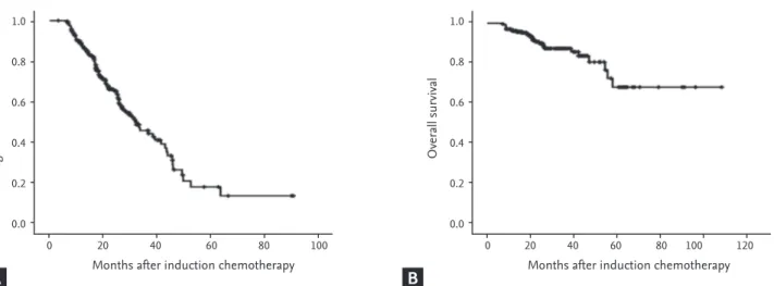
The median follow-up duration was 24.5 months (range, 6.9 to 108.9). The median PFS was 31.93 months (range, 25.1 to 38.8), and the median OS was not reached during the follow-up (Fig. 1).
Prognostic factors in patients with MM receiving thalidomide as induction chemotherapy
The following factors in the univariate analysis were associated with a greater than 2-year PFS (Table 2):
low β2MG (< 3.5 mg/L vs. ≥ 3.5 mg/L, 74.8% vs. 54.5%, p
= 0.022), normal LDH (normal vs. abnormal, 67.4% vs.
50.5%, p = 0.009), low cytogenetic risk (standard vs. in- termediate vs. high, 68.5% vs. 48.5% vs. 61.5%, p = 0.018), and having fewer than two combined abnormal inflam- matory factors (< 2 vs. ≥ 2, 72.1% vs. 53.3%, p = 0.004) (Fig.
2A). The following factors were associated with a great- er than 2-year OS (Table 2): higher hemoglobin level (<
10 g/dL vs. ≥ 10 g/dL, 84.6% vs. 96.1%, p = 0.042), higher platelet count (< 100 × 109/L vs. ≥ 100 × 109/L, 48.5% vs.
93.1%, p < 0.001), lower β2MG (< 3.5 mg/L vs. ≥ 3.5 mg/L, 98.7% vs. 84.7%, p < 0.001), normal LDH (normal vs. ab-
Progression-free survival Overall survival
Months after induction chemotherapy 0
1.0 0.8 0.6 0.4 0.2
0.0
20 40 60 80 100
Months after induction chemotherapy 0
1.0 0.8 0.6 0.4 0.2
0.0
20 40 60 80 100 120
Figure 1. (A) Median progression-free survival was 31.93 months (range, 25.1 to 38.8), and (B) median overall survival was not reached during the follow-up in patients treated with thalidomide induction chemotherapy and who underwent autologous stem cell transplantation.
A B
Table 2. Clinical and laboratory values associated with progression-free survival and overall survival in the univariate analysis
Characteristic Progression-free survival Overall survival
2-Year, % p value 2-Year, % p value
Age, yr 0.811 0.200
< 55 64.0 95.8
≥ 55 66.1 87.4
Gender 0.350 0.700
Male 59.4 88.5
Female 72.3 92.9
Hemoglobin, g/dL 0.390 0.042
< 10 60.8 84.6
≥ 10 69.3 96.1
Platelet count, × 109/L 0.286 < 0.001
< 100 44.3 48.5
≥ 100 66.7 93.1
Absolute lymphocyte count, × 109/L 0.224 0.467
< 1.0 59.7 91.0
≥ 1.0 66.2 90.3
Serum β2-microglobulin, mg/L 0.022 < 0.001
< 3.5 74.8 98.7
≥ 3.5 54.5 84.7
Serum albumin, g/dL 0.737 0.637
< 3.5 61.9 85.6
≥ 3.5 66.9 95.0
Lactate dehydrogenase, IU/L 0.009 < 0.001
Normal 67.4 95.2
Abnormal 50.5 73.4
Cytogenetic abnormalities 0.018 0.028
Standard 68.5 92.1
Intermediate 48.5 83.3
High 61.5 81.0
International Staging System 0.024
I 74.7 0.062 98.0
II 63.6 90.3
III 52.5 83.8
Abnormal inflammatory factors 0.004 < 0.001
< 2 72.1 98.1
≥ 2 53.3 81.1
Response before ASCT 0.103 0.617
CR or sCR 73.0 92.4
< CR 61.7 89.2
ASCT, autologous stem cell transplantation; CR, complete response; sCR, stringent complete response.
normal, 91.6% vs. 69.5%, p < 0.001), lower cytogenetic risk (standard vs. intermediate vs. high, 92.1% vs. 90.3%
vs. 83.8%, p = 0.028), lower ISS (I, II, and III, 98.0%, 90.3%, and 83.8%, respectively; p = 0.024), and having fewer than two combined abnormal inflammatory factors (< 2 vs. ≥ 2, 98.1% vs. 81.1%, p < 0.001) (Fig. 2B). The survival curves of 2-year PFS and OS according to the inflammatory fac- tors (Fig. 2A and 2B) are compared to 2-year PFS and OS according to ISS in Fig. 2C and 2D. The factors correlat- ed with longer survival in the univariate analysis were included in a multivariate analysis, excluding β2MG and LDH because they were already included as abnormal inflammatory factors. ISS was also excluded from the
multivariate analysis because β2MG and albumin were already included as abnormal inflammatory factors. In the multivariate analysis, abnormal inflammatory fac- tors (< 2) was the only independent prognostic factor for superior PFS (relative risk [RR], 0.618; 95% confidence interval [CI], 0.409 to 0.933; p = 0.022), and platelet count
> 100 × 109/L, and fewer than two abnormal inflammato- ry factors were the only independent prognostic factors for greater OS (RR, 4.793; 95% CI, 1.897 to 11.839; p = 0.001 and RR, 0.263; 95% CI, 0.113 to 0.612; p = 0.002, respec- tively) (Table 3).
Progression-free survival
Months after induction chemotherapy 0
1.0 0.8 0.6 0.4 0.2
0.0
20 40 60 80 100
Overall survival
Months after induction chemotherapy 0
1.0 0.8 0.6 0.4 0.2
0.0
20 40 60 80 100 120
Progression-free survival
Months after induction chemotherapy 0
1.0 0.8 0.6 0.4 0.2
0.0
20 40 60 80 100
Overall survival
Months after induction chemotherapy 0
1.0 0.8 0.6 0.4 0.2
0.0
20 40 60 80 100 120
ISS stage 12 3
ISS stage 12 3 Inflammatory factors
< 2
≥ 2 Inflammatory factors
< 2
≥ 2
Figure 2. The 2-year progression-free survival rate and 2-year overall survival rate were superior in patients with two or more combined abnormal inflammatory factors compared to those with fewer than two (A: 72.1% vs. 53.3%, p = 0.004; B: 98.1% vs.
81.1%, p < 0.001, respectively). The inflammatory factor survival curves in (A) and (B) were more significant than the Interna- tional Scoring System (ISS) stage survival curves in (C, p = 0.062) and (D, p = 0.024).
A
C
B
D
DISSCUSSION
In this study, various parameters were estimated to identify prognostic factors for survival in patients who were treated with thalidomide-containing chemother- apy and who underwent ASCT. In the univariate anal- ysis, higher β2MG (≥ 3.5 mg/L), abnormal LDH, poor cytogenetic risk, and two or more combined abnormal inflammatory factors were associated with a < 2-year PFS. Lower hemoglobin level (< 10 g/dL), lower platelet count (< 100 × 109/L), higher β2MG (≥ 3.5 mg/L), abnor- mal LDH, poor cytogenetic risk, higher ISS, and having two or more combined abnormal inflammatory factors were associated with < 2-year OS. Only two or more combined abnormal inflammatory factors was an inde- pendent prognostic factor for PFS in the multivariate analysis, and platelet count as well as combined abnor- mal inflammatory factors were independent prognostic factors for OS. Lower platelet count has been associated previously with a poor prognosis in a study reporting that low platelet count as well as low calcium, LDH, CRP, and performance status were consistent with rapid attrition in such patients due to disease aggressiveness or co-morbidities [25].
In the present study, the combined abnormal in- flammatory factors included LDH, β2MG, albumin,
and ALC. The reason for including albumin and ALC, which did not show significant results in the univariate analysis, was that the purpose of this study was not to show correlations between individual inflammatory fac- tors and prognosis or to create a new prognostic index but to determine if there was an association between a number of combined inflammatory parameters and prognosis for survival. We demonstrated that having two or more combined abnormal inflammatory factors was correlated with short survival. These results suggest that patients with MM who have multiple combined in- flammatory factors may show a poor prognosis. Some studies have reported mechanisms that may explain why inflammatory factors are associated with progno- sis. Low serum albumin level has also been associated with advanced age and poor performance status, which are known poor prognostic factors in patients with MM [26]. Serum albumin level was previously shown to neg- atively correlate with serum interleukin 6 (IL-6) levels and reflects IL-6 effects on the liver, indicating its role as a potent myeloma cell growth factor in vitro [26]. Se- rum IL-6 level is also correlated with disease severity in patients with plasma cell dyscrasias [27]. Serum soluble IL-6 receptor (sIL-6R) level is correlated with β2MG, CRP, ferritin, and LDH concentrations and is believed to be correlated with the duration of disease-free sur- Table 3. Multivariate analysis for survival
Variable Progression-free survival Overall survival
RR 95% CI p value RR 95% CI p value
Hemoglobin, g/dL
< 10
≥ 10 0.989 0.441–2.223 0.980
Platelet count, × 109/L
< 100
≥ 100 4.739 1.897–11.839 0.001
Abnormal inflammatory factors
< 2
≥ 2 0.618 0.409–0.933 0.022 0.263 0.113–0.612 0.002
Cytogenetic abnormalities Standard
Intermediate 0.737 0.350–1.551 0.422 0.762 0.216–2.689 0.673
High 1.240 0.541–2.842 0.611 1.347 0.346–5.251 0.668
RR, relative risk; CI, confidence interval.
vival [28,29].
Therefore, patients with two or more combined in- flammatory factors who were treated with thalidomide induction chemotherapy and who underwent ASCT showed significantly shorter survival than patients with fewer than two combined inflammatory factors. These results might be helpful for predicting prognosis in pa- tients with MM. However, the ORR before ASCT in this study was higher compared to those of other studies, in- cluding patients treated with thalidomide. This is likely because most hematologists consider ASCT only after patients achieve a response greater than PR in South Ko- rea. Moreover, this study had some limitations includ- ing the fact that CRP, ferritin, and other cytokines, such as IL-6 and sIL-6R, which were regarded as inflammato- ry factors, were not analyzed independently because of insufficient retrospective data. Therefore, further pro- spective studies are needed to confirm the correlations between prognosis and these inflammatory factors in transplantation-eligible or -ineligible patients with MM treated with novel agents, such as thalidomide, bortezo- mib, or lenalidomide.
Conflict of interest
No potential conflict of interest relevant to this article was reported.
Acknowledgments
This study was supported by a grant from Kosin Uni- versity College of Medicine. The authors are indebted to Chul Won Choi for advising of this manuscript. A spe- cial acknowledgment is extended to the Korean Multi- ple Myeloma Working Party (KMMWP) for supporting our data.
REFERENCES
1. Palumbo A, Facon T, Sonneveld P, et al. Thalidomide for treatment of multiple myeloma: 10 years later. Blood 2008;111:3968-3977.
2. Cavo M, Tacchetti P, Patriarca F, et al. Bortezomib with thalidomide plus dexamethasone compared with thalid- omide plus dexamethasone as induction therapy before, and consolidation therapy after, double autologous stem- cell transplantation in newly diagnosed multiple myelo- ma: a randomised phase 3 study. Lancet 2010;376:2075- 2085.
3. Morgan GJ, Davies FE. Role of thalidomide in the treat- ment of patients with multiple myeloma. Crit Rev Oncol Hematol 2013;88 Suppl 1:S14-S22.
4. Moreau P, Touzeau C. Initial treatment of transplant can- didates with multiple myeloma. Semin Oncol 2013;40:585- 591.
5. Ozaki S, Shimizu K. Autologous stem cell transplantation in elderly patients with multiple myeloma: past, present, and future. Biomed Res Int 2014;2014:394792.
6. Ahn SY, Jung SH, Joo YD, et al. Early response-based in- tensification of primary therapy in newly diagnosed mul- tiple myeloma patients who are eligible for autologous stem cell transplantation: phase II study. Ann Hematol 2014;93:1571-1577.
7. Minarik J, Sandecka V, Maisnar V, et al. 10 Years of experi- ence with thalidomide in multiple myeloma patients: re- port of the Czech Myeloma Group. Leuk Res 2013;37:1063- 1069.
8. Vande Broek I, Jacobs P. Continuous treatment in multi- ple myeloma: the future? Transfus Apher Sci 2013;49:147- 150.
9. Lokhorst HM, Schmidt-Wolf I, Sonneveld P, et al. Tha- lidomide in induction treatment increases the very good partial response rate before and after high-dose therapy in previously untreated multiple myeloma. Haematologi- ca 2008;93:124-127.
10. Copur S, Kus S, Kars A, Renda N, Tekuzman G, Firat D.
Lactate dehydrogenase and its isoenzymes in serum from patients with multiple myeloma. Clin Chem 1989;35:1968- 1970.
11. Engelhardt M, Terpos E, Kleber M, et al. European My- eloma Network recommendations on the evaluation and treatment of newly diagnosed patients with multiple my- eloma. Haematologica 2014;99:232-242.
KEY MESSAGE
1. Serologic inf lammatory markers have been re- ported to be correlated with the prognosis of patients with multiple myeloma.
2. The inf lammatory markers were independent prognostic factors for progression-free survival and overall survival in patients who were treated with thalidomide induction chemotherapy and who underwent autologous stem cell transplan- tation.
12. Kyrtsonis MC, Maltezas D, Tzenou T, Koulieris E, Brad- well AR. Staging systems and prognostic factors as a guide to therapeutic decisions in multiple myeloma. Se- min Hematol 2009;46:110-117.
13. Bataille R, Boccadoro M, Klein B, Durie B, Pileri A. C-re- active protein and beta-2 microglobulin produce a simple and powerful myeloma staging system. Blood 1992;80:733- 737.
14. Kyrtsonis MC, Vassilakopoulos TP, Kafasi N, et al. Prog- nostic value of serum free light chain ratio at diagnosis in multiple myeloma. Br J Haematol 2007;137:240-243.
15. Strasser-Weippl K, Ludwig H. Ferritin as prognostic marker in multiple myeloma patients undergoing au- tologous transplantation. Leuk Lymphoma 2014;55:2520- 2524.
16. Ege H, Gertz MA, Markovic SN, et al. Prediction of surviv- al using absolute lymphocyte count for newly diagnosed patients with multiple myeloma: a retrospective study. Br J Haematol 2008;141:792-798.
17. Greipp PR, San Miguel J, Durie BG, et al. Internation- al staging system for multiple myeloma. J Clin Oncol 2005;23:3412-3420.
18. Durie BG, Salmon SE. A clinical staging system for multi- ple myeloma: correlation of measured myeloma cell mass with presenting clinical features, response to treatment, and survival. Cancer 1975;36:842-854.
19. Lim JH, Seo EJ, Park CJ, et al. Cytogenetic classification in Korean multiple myeloma patients: prognostic sig- nificance of hyperdiploidy with 47-50 chromosomes and the number of structural abnormalities. Eur J Haematol 2014;92:313-320.
20. Palumbo A, Rajkumar SV, San Miguel JF, et al. Interna- tional Myeloma Working Group consensus statement for the management, treatment, and supportive care of pa- tients with myeloma not eligible for standard autologous stem-cell transplantation. J Clin Oncol 2014;32:587-600.
21. Mikhael JR, Dingli D, Roy V, et al. Management of new-
ly diagnosed symptomatic multiple myeloma: updated Mayo Stratification of Myeloma and Risk-Adapted Thera- py (mSMART) consensus guidelines 2013. Mayo Clin Proc 2013;88:360-376.
22. Rajkumar SV. Multiple myeloma: 2013 update on diagno- sis, risk-stratification, and management. Am J Hematol 2013;88:226-235.
23. Oh S, Koo DH, Kwon MJ, et al. Chromosome 13 dele- tion and hypodiploidy on conventional cytogenetics are robust prognostic factors in Korean multiple myeloma patients: web-based multicenter registry study. Ann He- matol 2014;93:1353-1361.
24. Cavo M, Rajkumar SV, Palumbo A, et al. International Myeloma Working Group consensus approach to the treatment of multiple myeloma patients who are can- didates for autologous stem cell transplantation. Blood 2011;117:6063-6073.
25. Barlogie B, Bolejack V, Schell M, Crowley J. Prognostic factor analyses of myeloma survival with intergroup trial S9321 (INT 0141): examining whether different variables govern different time segments of survival. Ann Hematol 2011;90:423-428.
26. Kim JE, Yoo C, Lee DH, Kim SW, Lee JS, Suh C. Serum albumin level is a significant prognostic factor reflecting disease severity in symptomatic multiple myeloma. Ann Hematol 2010;89:391-397.
27. Bataille R, Jourdan M, Zhang XG, Klein B. Serum levels of interleukin 6, a potent myeloma cell growth factor, as a reflect of disease severity in plasma cell dyscrasias. J Clin Invest 1989;84:2008-2011.
28. Papadaki H, Kyriakou D, Foudoulakis A, Markidou F, Alexandrakis M, Eliopoulos GD. Serum levels of soluble IL-6 receptor in multiple myeloma as indicator of disease activity. Acta Haematol 1997;97:191-195.
29. Lauta VM. A review of the cytokine network in multiple myeloma: diagnostic, prognostic, and therapeutic impli- cations. Cancer 2003;97:2440-2452.