This is an Open Access article distributed under the terms of the Creative Commons Attribution Non-Commercial License (http: //creativecommons.org/licenses/by- nc/4.0/) which permits unrestricted non-commercial use, distribution, and reproduction in any medium, provided the original work is properly cited.
RESEARCH NOTE
원목 및 톱밥배지 버섯 재배사 내 실내 공기서 분리한 미기록 진균 보고
안금란
1, 김지은
1, 김준영
1, 김성환
1,2*1
단국대학교 미생물학과,
2단국대학교 생물다양성연구소
Unrecorded Fungal Species Isolated from Indoor Air in the Log Bed- and Sawdust
Media-based Mushroom Cultivation Houses
Geum Ran Ahn
1, Ji Eun Kim
1, Jun Young Kim
1, Seong Hwan Kim
1,2*1
Department of Microbiology, Dankook University, Cheonan 31116, Korea
2
Institute of Biodiverstiy, Dankook University, Cheonan 31116, Korea
*
Corresponding author: [email protected]
ABSTRACT
Oak mushroom is cultivated using logs and sawdust media as substrates. In this study, fungi were isolated during a monitoring of indoor air in the oak mushroom cultivation houses located in Cheongyang-gun of Chungnam, Geoje-gun of Gyeongnam, Gumi-si of Gyeongbuk, Jangheung-gun of Jeonnam and Yeoju-si of Gyeongggi-do. Identification of the fungi based on morphology and molecular analysis of the internal transcribed spacer region and 28S rDNA, translation elongation factor translation elongation factor 1 a gene, and β-tubulin gene revealed that six fungi, Cenangium acuum, Neopestalotiopsis surinamensis, Metarhizium marquandii, Periconia macrospinosa, Trichoderma petersenii, and Trichoderma paratroviride that have not been recorded previously in Korea.
Keywords: Indoor air, Oak mushroom cultivation houses, Unrecorded fungi
표고버섯은 약용물질로서 항암작용이 있는
lentinan
과 혈중 콜레스트롤의 함량을 낮추는eritadenine
등을 포함하고 있으며[1
-3
] 다이어트에도 도움이 되는 버섯이다[4
]. 또한 탄수화물, 단백질, 지방, 비타민, 미네랄 등 다양한 성분을 함유하고 있는[5
] 맛과 풍미가 뛰어난 식용버섯이다. 이렇듯 맛 과 생물학적 기능을 담고 있는 표고버섯은 재배하는 임가의 증가에 더하여 재배면적, 생산량이 매 년 증가하고 있다[6
]. 표고버섯 생산은 현재 원목재배와 톱밥배지를 이용하고 있다. 최근에는 재배 자의 노령화에 따라 원목재배보다 톱밥배지를 이용한 버섯 생산을 선호하고 있다. 기존에 발표된 논문에 의하면 원목재배와 톱밥배지를 이용하여 표고버섯을 재배하는 재배사 내의 환경에서 다양 한 미생물 상이 존재하는 것으로 보고되었다[7
-10
]. 표고버섯 재배사 내 환경에서 분리된 진균들은OPEN ACCESS
© 2018 THE KOREAN SOCIETY OF MYCOLOGY.
https://doi.org/10.4489/KJM.20180054
Accepted: November 19, 2018 Revised: November 19, 2018 Received: November 12, 2018 Kor. J. Mycol. 2018 December, 46(4): 495-503 pISSN : 0253-651X
eISSN : 2383-5249
대체로 포자가 바람에 의해 쉽게 부유하여 전파되기 쉬운 종들이었고 이중 몇몇 종은 인체에 유해 한 종으로 보고되었다. 더불어 국내에 보고되지 않았던 미기록 진균들도 발굴되었다. 이에 따라 본 연구에서는 지속적으로 미기록 진균을 발굴하기 위해서 표고버섯 재배사의 실내공기로부터 진균 모니터링을 수행하여 진균을 포집하고 분리하였다. 분리된 진균을 동정한 결과 국내에 보고되지 않은 미기록 진균
6
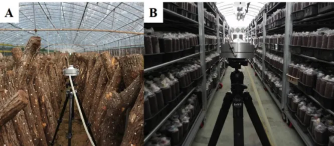
종이 검출되어 이들에 대해 보고하고자 한다.공기시료 채취는
2016
년4
월 전라남도 장흥군,5
월 경상북도 구미시, 경상남도 거제시,6
월 경기 도 여주시 소재의 원목으로 버섯을 재배하는 임가4
곳 (Fig
.1A
)과 톱밥으로 버섯을 재배하는 임가1
곳(2016
년6
월 충청남도 청양군) (Fig
.1B
)의 버섯재배사 내서 실내공기를 포집하였다. 실내공기 채집 방법은ISO 16000
-18
에 기반한 충돌법에 따라Andersen sampler
(KAS
-110
,Kemik Corporation
,Seongnam
,Korea
)를 사용하여28
.3 L
/min
유량으로1
분간 포집하였다[8
-10
]. 공기 채집은 표고버섯 재배사의 가운데 지점에서 진행하였고1
.3 m
높이에서 수평을 맞추어 수행하였다. 배양 배지로서 는 암피실린(ampicillin
)을100 µg
/mL
농도로 첨가한malt extract agar
(Difco
,Detroit
,MI
,USA
)를 사용 하였다. 실내공기를 채집한 배지를5
일 동안25
℃ 배양기에서 배양하였고, 자라난 진균은 단포자 분리를 통해 순수 분리하였다. 형태적 동정을 위해서는PDA
에 순수 배양된 진균의 포자와 균사를 대상으로 광학현미경 (Axioskop40
;Carl Zeiss
,Oberkochen
,Germany
)을 이용하여 미세구조를 관찰 기 록하였다. 배지에 나타난 균총의 형태는 육안으로 관찰하고 사진으로 촬영 기록하였다. 분자생물 학적 동정을 위해서는Kim
의drilling
방법[11
]을 참조하여 진행 후Qiagen
사의Plant genomic DNA extraction kit
(69106
,Germany
)을 이용하여genomic DNA
를 추출하였다.1
차 동정으로internal transcribed spacer
(ITS
)rDNA region
을 증폭하기 위해서는ITS1
(5
'-TCCGTAGGTGAACCTGCG
-3
')/ITS4
(5
'-TCCTCCGCTTATTGATATGC
-3
')[12
] 프라이머를 이용하여 중합효소연쇄반응 (polymerase chain reaction
,PCR
)을 수행하였다.PCR
증폭된DNA
산물은1
% 아가로스겔에서 전기영동을 수행 하여 기대되는 밴드의 크기를 확인한 후NAVIGen
TMPCR Purification Kit
(NAVIBIOTECH
,Korea
)를 사용하여 정제하고 마크로젠사 (Seoul
,Korea
)에 의뢰하여 염기서열을 분석하였다. 분석된 염기서 열의 분자동정을 위해서는 미국National Center for Biotechnology Information
(http
://www
.Fig. 1. Example of indoor air uptake using air samplers in two different oak mushroom cultivation houses. A, log bed-based
mushroom cultivation houses; B, sawdust media-based mushroom cultivation houses.ncbi
.nlm
.nih
.gov
)의 웹에 있는BLAST
프로그램을 사용하여DNA
데이터베이스에 등록되어 있는 진균들의DNA
와 상동성을 비교하였다.ITS rDNA region
염기서열 결과를1
차적으로 검토 후 알려 진 진균 종과 염기서열의 상동성이 낮은 균주는 논문검색을 통하여 그 종이 속한 속을 동정할 때 사용된 여러 다른 유전자 특이적 프라이머를 이용하여 증폭하였다. 사용된 증폭용 특이적 프라이머로 는28S
를 증폭하는 프라이머LR0R
(5
'-ACCCGCTGAACTTAAGC
)/LR7
(5
'-GCAGATCTTGGTGGTAG
-3
') [13
],β
-tubulin gene
을 증폭하는 프라이머BT12
(5
'-GTTGTCAATGCAGAAGGTCTC
-3
')/T10
(5
'-ACGATAGGTTCACCTCCAGAC
-3
')[12
],translation elongation factor 1 a
(tef
-1a
)gene
을 증폭하는 프 라이머TEF728
(5
’-CATCGAGAAGTTCGAGAAGG
-3
’)/TEF1
(5
’-GCCATCCTTGGAGATACCAGC
-3
’)[14
] 를 이용하였다. 계통분석을 위해서는 분리 균주와 관련된taxon
의 염기서열을NCBI
의GenBank
에서 다 운받아 다중정렬 한 후MEGA 6
프로그램[15
]을 이용하여 염기서열의 유사도 및phylogenetic analysis
를 수행하였다. 계통도는neighbor
-joining
방법[16
]으로 분석하였고 계통도 가지의clade
신뢰도는1
,000
번의bootstrap resampling
을 수행하여 분석하였다. 동정된 진균 중 미기록 균주인DK12
-2
,DK12
-3
,DK12
-4
,DK12
-5
,DK12
-6
,DK12
-7
번은 국립생물자원관(National Institute of Biological Resource
)에 기탁하여 등록번호를 받았다.DK12
-2
는NIBRFGC000499879
,DK12
-3
은NIBRFGC000499880
,DK12
-4
는NIBRFGC000499881
,DK12
-5
는NIBRFGC000499882
,DK12
-6
은NIBRFGC000499883
,DK12
-7
은NIBRFGC000499884
번으로 등록번호를 받았다. 또한 이들 균주의Fig. 2. Unrecorded fungal species isolated from mushroom cultivation houses in this study. The fungi were grown on potato
dextrose agar at 25℃ for 7 days. A, Cenangium acuum; B, Periconia macrospinosa; C, Neopestalotiopsis surinamensis; D, Metarhizium marquandii; E, Trichoderma petersenii; F, Trichoderma paratroviride.분석된 염기서열은
NCBI
의GenBank DNA database
에 등록하였고 등록번호는Table 1
에 제시하였 다.균주 DK12-2 (Cenangium acuum)
2016
년5
월 경상북도 구미시에 위치한 표고 원목 재배사 내 실내공기에서 분리한 균을PDA
에서10
일간 배양하였을 때 흰색의 균사체로 생장하며 중앙에 균사가 올리브색으로 부분적으로 변하였다 (Fig
.2
-A
). 광학현미경으로 관찰하였을 때, 균사 끝에5
-7
×2
.5
-3 μm
크기의 곤봉 모양의 포자를 가지고 있었다(Fig
.3
-A
).1
차적으로ITS region
을 분석하였을 때Cenangium
속으로 동정이 되었으 며, 추가로LSU rDNA region
을 분석한 결과Cenangium accuum Cooke
&Peck
,Grevillea
(KX090828
) 과98
% 상동하였다(Fig
.4
-A
).Cennangium
종 중에C
.ferruginosum
은 소나무류의 피목가지마름병을 유발시키는 것으로 알려져 있다[17
].Table 1. List of the fungal species isolated and identified in this study from mushroom cultivation houses in this study Cultivation type Sampling site Isolate from this study Identified species Sequence similarity of
analyzed DNA region Accession no.
Log Bed Jangheung DUCC5186 Neopestalotiopsis surinamensis β-tubulin MH844695
Gumi DUCC5184 Cenangium acuum LSU MH819183
Geoje DUCC5197 Periconia macrospinosa ITS MH809377
Yeoju DUCC5183 Trichoderma petersenii tef1α MK178278
DUCC5181 Trichoderma paratroviride tef1α MK178277
Sawdust media Cheongyang DUCC5194 Metarhizium marquandii β-tubulin MH844693 LSU, large subunit; ITS, internal transcribed spacer; tef1α, translation elongation factor 1-alpha.
Fig. 3. Microscopic image of unrecorded fungal species isolated from mushroom cultivation houses in this study. A, Cenangium
acuum; B, Periconia macrospinosa; C, Neopestalotiopsis surinamensis; D, Metarhizium marquandii; E, Trichoderma petersenii; F, Trichoderma paratroviride. Red arrow is important feature (scale bar = 2 μm).균주 DK12-3 (Periconia macrospinosa)
2016
년5
월 경상남도 거제시의 표고 원목 재배사 내 실내공기에서 분리한 균을PDA
에서6
일간 배 양하였을 때 균사가 동심 윤문을 그리며 생장하였으며 짙은 카키색 또는 녹회색의 균사로 생장하 였다(Fig
.2
-B
). 광학현미경으로 관찰한 결과 균사 끝에1
–1
.5
×1
–1
.5 μm
크기의 구형 모양의 포 자가 체인형태로 존재하였다(Fig
.3
-B
).ITS region
을 분석한 결과Periconia macrospinosa Lefebvre
&Aar
.G
.Johnson
(JQ781723
)과100
% 상동하였다(Fig
.4
-B
).Periconia macrospinosa
균은 염소를 포함한 대사과정에서 생합성을 하는 균으로 보고되었으며[18
], 미소균핵과 후막포자를 형성하는 균으로 알려져 있다[19
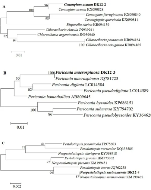
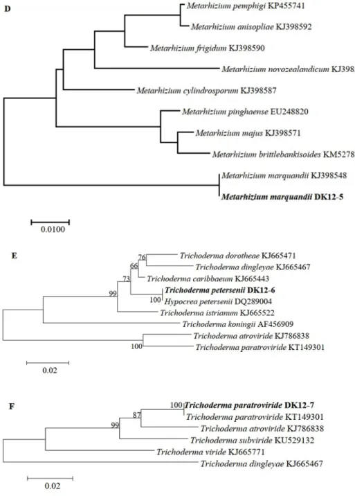
].Fig. 4-1. Phylogenic relationships of the six fungal isolates inferred by the neighbor joining analysis
based on 28S rDNA (A), internal transcribed spacer rDNA (B, D), β-tubulin gene (C) or the translation elongation factor 1a gene (E, F) sequences. A, Cenangium acuum (NIBRFGC000499879); B, Periconia macrospinosa (NIBRFGC000499880); C, Neopestalotiopsis surinamensis (NIBRFGC000499881);균주 DK12-4 (Neopestalotiopsis surinamensis)
2016
년4
월 전라남도 장흥군의 표고 원목 재배사 내 실내공기에서 분리한 균은PDA
에서6
일간 배 양하였을 때 흰색의 균사체가 조밀하며 솜처럼 약간 부유하여 생장하였다(Fig
.2
-C
). 광학현미경 관 찰하였을 때2
개의 편모와3
개의 격벽을 가지고 있는7
-8
×3
–3
.5 μm
크기의 포자를 확인하였다 (Fig
.3
-C
).1
차적으로ITS region
을 분석하였을 때Neopestalotiopsis
속으로 동정되었으며,β
-tubulin
Fig. 4-2. Phylogenic relationships of the six fungal isolates inferred by the neighbor joining analysis
based on 28S rDNA (A), internal transcribed spacer rDNA (B, D), β-tubulin gene (C) or the translation elongation factor 1a gene (E, F) sequences. D, Metarhizium marquandii (NIBRFGC000499882); E, Trichoderma petersenii (NIBRFGC000499883); F, Trichoderma paratroviride (NIBRFGC000499884).region
을 분석하여Neopestalotiopsis surinamensis Maharachch
.,K
.D
.Hyde
&Crous
(KM199465
)와100
% 상동하였다(Fig
.4
-C
).Neopestalotiopsis
속의 다른 종들은 토마토에 무름병[20
] 또는 포도덩굴의 잎에 반점을 야기시키는 것으로 알려져 있다[21
].균주 DK12-5 (Metarhizium marquandii)
2016
년6
월 충청남도 청양군에 있는 표고 톱밥 재배사 내 실내공기에서 분리한 균을PDA
에서6
일 간25
℃에서 배양하였을 때 아래Fig
.2
-D
와 같이 생장이 매우 느렸다. 균사는 연보라색 또는 핑크 색의 균사로 생장하였다. 광학현미경으로 균사를 관찰한 결과 분생포자경 정단에0
.5
-1
×0
.5
-1 μm
크기의 구형 모양의 작은 분생포자를 가지고 있었다(Fig
.3
-D
). 청양 톱밥 재배사 내 실내공기에 서 분리한 균은β
-tubulin region
을 분석하였을 때Metarhizium marquandii
(Massee
)Kepler
,S
.A
.Rehner
&
Humber
(KJ398548
)와99
% 상동하였다(Fig
.4
-D
).Metarhizium
속의M
.viride
는 카멜레온, 턱수염도 마뱀 등에 진균증을 일으키는 종으로 보고되어있다[22
].균주 DK12-6 (Trichoderma petersenii)
2016
년6
월 경기도 여주시의 표고 원목 재배사 내 실내공기에서 분리한 균을potato dextrose agar
(PDA
)에서3
일 간 배양하였을 때 아이보리 또는 연한 회색의 균사가 빠르게 생장하였다(Fig
.2E
).광학현미경으로 균사를 관찰한 결과 삼지창 모양의 분생포자경 정단에
2
~2
.5
×2
~2
.5 μm
크기의 구형 모양의 분생포자를 가지고 있었다(Fig
.3E
).tef
-1a region
을 분석하였을 때Trichoderma petersenii Samuels
,Dodd
&Schroers
(FJ860670
)과100
% 상동하였다(Fig
.4E
).Trichoderma
속은 기존 다른 논문 에 의하면 표고버섯 재배사 내에서 자주 발견되는 진균이다[7
,23
]. 본T
.petersenii
와 더불어 다음의T
.paratroviride
균에 대한 정보는 아직 분류학적 정보를 제외하고는 연구된 바가 없다. 본 연구에서 도 표고버섯 재배사에서 분리된바 향후 버섯재배에 대해 어떠한 영향을 줄지에 대한 연구가 시급 히 필요하다고 생각된다.균주 DK12-7 (Trichoderma paratroviride)
2016
년6
월 경기도 여주시의 표고 원목 재배사 내 실내공기에서 분리한 균을PDA
에서3
일 간 배양 하였을 때 아이보리 또는 연한 회갈색의 균사가 빠르게 생장하였다(Fig
.2F
). 광학현미경으로 균사 를 관찰한 결과 삼지창 모양의 분생포자경 정단에3
~3
.5
×3
~3
.5 μm
크기의 구형 모양의 분생포자 를 가지고 있었다(Fig
.3F
).tef
-1a region
을 분석하였을 때Trichoderma paratroviride Jaklitsch
&Voglmayr
(KT149301
)과99
% 상동하였다(Fig
.4F
).적요
2016
년4
월부터6
월까지 표고 원목재배 임가4
곳과 표고 톱밥재배 임가1
곳의 재배사 내 실내 공기 에서 진균을 포집 및 분리 동정하였다. 총6
개의 국내 미기록 진균(Cenangium acuum
,Periconia
macrospinosa
,Neopestalotiopsis surinamensis
,Metarhizium marquandii
,Trichoderma petersenii
,Trichoderma
paratroviride
)을 분리하였으며 이들 균류에 대하여 형태학적 특성 및internal transcribed spacer rDNA
region
,large subunit region
,β
-tubulin
또는translation elongation factor 1α
유전자 염기서열에 기반하여 계 통학적 분석 결과를 기술하였다.ACKNOWLEDGEMENTS
This work was carried out with the support of National Institute of Biological Resource
(NIBR
).REFERENCES
1. Jong SC, Birmingham JM. Medicinal and therapeutic value of the shiitake mushroom. Adv Appl Microbiol 1993;39:154-84.
2. Suzuki S, Oshima S. Influence of shiitake on human serum cholesterol. In: Proceedings of the 9th International Scientific Congress on the Cultivation of Edible Fungi; 1974; Tokyo, Japan and Taipei, Taiwan. Tokyo: Mushroom Research Institute in Japan; 1977. p. 463-7.
3. Lee MR, Oh DS, Wee AJ, Yun BS, Jang SA, Sung CK. Anti-obesity effects of Lentinus edodes on obese mice induced by high fat diet. J Korean Soc Food Sci Nutr 2014;43:194-9.
4. Nanba H, Mori K, Toyomasu T, Kuroda H. Antitumor action of shiitake (Lentinus edodes) fruit bodies orally administered to mice. Chem Pharm Bull (Tokyo) 1987;35:2453-8.
5. Joo MC. The analysis of management and the management of cultivation of Lentinus edodes I. for full-development of mycelium in bed logs. J Korean Forest Soc 1996;85:596-604.
6. Kang MY, Kim S, Yun HJ, Nam SH. Antioxidative activity of the extracts from browned oak mushroom (Lentinus edodes) with unmarketable quality. Korean J Food Sci Technol 2004;36:648-54.
7. Kim CS, Park MS, Kim SC, Maekawa N, Yu SH. Identification of Trichoderma, a competitor of shiitake mushroom (Lentinula edodes), and competition between Lentinula edodes and Trichoderma species in Korea. Plant Pathol J 2012;28:137-48.
8. Kwon HW, Yun YH, Kim JY, Kim SH, Ko HK. New records of fungi isolated from indoor air of greenhouse used for shiitake cultivation in Korea. Kor J Mycol 2015;43:58-63.
9. Ahn GR, Kwon HW, Ko HK, Kim SH. Unrecorded fungal species isolated from greenhouses used for shiitake cultivation in Korea. Kor J Mycol 2016;44:8-15.
10. Ahn GR, Ahn HS, Kwon HW, Ko HK, Kim SH. Unrecorded fungi isolated from indoor air of cultivation houses used for field test of a newly bred domestic shiitake cultivar. J Mushrooms 2016;14:168-78.
11. Kim SH, Uzunovic A, Breuil C. Rapid detection of Ophiostoma piceace and O. quercus in stained wood by PCR. Appl Environ Microbiol 1999;65:287-90.
12. White TJ, Bruns T, Lee S, Taylor J. Amplification and direct sequencing of fungal ribosomal RNA genes for phylogenetics. In: Innis MA, Gelfand DH, Sninsky JJ, White TJ, editors. PCR protocols: a guide to methods and applications. San Diego: Academic Press; 1990. p. 315-22.
13. Glass NL, Donaldson GC. Development of primer sets designed for use with the PCR to amplify conserved genes from filamentous ascomycetes. Appl Environ Microbiol 1995;61:1323-30.
14. Evidente A, Ricciardiello G, Andolfi A, Sanbatini MA, Ganassi S, Altomare C, Favilla M, Melck D. Citrantifidiene and citrantifidiol: bioactive metabolites produced by Trichderma citrinoviride with potential antifeedant activity toward aphids. J Agric Food Chem 2008;56:3569-73.
15. Tamura K, Stecher G, Peterson D, Filipski A, Kumar S. MEGA6: Molecular Evolutionary Genetics Analysis version 6.0. Mol Biol Evol 2013;30:2725-9.
16. Saitou N, Nei M. The neighbor-joining method: a new method for reconstructing phylogenetic trees. Mol Biol Evol 1987;4:406-25.
17. Kim MJ, Kim KH. Cenangium dieback associated with Cenangium ferruginosum. Korean Turfgrass Sci 2009;23:361-8.
18. Henderson GB, Hill RA. The biosynthesis of chlorine-containing metabolites of Periconia macrospinosa. J Chem Soc Perkin 1 1982;0:3037-9.
19. Hall G. Demonstration of chlamydospores and evidence for microsclerotia in Periconia macrospinosa. Trans Br Mycol Soc 1986;86:347-9.
20. Ayoubi N, Pari SS. Morphological and molecular identification of Neopestalotiopsis mesopotamica causing tomato fruit rot. J Plant Dis Prot 2016;123:267-71.
21. Jayawardena RS, Liu M, Maharachchikumbura SS, Zhang W, Xing Q, Hyde KD, Nilthong S, Li XH, Yan JY. Neopestalotiopsis vitis sp. nov. causing grapevine leaf spot in China. Phytotaxa 2016;258:063-074.
22. Schmidt V, Klasen L, Schneider J, Hübel J, Pees M. Characterization of Metarhizium viride mycosis in veiled chameleons (Chamaeleo calyptratus), panther chameleons (Furcifer pardalis), and inland bearded dragons (Pogona vitticeps). J Clin Microbiol 2017;55:832-43.
23. Kim JY, Kwon HW, Yun YH, Kim SH. Identification and characterization of Trichoderma species damaging shiitake mushroom bed-logs infested by Camptomyia pest. J Microbiol Biotechnol 2016;26:909-17.