서론
수직적, 수평적 골흡수가 심한 환자에서 임플란트를 이용한 전악 수복은 임플란트의 위치 제한과 치주적인 문제을 포함하기 때문에 광범위한 외과적 재건이 필요 할 수 있다. 많은 경조직과 연조직 이식을 통해서 임플란 트를 원하는 위치에 식립하므로써 상실된 조직을 재건하 는 것이 가능하지만, 치은과 치아의 외형을 심미적이면 서도 기능적으로 회복하기 위해서는 보철물의 제작 방법 과 재료 선택에 있어 신중을 기해야 한다.1-3 이에 금속 도 재 수복물, 전부 도재 수복물을 포함한 다양한 재료의 조 합이 수복물의 제작에 사용 가능하다.4,5 금속-도재 수복 물은 주조 금속의 내구성과 좋은 마진 적합성을 결합시
킨 우수한 수복물이고, 1950년대 초에 만들어진 후 임상 에서 폭넓게 사용되어 왔다. 그러나,주조 방법을 이용한 기존의 금속 도재 수복물은 주조 과정 중 발생할 수 있는 손상과 변형 가능성을 가지고 있다.4,6 전부 도재 수복물 은 심미성이 높은 수복물 제작이 가능하지만 구치부 적 용시 낮은 강도로 인한 연결부의 파절 및 비니어링 도재 의 chipping과 같은 문제가 보고된다.5,7
최근 Computer-Aided Design/Computer-Aided Manufacturing (CAD/CAM) 시스템에 대한 관심이 증 가하고 보철물 제작에 사용하는 경우가 증가되고 있다.
CAD/CAM 시스템은 보철물의 길이와 관계없이 전악 수복 증례에서도 높은 정확성을 가지는 보철물 제작이 가능하다고 보고되고 있다.8 이런 CAD/CAM 시스템을
Case Report
http://dx.doi.org/10.14368/jdras.2014.30.1.71
*Correspondence to: Yu-Sung Choi, PhD
Department of Prosthodontics, College of Dentistry, Dankook University 119 Dandae-ro, Dongnam-gu, Cheonan, 330-714, Republic of Korea Tel: +82-41-550-1979, Fax: +82-41-550-1859, E-mail: [email protected] Received: January 2, 2014/Last Revision: March 2, 2014/Accepted: March 11, 2014
Maxillary cement retained implant supported fixed prosthesis using the millingable Pd-Ag alloy generated by CAD/CAM system: clinical report after two years in service
Jun-Sik Lee
1, Se-Jin Han
2, Yu-Sung Choi
1*
1
Department of Prosthodontics, College of Dentistry, Dankook University, Cheonan, Republic of Korea
2
Department of Oral and Maxillofacial Surgery, College of Dentistry, Dankook University, Cheonan, Republic of Korea
Full-mouth reconstruction of a patient using dental implants is a challenge if there is vertical and horizontal bone resorption.
Therefore, it is should be cautious in making the fixed prostheses that restore the function and the esthetics of the gingiva and teeth. In full mouth rehabilitation, CAD/CAM system makes it possible to fabricate restorations with high precision, regardless of span of the restoration. Recently, Palladium-silver (Pd-Ag) alloy which is highly biocompatible and millingable has been developed to compensate for the shortcomings of the titanium or zirconia. This clinical report presents the reconstruction of a maxillary arch with a cement retained implant supported fixed prosthesis using a Pd-Ag alloy generated by CAD/CAM system on eleven osseointegrated implants. The occluding surfaces were made of Pd-Ag alloy, to decrease the risk of chipping or fracture. The prostheses were esthetically pleasing, and no clinical complications have been reported after two years. (J Dent Rehabil Appl Sci 2014;30(1):71-80)
Key words: Pd-Ag alloy; CAD/CAM system; cement-retained prosthesis; implant supported prosthesis
Copyright© 2014 The Korean Academy of Stomatognathic Function and Occlusion.
It is identical to Creative Commons Non-Commercial License.
cc
템을 이용하여 제작할 수 있는 팔라듐-은(Pd-Ag) 합금 (Innovium®, CeragemBiosys, Ilsan, Korea)이 최근에 개 발되었다. 1970년 이래로, Pd-Ag 합금이 사용되어 왔 고, 팔라듐에 관한 수많은 연구에서 생체내에서 높은 함 량의 팔라듐은 22 K 금과 비슷한 반응을 보인다고 보 고되었다.12,13 Pd-Ag 합금(Innovium®)은 금, 은, 팔라듐 등의 귀금속 함량 72.98%의 신소재 보철 합금으로 충 분한 항복 강도(390 MPa)와 팔라듐의 높은 비율로 인 한 낮은 밀도(10.93 g/cm3)를 보여준다.14 Pd-Ag 합금 (Innovium®)은 귀금속 합금으로 티타늄보다 도재와 의 결합력이 높고, 지르코니아보다 높은 강도를 보이 고, 주조되는 합금보다 높은 정확성을 보이는 우수한 합 금이라고 보고된다.12-14 그리하여, 본 임상 증례에서는 CAD/CAM 시스템으로 제작되는 Pd-Ag 합금을 이용 한 고정성 임플란트 전악 수복에 대해 보고하고자 한다.
증례 보고
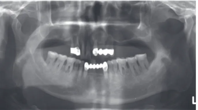
2009년 10월에 단국대학교 치과병원 치과보철과에 52세의 남성이 ‘임플란트를 하고싶다’는 주소로 내원 하였다. 전반적인 임상적, 방사선학적 검사에서 진행중 인 치주염으로 인한 상악에 심한 치조골의 흡수를 관찰 할 수 있었고, 전신 건강에는 특이할 만한 문제가 없었 다(Fig. 1). 골량과 골질 분석을 위해 컴퓨터 단층 방사 선 사진 촬영을 시행하였고, 잔존 치조골에 임플란트 식 립이 가능한지에 대해서 평가하였고, 환자의 요구, 심미 성, 악간 공간, 치조골 흡수 정도, 치조골 형태, 골질 등 을 고려하여 임플란트에 의해 지지되는 고정성 보철물 을 계획하였다. 치료는 환자의 기능과 심미성을 조절하 기 위해 여러 단계로 구분되어 계획하였다. 환자에게 하 악의 치조골 상태도 예후가 좋지 않다고 설명했으나 일 단 상악 치료 종료 후 하악의 치료를 원하셔서 상악의 치료만 진행하기로 계획하였다. 첫 번째 단계에서 본원 구강악안면외과에서 심한 치조제 흡수를 보이는 상악
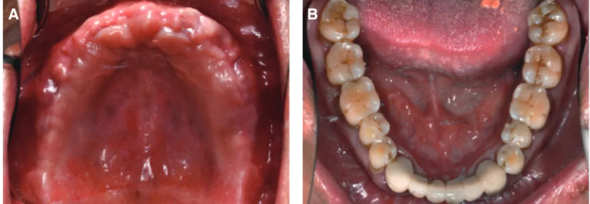
에 iliac bone graft를 동반한 골이식을 전신마취 하에 시 행하였다. 교합 수직고경을 회복하고 심미적인 부분을 해결하기 위해서 즉시 임시 의치를 장착하였다. 임시의 치의 유지를 위해 #11, 12, 13, 21는 남기고 #14, 23, 24 의 발치를 시행하였다. 골이식 후 5개월 뒤에 잔존 치 아 #11, 12, 13, 21를 발치하고 상악에 11개의 임플란트 (USII RBM fixture, Osstem Co., Seoul, Korea)를 식립 하였고 새로 제작한 임시 총의치를 soft liner (Sofreliner Tough®,Tokuyama Dental Co., Tokyo, Japan)를 이용하 여 재이장하고 장착하였다(Table 1, Fig. 2, 3). 임시 의 치를 복제하여 방사선 촬영용 스텐트를 제작하고 골량 과 골질 분석을 위해 컴퓨터 단층 방사선 사진 촬영을 재시행하였고, 스텐트를 변형하여 수술용 가이드를 제 작하여 사용하였다. 임플란트 식립 후 6개월 뒤 이차수 술을 시행하고, 봉합을 시행한 후에 임시 총의치를 조
Table 1. Implant distribution, diameters, and lengths
Implant
distribution Implant diameter
(mm) Implant length (mm)
#17 5.00 11.50
#16 5.00 11.50
#15 5.00 11.50
#13 4.00 13.00
#12 3.75 13.00
#22 3.30 13.00
#23 4.00 11.50
#24 4.00 11.50
#25 4.00 11.50
#26 4.00 11.50
#27 4.00 11.50
Fig. 1. Residual dentition and bone loss due to advanced
periodontal disease.정하였고, 골유착 기간 동안 healing abutment를 건드 리지 않도록 healing aubtment가 있는 부위의 acrylic resin은 제거하였고, soft liner를 이용하여 재이장하였 다(Fig. 4). 이차수술시 초기고정에 실패한 #17 임플 란트는 후방부에 재식립하고 4개월 경과 후 최종 보철 물 제작시 포함시켜 진행하기로 결정하였다. 2주 뒤 초 기고정에 실패한 #17 임플란트를 제외하고 10개의 식 립된 임플란트에 fixture transfer impression coping (Osstem Co.)을 장착하고, 알지네이트(Alginoplast®, Heraeus Holding GmbH, Hanau, Germany)를 이용하 여 예비인상을 채득한 후 제작된 작업모형에 트레이 레 진(Quicky Powder/Liquid, Nissin Dental Products Inc., Kyoto, Japan)을 이용하여 개별 트레이를 제작하였다.
최종인상은 Open tray를 이용한 fixture level impression 이 Polysiloxane 인상재(Honigum®, DMG, Hamburg, Germany)를 이용하여 채득하였다. 인상체 내의 연조직 은 vinylpolysiloxane (Dura Base resin, Reliance Dental Mfg. Co., Worth, IL, USA)으로 재현되었고, 상악의 주
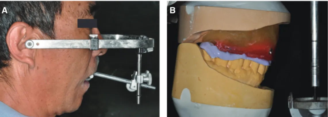
모형은 제4형 경석고(GC Fujirock® EP, GC Europe N.V., Leuven, Belgium)로 제작하였다(Fig. 5). 임의형 페 이스보우(HanauTM Spring-Bow, Waterpik Technologies Inc., Fort Collins, CO, USA)를 이용하여 안궁이전을 시 행하였다. Third reference point로 infraorbital foraman 을 사용하였다. 교합 수직고경과 중심위 악간관계를 교
Fig. 2. Five months after extractions and iliac bone graft in maxilla. (A) Maxilla, (B) Mandible.
A B
Fig. 3. Implant placement in maxilla for conventional
loading.Fig. 4. Implant placement of healing abutments for
secondary surgery.Fig. 5. Master cast for final prostheses.
크릴릭 인공치(Endura Anterior/Posterior, Shofu Inc., Kyoto, Japan)를 이용하여 진단용 납의치를 제작하였 다. 교합기의 교두 경사의 경우 시상면에서 33도, lateral condyle path inclination은 15도의 평균값으로 설정하였 다.15 이후 구내에서 verification device가 주모형의 정확 성을 평가하기 위해 제작하였다. 인상용 코핑을 fixture 에 연결하고, 아크릴릭 레진(Duralay Reliance, Dental Mfg. Co.)을 이용하여 스플린팅 하였다. Verification jig 를 절단하고 재연결한 후 구강내에서 분리하고, 주모 형으로 옮겼다. 주모형에서 index의 수동적 적합을 평
를 이용하여 복제하고, 자가 중합형 아크릴릭 레진(Jet Tooth ShadeTM Powder & Liquid Shade 60/A1, Lang Dental Mfg. Co., Inc., Wheeling, IL, USA)를 이용하 여 제작하였다(Fig. 8). 상악의 fixed detachable interim prostheses의 수동적 적합은 다양한 방법들로 평가하
였다.16,17 먼저, 한 쪽 끝의 지대주에 먼저 압력을 가하
고 반대편에서 보철물의 움직임이 있는지를 평가하였 다.시각적인 평가 후에 explorer를 이용한 적합을 평가 하였다. 수동적 적합은 양 쪽 끝에 있는 지대주의 나사 에 의해 확인하였다. 수복물의 움직임 없다는 것이 확
Fig. 7. Diagnostic wax coping try-in for determination of standard esthetic parameters. (A) Wax
coning try-in, (B) Analysis of esthetic parameters.A B
Fig. 6. Vertical dimension & interocclusal centric relation were transferred to a semiadjustable
articulator. (A) Facebow transfer, (B) Mounting used occlusal rims.A B
인되고 수복물이 반대편 지대주에 위치해 있다면, 보 철물과 모든 지대주의 삼차원적인 적합을 임상적으 로 확인하는 것이고, 이차원적으로는 치근단 방사선 사진을 통해 확인할 수 있다고 보고된다. 이렇게 제작 된 fixed detachable interim prostheses를 통해서, 심미 와 기능의 요소들을 평가하였다. 환자의 안모 및 미소 의 분석을 통해 상악 전치부의 길이가 약간 긴 것을 확 인할 수 있었고 길이 수정을 interim fixed detachable prostheses에서 수행하였다. 상악 중절치의 길이는 hign speed diamond bur (Komet 5850.314.016; Komet USA LLC, Rock Hill, SC, USA)를 이용해 구강 내에서 조정 하였고, 환자의 smile line을 하순과의 관계에서 기록 하였다. 모든 심미적, 기능적 요소에 대해 환자가 만족 하고 난 후, 알지네이트(Alginoplast®)를 이용하여 상 악 interim fixed detachable prostheses의 인상을 채득 하였고, 제3형 경석고(Snowrock, Munkyo Gypsum &
Engineering Co., Busan, Korea)를 이용하여 임시 보철 물을 복제한 모델을 제작하였다. 최종인상은 open tray 를 이용한 fixture level impression이 Polysiloxane 인
상재(Honigum®, DMG)를 이용하여 채득하였다. 인상 체 내의 연조직은 vinylpolysiloxane (Dura Base resin, Reliance Dental Mfg. Co.)으로 재현되었고, 상악의 주 모형은 제4형 경석고(GC FujirockⓇEP, GC Europe N.V., Leuven, Belgium)로 제작하였다. 그 후, 환자의 fixed detachable interim prostheses를 제거하여 상악 주 모형과 연결시킨 후, 이전에 임시 보철물 제작시 사용 했던 동일한 교합기에 마운팅된 하악 대합치 모형에 같 은 수직고경으로 대합되도록 마운팅하였다. 이러한 마 운팅 방법을 이용하여 하악 대합치에 맞춰서 교합면, 중심선, 미소선을 확인하였다. 환자의 fixed detachable interim prostheses를 복제한 full contour wax up 모형 을 제작한 후, 교합과 심미를 평가하기 위해 환자의 구 강에 장착하였다. Abutment는 temporary abutment (US II UCLA Temporary abutment, Osstem Co.)를 이용 하여 제작된 개별맞춤형 티타늄 abutment (MyplantTM, RaphaBio, Seoul, Korea)를 사용하였다(Fig. 9). 최종 보철물은 3개의 고정성 보철물, #17 = 14, #13 = 23,
#24 = 27로 제작되었다. 상악 6전치는 보다 나은 심
Fig. 8. Fixed detachable interim prostheses duplicated from wax coping. (A) Frontal view, (B)
Occlusal view.
A B
Fig. 9. Try-in of prostheses to customized titanium abutment. (A) Frontal view, (B) Occlusal view.
A B
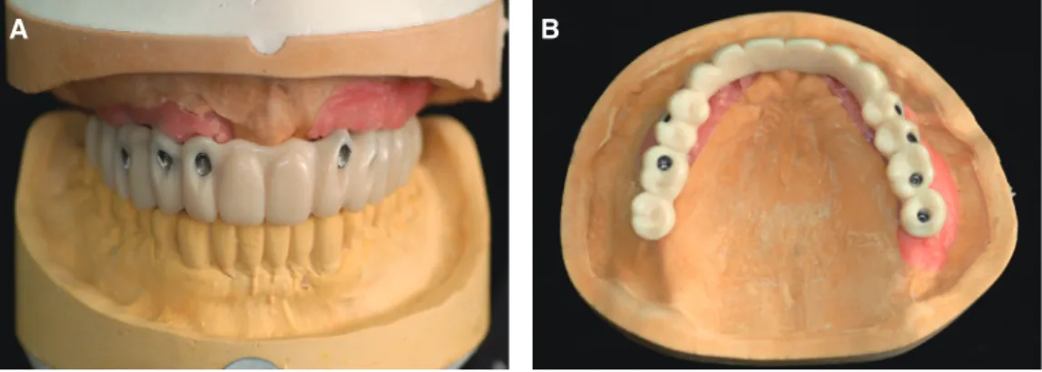
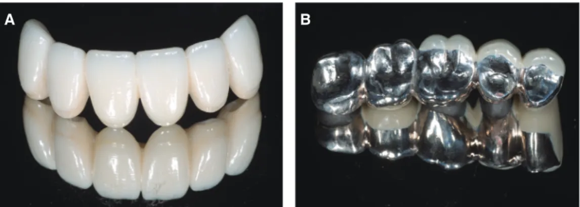
여 zirconium oxide (Ytrium-partially stabilized with tetragonal polycrystalline structure)(Prettau Zirconia 16er XH40, Zirkonzahn GmbH) 블록을 이용하여 지르 코니아 코핑을 제작하였다. 구치부도 full contour wax up 모형을 cut-back하여 white resin prostheses (Frame®) 를 제작하고 copy-milling unit을 이용하여 Pd-Ag 합금 (Innovium®) 블록을 이용하여 코핑을 제작하였다. 상 부 코핑에 비니어링 도재의 축성을 시행하였고, 최종적 으로 글레이징 및 연마를 시행하였다(Fig. 10). 최종 보 철물의 적합은 치근단 방사선 사진을 통해 확인하였 다. 상악 티타늄 abutment의 abutment screw (Ti screw, Osstem Co.)는 20 Nm의 torque로 조였고, access hole 은 G-P (Gutta Percha, Henry Schein, Inc., Melville, NY, USA)로 채운 후 광중합형 레진(FiltekTM Z-250, 3M ESPE, Seefeld, Germany)으로 막아주었다. 티타 늄 abutment와 상부 보철물은 임플란트 전용 시멘트인 IntercemTM (SCI-PHARM, Pomona, CA, USA)을 이용 하여 접착하였다(Fig. 11, 12). 장착 초기의 파노라마 방 사선 사진은 시멘트 유지형 임플란트 고정성 보철물이 fixture에 잘 적합된 것을 확인할 수 있었고, 임플란트 주 변의 bone level을 보여주었다(Fig. 13). 2년 후에 치근
로 제작된 임플란트 고정성 보철물은 환자의 저작 기능 과 심미적 외형을 증진해주었다고 사료된다.
고찰
기술적 진보에도 불구하고, 전악 수복과 같은 복잡한 임상 증례에서는 치료계획을 철저히 세우고, 보철물의 제작 방법과 재료 선택에 있어 신중을 기할 필요가 있 다. 최종 보철물의 디자인은 기본적인 수복 원칙(기능, 발음, 심미, 재료물성)을 고려해야 한다. 본 증례에서는 상악 무치악 환자의 보철적 수복에서 CAD/CAM 시스 템으로 제작할 수 있는 특징을 가지는 귀금속 Pd-Ag 합 금으로 제작된 시멘트 유지형 임플란트 지지 고정성 보 철물의 사용 가능성을 제시하고자 하였다.
전부 무치악 환자에서 도재 수복물을 이용한 임플 란트 지지 고정성 보철물 수복에 관한 많은 다양한 합 병증들이 발생되었다.1-3 금속 도재 수복물에서 도재의 chipping이나 파절은 피로 하중, 교합력, 열팽창계수의 차이, 금속의 낮은 탄성 계수, 부적절한 디자인, 미세결 함, 외상과 같은 다양한 인자에 의해서 발생한다고 보고 되고 있다.4,5,18 2008년 Denry와 Kelly는 전부 도재 수복
Fig. 10. Final prostheses to Pd-Ag Alloy and zirconia after being veneered. (A) Zirconia, (B) Pd-
Ag alloy.A B
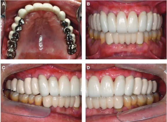
Fig. 11. Esthetics and fuction were restored with the Pd-Ag alloy and zirconia restorations.
(A) Occlusal view (maxilla), (B) Frontal view, (C) Lateral view (right side), (D) Lateral view (left view).
A B
C D
Fig. 12. Esthetics and fuction were restored with the
restorations in extraoral view.Fig. 13. Initial X-ray of the full zirconia fixed detachable
restorations.Fig. 14. Radiographic control after 2 years in use showing stable marginal bone at the level of the implants.
를 보이고, 주조되는 합금보다 높은 정확성을 보이는 우 수한 합금이라고 보고된다.12 Pd-Ag 합금의 탄성 계수, 항복 강도와 같은 기계적인 특성은 티타늄 합금, Co-Cr 합금보다 우수하다고 보고되었다.13,14 또한 높은 열팽창 계수로 인해 Pd-Ag 합금이 금속 도재 수복물 제작시 도 재와의 결합력이 낮을 가능성이 있지만, 이전 연구에서 는 임상적으로 사용하는데 도재와의 결합력에 큰 차이 를 보이지 않는다고 보고하였다.20,21 부가적으로 Pd-Ag 합금의 밀도는 10.93 g/cm3으로 다른 귀금속합금보다 상대적으로 낮다고 보고되며, 최종 보철물을 제작시 환 자에게 가벼움을 느낄 수 있게 하는 장점이 있다.22 우수 한 생체 적합성을 보이는 티타늄 합금의 밀도는 4.5 g/
cm3으로 Pd-Ag 합금보다 낮지만 관리가 까다로우며, 포 세린에 낮은 결합 강도를 가지는 문제점이 있다. 심미적 이면서 높은 강도와 정확성을 가지는 지르코니아의 밀 도도 6.1 g/cm3으로 Pd-Ag 합금보다 낮지만 연결부 파 절이나 비니어링 도재의 chipping과 같은 문제점을 가진 다고 보고된다.20-22
CAD/CAM 시스템으로 제작할 수 있는 특징을 가 지는 귀금속 Pd-Ag 합금(Innovium®)은 경제적인 이유 로 금사용을 대체하며 사용이 증가되고 있으며, 높은 생 체 적합성과 좋은 물리적 성질을 나타내고, CAD/CAM 시스템으로 보철물이 만들어지기 때문에 주조 시 발생 할 수 있는 손상과 변형 가능성이 적다고 보고된다.12-
14 CAD/CAM 시스템으로 제작된 Pd-Ag 합금 보철물 의 주요한 장점은 균일한 강도, 정확성, 비니어링 도재 의 chipping 최소화, 그리고 교합 조정의 최소화이다. 최 근 들어Pd-Ag 합금으로 금속 도재 수복물을 제작하면 강도 및 파절 저항성이 높아 임상적으로 기계적 안정성 을 높일 수 있어 수복물의 파절로 인한 실패가 감소하 고, 적응증의 범위도 넓어질 것이라는 임상 보고가 발 표되고 있다.20-23 하지만, Pd-Ag 합금은 아직은 전치부 에 사용하기에 심미성이 부족하고, 기존 금속 도재 합 금에 비하여 높은 열팽창 계수(coefficient of thermal
본 증례는 상악 전부 무치악 환자에서 CAD/CAM을 이용하여 전치부는 지르코니아, 구치부는 Pd-Ag 합금로 제작된 임플란트 지지 고정성 보철물로 전악 수복하였 고, 이에 기능적, 심미적 회복을 도모하여 환자와 술자 모두에게 만족스러운 결과를 얻었기에 보고하는 바이 다. 최종 보철물을 장착 후 6개월 단위로 2년 간의 주기 적인 정기검진을 통해 좋은 예후를 보이고 있으며, 추후 지속적인 경과 관찰이 필요할 것으로 본다. 현재 CAD/
CAM을 이용하여 Pd-Ag 합금으로 제작된 임플란트 고 정성 보철물의 임상적 보고는 거의 없는 것으로 보인다.
앞으로 이런 유형의 보철물을 이용한 장기간의 연구가 필요할 것으로 생각되고, 이 재료를 치과계에서 사용되 는 다른 재료와 비교하는 것이 필요하며, 이번 임상 보 고에서 논의된 장, 단점에 대해 더 많은 연구가 진행되 어야 할 것으로 사료된다.
References
1. Purcell BA, McGlumphy EA, Holloway JA, Beck FM. Prosthetic complications in mandibular metal- resin implant-fixed complete dental prostheses: a 5-to 9-year analysis. Int J Oral Maxillofac Implants 2008;23:847-57.
2. Duncan JP, Nazarova E, Vogiatzi T, Taylor TD.
Prosthodontic complications in a prospective clini- cal trial of single-stage implants at 36 months. Int J Oral Maxillofac Implants 2003;18:561-5.
3. Bozini T, Petridis H, Garefis K, Garefis P. A meta- analysis of prosthodontics complication rates of implant-supported fixed dental prostheses in eden- tulous patients after an observation period of at least 5 years. Int J Oral Maxillofac Implants 2011;
26:304-18.
4. Ortorp A, Jemt T. Early laser-welded titanium
frameworks supported by implants in the edentu- lous mandible: a 15-year comparative follow-up study. Clin Implant Dent Relat Res 2009;11:311-22.
5. Larsson C, Vult von Steyern P, Nilner K. A pro- spective study of implant-supported full-arch yttria-stabilized tetragonal zirconia polycrystal man- dibular fixed dental prostheses: three-year results.
Int J Prosthodont 2010;23:364-9.
6. Ortorp A, Jemt T. Clinical experiences of CNC- milled titanium frameworks supported by implants in the edentulous jaw: 1-year prospective study. Clin Implant Dent Relat Res 2000;2:2-9.
7. Linkevicius T, Vladimirovas E, Grybauskas S, Puisys A, Rutkunas V. Veneer fracture in implant- supported metal-ceramic restorations. Part I: Over- all success rate and impact of occlusal guidance.
Stomatologija 2008;10:133-9.
8. Katsoulis J, Mericske-Stern R, Rotkina L, Zbären C, Enkling N, Blatz MB. Precision of fit of implant- supported screw-retained 10-unit computer- aided-designed and computer-aided-manufactured frameworks made from zirconium dioxide and titanium: an in vitro study. Clin Oral Implants Res 2014;25:165-74.
9. Gilbert JL, Covey DA, Lautenschlager EP. Bond characteristics of porcelain fused to milled tita- nium. Dent Mater 1994;10:134-40.
10. Reich S, Wichmann M, Nkenke E, Proeschel P.
Clinical fit of all-ceramic three-unit fixed partial dentures, generated with three different CAD/
CAM systems. Eur J Oral Sci 2005;113:174-9.
11. Bachhav VC, Aras MA. Zirconia-based fixed partial dentures: a clinical review. Quintessence Int 2011;
42:173-82.
12. Huget EF, Civjan S. Status report on palladium- silver-based crown and bridge alloys. J Am Dent Assoc 1974;89:383-5.
13. Goodacre CJ. Palladium-silver alloys: a review of the literature. J Prosthet Dent 1989;62:34-7.
14. Kansu G, Aydin AK. Evaluation of the biocompat- ibility of various dental alloys: Part I-Toxic poten- tials. Eur J Prosthodont Restor Dent 1996;4:129- 36.
15. Henry PJ. An alternate method for the production of accurate casts and occlusal records in the osseo- integrated implant rehabilitation. J Prosthet Dent 1987;58:694-7.
16. Jemt T, Book K. Prosthesis misfit and marginal bone loss in edentulous implant patients. Int J Oral Maxillofac Implants 1996;11:620-5.
17. Tan KB, Rubenstein JE, Nicholls JI, Yuodelis RA.
Three-dimensional analysis of the casting accuracy of one piece, osseointegrated implant-retained prostheses. Int J Prosthodont 1993;6:346-63.
18. Roberts DH. The failure of retainers in bridge prostheses. An analysis of 2,000 retainers. Br Dent J 1970;128:117-24.
19. Denry I, Kelly JR. State of the art of zirconia for dental applications. Dent Mater 2008;24:299-307.
20. Barghi N, McKeehan-Whitmer M, Aranda R. Com- parison of fracture strength of porcelain-veneered- to-high noble and base metal alloys. J Prosthet Dent 1987;57:23-6.
21. Warpeha WS Jr, Goodkind RJ. Design and technique variables affecting fracture resistance of metal- ceramic restorations. J Prosthet Dent 1976;35:291- 8.
22. Anusavice KJ, Dehoff PH, Gray A, Lee RB. De- layed crack development in porcelain due to incom- patibility stress. J Dent Res 1988;67:1086-91.
23. Rosenstiel SF, Land MF, Fujimoto J. Contemporary fixed prosthodontics. 4th ed. New York; Elsevier Inc.; 2006. p. 743-50.
*교신저자: 최유성
(330-714) 충남 천안시 동남구 단대로 119 단국대학교 치과대학 치과보철학교실 Tel: 041-550-1979|Fax: 041-550-1859|E-mail: [email protected] 접수일: 2014년 1월 2일|수정일: 2014년 3월 2일|채택일: 2014년 3월 11일
골흡수가 심한 환자에서 임플란트를 이용한 전악 수복의 경우 치은과 치아의 외형을 심미적이면서도 기능적으로 회복 하기 위해서는 보철물 제작에 있어 신중을 기해야 한다. 이러한 전악 수복 증례에서 CAD/CAM 시스템이 높은 정확성 을 가지는 보철물 제작을 가능하게 함으로써 사용이 증가하고 있다. 최근에 임상에서 티타늄이나 지르코니아의 단점을 보완하면서 높은 생체적합성을 가지고 CAD/CAM 시스템으로 제작할 수 있는 팔라듐-은(Pd-Ag) 합금이 개발되어 사 용되고 있다. 본 증례에서는 상악 전부 무치악 환자에서 임플란트를 식립하고 도재의 chipping이나 파절의 위험을 감소 시키기 위해 Pd-Ag 합금을 이용해 제작된 임플란트 고정성 보철물로 수복한 환자에 대해 보고하고자 한다.
(구강회복응용과학지 2014;30(1):71-80)
주요어: Pd-Ag 합금; CAD/CAM 시스템; 시멘트 유지형 보철물; 임플란트 지지 보철물