Protective Effects of an Ethanol Extract of Elaeagnus umbellata Leaves on α-MSH-induced Melanin Production in B16-F0 Cells and UVB-induced Damage in CCD-986sk Cells
Se-Ho Park1,2, Kwang-Hwan Jhee2 and Seun-Ah Yang3*
1Institute of Natural Science, Keimyung University, 1095 Dalgubeol-daero, Daegu 42601, Korea
2Department of Applied Chemistry, Kumoh National Institute of Technology, Gumi 39177, Korea
3Department of Food Science and Technology, Keimyung University, Daegu 42601, Korea Received February 11, 2019 /Revised March 20, 2019 /Accepted April 2, 2019
This study was undertaken to investigate the effect of an ethanol extract of Elaeagnus umbellata leaves (EUL-EE) on skin-related biological activities. Previously, we have reported that gallic acid was the major phenolic compound in EUL-EE through quantitative analysis and that EUL-EE had an in- hibitory effect against the proliferation of liver cancer HepG2 cells. In the present study, the inhibitory effects of EUL-EE on melanin production and tyrosinase activity in α-melanocyte-stimulated hor- mone-stimulated B16-F0 cells were determined to assess the effects of EUL-EE on skin whitening. The anti-wrinkle effect using UVB-irradiated CCD-986sk cells was examined by the expression of type I procollagen and metalloproteinase (MMP)-1 release. The EUL-EE significantly decreased intracellular melanin production (33.0% inhibition at 100 μg/ml) when compared with untreated B16-F0 cells.
Tyrosinase activities in the stimulated B16-F0 cells were also decreased by EUL-EE (47.8% inhibition at 100 μg/ml). The EUL-EE also dose-dependently increased the production of type I procollagen (up to 1.74-fold at 250 μg/ml) in CCD-986sk cells when compared with UVB-irradiated controls. EUL-EE showed no cytotoxicity at concentrations up to 500 μg/ml. In addition, EUL-EE at 10-500 μg/ml in- hibited the release of MMP-1 to the medium from UVB-irradiated CCD-986sk cells. Taken together, these observations indicate that EUL-EE has high potential for use as inner beauty and cosmetic mate- rials due to its whitening and anti-wrinkle effects.
Key words : Anti-wrinkle, Elaeagnus umbellata leaves, melanin production, reactive oxygen species, skin whitening
*Corresponding author
*Tel : +82-53-580-5117, Fax : +82-53-580-5372
*E-mail : [email protected]
This is an Open-Access article distributed under the terms of the Creative Commons Attribution Non-Commercial License (http://creativecommons.org/licenses/by-nc/3.0) which permits unrestricted non-commercial use, distribution, and reproduction in any medium, provided the original work is properly cited.
ISSN (Online) 2287-3406 Journal of Life Science 2019 Vol. 29. No. 5. 555~563 DOI : https://doi.org/10.5352/JLS.2019.29.5.555
Introduction
As the aging of human skin progresses, the secretion of various hormones that regulate metabolism decrease and the activities of immune cells and skin cells are diminished, and thus, the biosynthesis of immune proteins and biologic pro- teins are reduced. Furthermore, it is known that wrinkles, skin dryness, and skin inelasticity are diminished and stain, freckles and age spot are produced due to intrinsic aging and extrinsic aging caused photo-aging and environmental pollutants [22, 23]. Melanin, a complex of black pigment and protein is responsible for the color of human hair. When
melanin biosynthesis increased in skin, the color of skin changes from yellowish brown to dark brown. This process may cause spots, freckles, skin spots, and other skin prob- lems, or even cell death and skin cancer due to the toxic effects of melanin precursors produced in melanosomes lo- cated, which include tyrosine, 3,4-dihydroxyphenylalanine (DOPA), and dopaquinone [16]. Skin whitening agents are developed in three ways: by inhibiting melanin production in melanocytes, by controlling melanocyte stimulating sub- stances, and by promoting melanin excretion [7]. In general, melanogenesis inhibitors reduce melanin production in skin through a series of redox reactions [9]. In the dermis, colla- gen affects skin moisturization and elasticity, and with in- creasing age, amounts of structural proteins such as dermal fibroblasts, collagen and elastin decrease and tissues loosen.
Exposure to ultraviolet light, especially UVB, induces the production of reactive oxygen species (ROS) and that pro- mote photo-aging [4]. ROS directly break down collagen and deactivate matrix metalloproteinase (MMP), which results in
increase MMP production and collagen break down [2]. In addition, ultraviolet rays penetrate deeply into skin and can destroy or mutate DNA in skin cells [20]. The MMPs are a family of enzymes that function by degrading extracellular matrix and basement membranes, and are divided into four subfamilies, that is, interstitial collagenase, stromelysin, ge- latinase, and membrane-type MMPs [12]. Therefore, to re- duce skin wrinkling, it is important to inhibit the actions of collagenase and ROS and to enhance collagen synthesis.
Elaeagnus umbellata (Thunb.) is a deciduous shrub belong- ing to the Elaeagnus family and is native to Pakistan, China, India, Japan, and Korea [6]. The fruits and seeds of E. umbel- lata are edible, and in Korea were recently approved as an edible material. Its fruits have 7 to 17 fold more lycopene than tomatoes [3], and it is well known that lycopene con- sumption prevents various cancers, coronary heart disease, type 2 diabetes, and osteoporosis [5, 19, 24]. The fruits of E. umbellata contain high levels of rutin and gallic acid, while its seeds contain high levels of chlorogenic acid and gallic acid [18]. On the other hand, little is known of the phenolic components of the leaves of E. umbellata. Previously, we re- ported that E. umbellata leaves ethanol extract (EUL-EE) has a potent anti-proliferative effects on liver cancer cells [8], but little is known of the biological properties of EUL-EE. To evaluate the skin-related efficacy of EUL-EE with respect to skin whitening and anti-wrinkle activities, we investigated its effects on melanin production and tyrosinase activity in a melanocyte cell-line. In addition, the effect on collagen bio- synthesis and MMP-1 inhibition, which were viewed as sur- rogate anti-UVB-induced skin wrinkling, were also examined. Thus, this preliminary biological study was un- dertaken to investigate the effects of EUL-EE on skin health.
Materials and Methods
Plant sample
EUL-EE was purchased from Bosamo Agricultural Associ- ation Corporation (Changnyeong-gun, Gyeongsangnam-do, Korea), and were stored at ambient temperature in the dark after dry of 30℃ for 24 hr.
Chemicals and reagents
α-Melanocyte stimulated hormone (α-MSH), 3-isobutyl-1- methylxanthine (IBMX), melanin, tyrosinase from mush- room (5,771 units/mg), Triton X-100, 4-phenylazobenzylox- ycarbonyl-pro-leu-gly-pro-d-ar, collagenase, 4’,6-diamidino-
2-phenylindole (DAPI), 2,7-dichlorodihydrofluorescein diac- etate (DCFDA), and L-DOPA were purchased from Sigma- Aldrich Inc. (St. Louis, MO, USA). Dimethyl sulfoxide, pre- tanol A, and ethanol were purchased from Duksan Science (Seoul, Korea). 3-(4,5-dimethyl-2-thiazolyl)p-2,5-diphenyl-2H- tetrazolium bromide (MTT) was purchased from AMRESCO Chemical (Solon, USA). And sodium dihydrogen phosphate and disodium hydrogen phosphate were purchased from Junsei Chemical (Tokyo, Japan).
Preparation of E. umbellata extract
Air-dried powder of EUL was extracted with 70% ethanol.
Briefly, the ethanol extract (10 g) was extract with 2 l of sol- vent mixture at room temperature for 24 hr (two replicates).
EUL was filtered and concentrated using a rotary evaporator (EYELA N-3010, Tokyo Rikakikai, Japan). And then the ex- tract was lyophilized using a freeze dryer (FDU-2100, Tokyo Rikakikai, Japan).
Cell culture
B16-F0, a mouse melanoma cell lines was cultured in Dulbecco’s modified eagle’s medium (DMEM) supple- mented with 10% fetal bovine serum, 1% non-essential ami- no acids (100×), 1% L-glutamine (200 mM), 1% vitamins (100×), 1% penicillin (10,000 μm/ml) and 1% sodium pyr- uvate 100 mM. For all experiments, the cells were grown to 80~90% confluence, and were subjected to no more than 20 cell passages. CCD-986sk human fibroblasts (American Tissue Collection Center, ATCC, USA) were cultured in Iscove’s modified Dulbecco’s medium (IMDM) containing 10% fetal bovine serum, 1% antibiotics at 37℃ in a humidi- fied atmosphere of 5% CO2.
Cytotoxicity of B16-F0 melanoma cells and CCD- 986sk cells
Briefly, B16-F0 cells (5×10⁵ cells per well) were seeded into a 24-well cell culture plate and incubated for 24 hr. The cells were treated with various concentration of EUL-EE (0, 25, 50, 100, and 250 μg/ml), and then incubated for 48 hr. After incubation, 100 μl of the MTT reagent in PBS (5 mg/ml) was added to each well. After 4 hr of incubation, the reacting color was measured at 550 nm using a microplate spec- trophotometer (Molecular Devices, CA, USA).
Also, CCD-986sk cells (1×10⁴ cells per well) were seed into a 24-well cell culture plate and incubated for 24 hr. The me- dium of the experimental groups was treated with varied
concentrations of EUL-EE (0, 10, 100, 250, and 500 μg/ml), followed by incubation for 24 hr within or without UVB irra- diation (15 mJ/cm2). After incubation, 100 μl of the MTT reagent in PBS (5 mg/ml) was added to each well. After the incubation continued for another 4 hr, the resulting color was assayed at 550 nm using a microplate spectropho- tometer (Molecular Devices, CA, USA).
Melanin production in α-MSH treated B16-F0 melanoma cells
Briefly, B16-F0 cells (5×10⁵ cells per well) were seeded into a 6-well cell culture plate and incubated for 24 hr. After 24 hr of incubation, the cells were treated with various concen- tration of EUL-EE (0, 25, 50, 100, and 250 μg/ml), containing 100 nM α-MSH and 100 mM IBMX. After incubation for 48 hr at 37℃, each medium were removed and washing the cells with PBS, the cell pellet was dissolved in 150 μl of 1 N NaOH with 10% DMSO, incubated at 60℃ for 1 hr. After incubation, the resulting absorbance was measured at 405 nm using a microplate spectrophotometer. The amount of melanin production was determined by measuring sample absorbance at 405 nm against a synthetic melanin standard.
Cellular tyrosinase activity
Briefly, B16-F0 cells (5×10⁵ cells per well) were cultured in 6-well plates, incubated with α-MSH (100 nM), IBMX (100 μM), and various concentrations of EUL-EE (0, 25, 50, 100, and 250 μg/ml) for 2 days, and then washed with ice-cold PBS, lysed with phosphate buffer (pH 6.8) containing Triton X-100.
Extracellular tyrosinase activity
The tyrosinase inhibitory activities of the EUL-EE was de- termined by the L-DOPA oxidation assay. Briefly, 80 μl of L-DOPA (2.5 mM), and various concentrations of EUL-EE (100 μl) by using sodium phosphate buffer (100 mM, pH 6.8) were added to 96-well plates. The mixture was in- cubated for 10 mins at 37℃. The mixtures were injected to 20 μl of tyrosinase from mushroom (1,500 units/ml). The solution was measured by the DOPAchrome formation in optical density at 475 nm using UV-Vis spectra. The percent inhibition of tyrosinase activity was calculated as follows:
% Inhibition = [(A - B) / A] ×100
Where A = absorbance at 475 nm without test sample [10 μl of solution (%, deionized water) and B = absorbance at 475 nm with test sample (10 μl of plant extract)].
Assay of collagen type I synthesis and collagenase inhibition
Collagen synthesis was evaluated using the Takara MK101 kit (Takara Bio Inc., Japan). CCD-986sk human fibro- blasts were incubated into 24-well cell culture plate (1×10⁴ cells per well) and cultured at 37℃ in 5% CO2. After UVB irradiation (15 mJ/cm2), cells were treated with the sample at a concentration of 0, 10, 100, 250 and 500 μg/ml for 24 hr. After culturing, the supernatant was collected from each well, and the amount of pro-collagen type I was measured with a procollagen type I C-peptide assay kit (Takara Bio, Japan). Briefly for collagenase inhibition assay, 0.15 ml of collagenase (1 mg/ml) was added to the mixed solutions consisted of 2 mM 4-phenylazobenzyloxycarbonyl-pro-leu- gly-pro-d-ar 0.25 ml and 0.1 ml of EUL-EE (10, 100, 250, 500, 1000, 1500, 2000, and 3000 μg/ml) in 0.1 M Tris-HCl buffer (pH 7.5) and then reacted for 20 mins at 37℃. After that, the reactions were stopped by adding 6% citric acid 0.5 ml.
Absorbance was measured at 320 nm with a UV-visible spec- trophotometer after addition of ethyl acetate 1.5 ml. The per- centage of collagenase inhibitory activity was calculated from the following equation.
The percentage of collagenase inhibition (%) = (1- S / C) ×100
Where ‘S’ is the corrected absorbance of the samples con- taining collagenase inhibitor (the enzyme activity in the presence of the samples), and ‘C’ is the corrected absorbance of controls (the enzyme activity in the absence of the sam- ples).
Matrix metalloproteinase-1 (MMP-1) inhibition assay CCD-986sk cells were seeded at a density of 1×10⁴ cells/well in 96-well culture plates and were cultured for 24 hr. After UVB irradiation (15 mJ/cm2) and EUL-EE treat- ment, the supernatants were centrifuged at 12,000×g for 5 min, and stored at -80℃ in fresh tubes. The activity of colla- genase was measured with a MMP-1 human biotrak ELISA system (Amersham life science, USA).
4’,6-Diamidino-2-phenolindole (DAPI) staining and intracellular reactive oxygen species (ROS) assay
For 4’,6-diamidino-2-phenolindole (DAPI) staining, a 500×
stock solution (5 mM) was diluted to 10× in PBS, and then 50 μl of this 10x DAPI solution was added to each well of cells to achieve a final DAPI loading solution of 10 μM.
CCD-986sk cells were seeded at a density of 1×10⁴ cells/well
Fig. 1. Effects of EUL-EE on melanin production and tyrosinase activities in α-MSH-stimulated B16-F0 melanocytes. (A) The effects on cell viability, (B) melanin synthesis, and (C) inhibition on tyrosinase activity. Results were ex- pressed as % of control and data were means ± SD.
Values with different superscript letters are significantly different (p<0.05) by Duncan’s multiple comparison test (n=3).
in 24-well culture plates. After UVB irradiation (15 mJ/cm2) and treatment EUL-EE, the cells were washed. Cells were loaded with DCFH-DA (5 μM) and DAPI (10 μM) for 30 min, and then cells were washed twice with culture medium and images acquired by using fluorescence microscopy. The distribution of fluorescence was analyzed by fluorescence microscopy (Eclipse; Nikon, Tokyo, Japan) and analyzed by NIS-Elements software (Nikon). Fluorescence was measured with a spectrophotometer microplate reader where an ex- citation wavelength of 480 nm and an emission wavelength of 530 nm were used. Images captured using FITC and DAPI filters to determine the localization of intracellular ROS gen- eration and nucleus, respectively.
Statistical analysis
Data were expressed as mean ± SD determined from trip- licate analysis. The results were subjected to analysis of var- iance (ANOVA) using Duncan’s multiple comparison test to analyze differences, and p<0.05 was considered to be significant.
Results and Discussion
Skin-whitening effects on α-MSH-stimulated B16-F0 melanoma cells
In the present study, the inhibitions of melanin pro- duction and tyrosinase activity in B16-F0 melanocytes were used as surrogates of skin whitening ability due to their in- volvements in the mechanism of whitening. The cytotoxicity of EUL-EE on B16-F0 melanocytes were investigated using a MTT assay. As shown in Fig. 1A, EUL-EE was not cytotoxic to α-MSH-stimulated B16-F0 cells in the concentration range 25~100 μg/ml. In addition, arbutin (250 μM; a positive con- trol) was also showed no cytotoxic effect. To determine the effect of EUL-EE on melanin synthesis, B16-F0 cells were treated with EUL-EE at different concentrations and then melanin contents were determined by spectrophotometry.
EUL-EE significantly decreased the production of intra- cellular melanin (133.8 μg per 5×10⁵ cells at 100 μg/ml) as compared with untreated cells (199.7 μg per 5×10⁵ cells, Fig.
1B). Also, arbutin strongly suppressed melanin production (70.5 μg per 5×10⁵ cells). Arbutin is well known to inhibit melanin production in B16 cells induced with α-MSH and to reduce tyrosinase activity [14]. We next measured the ty- rosinase activity by dopaquinone production in the L-DOPA present, which the tyrosinase is the main regulatory melano-
genic enzyme in melanin synthesis pathway. Fig. 1C shows that tyrosinase activities in α-MSH-stimulated B16-F0 cells were dose-dependently reduced by EUL-EE (47.8% inhibi- tion at 100 μg/ml). The whitening activity of a number of active components isolated from plants has been previously reported, as have the anti-wrinkle effects of many bioactive compounds [15, 26]. Especially, kaempferol and gallic acid, which were found to present in EUL-EE, are known to in- hibit tyrosinase activity and melanin inhibition, and to in- hibit wrinkle formation [13]. It is, however, unclear whether
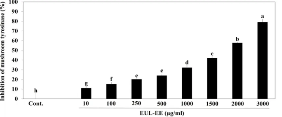
Fig. 2. Inhibition of EUL-EE on mushroom tyrosinase. L-DOPA (2.5 mM), and various concentrations of EUL-EE (0, 10, 100, 250, 500, 1,000, 1,500, 2,000, and 3,000 μg/ml) with 100 mM sodium phosphate buffer (pH 6.8) were added to 96-well plates.
The mixture was incubated for 10 mins at 37℃, and then, the mixtures were injected to 20 µl of tyrosinase from mushroom (1,500 units/ml). Absorbance was measured at 475 nm. Values with different superscript letters are significantly different (p<0.05) by Duncan’s multiple comparison test (n=3).
Fig. 3. Inhibition of EUL-EE on the collagenase activity. The collagenase (1 mg/ml) was added to the mixed solutions consisted of 2 mM 4-phenylazobenzyloxycarbonyl-pro-leu-gly-pro-d-ar and EUL-EE (0, 10, 100, 250, 500, 1,000, 1,500, 2,000, and 3,000 μg/ml) in 0.1 M Tris-HCl buffer (pH 7.5) and then reacted for 20 mins at 37℃. After reaction, the reactions were stopped by adding 6% citric acid 0.5 ml. Absorbance was measured at 320 nm after addition of ethyl acetate. Values with different superscript letters are significantly different (p<0.05) by Duncan’s multiple comparison test (n=3).
the key active components of the extract for its anti-melano- genesis activity are kaempferol and gallic acid, and a clear identification is needed. We then investigated the direct ef- fect of EUL-EE on mushroom tyrosinase activity [6]. EUL-EE weakly inhibited mushroom tyrosinase activity with an IC50
value of 1.717 mg/ml (Fig. 2). The tyrosinase inhibitory ac- tivity of EUL-EE (34.5% at 1 mg/ml) was higher than that of the Rhododendron mucronulatum Turcz extracts (24% at 1 mg/ml) [1].
Anti-wrinkle effects on UVB-irradiated CCD-986sk human dermal cells
The inhibitory effects of EUL-EE on collagenase activities were examined. Upon UVB irradiation, collagenase activity was dramatically increased and this was strongly sup- pressed by EGCG (10 μM, a positive control). As shown in Fig. 3, collagenase activity was also significantly inhibited by EUL-EE (IC50 value of 1.818 mg/ml). These results show EUL-EE significantly inhibited collagenase activity. Su et al.
reported kaempferol and gallic acid are both strong in- hibitors of collagenase [21]. Moreover, kaempferol showed good anti-oxidative and anti-inflammatory activities, which suggested its use in anti-aging cosmetic products [17, 25].
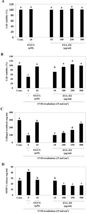
The effects of EUL-EE on cell viability were tested using
a MTT assay using within or without UVB-irradiated CCD- 986sk cells. EUL-EE was not cytotoxic to CCD-986sk cells in the concentration range 10~500 μg/ml (Fig. 4A). Exposure to UVB dramatically reduced cell viability by >50%. As shown in Fig. 4B, treatment with EUL-EE (10, 100, 250, and 500 μg/ml) reduced UVB-induced cell death. To evaluate collagen synthesis, we used a procollagen type I C-peptide assay kit. As procollagen type I amino- and carboxy terminal propeptide (procollagen type I amino propeptide and PICP) are cleaved from procollagen molecules to produce mature collagen fibers, amount of PICP secreted into culture me- dium provide a measure of collagen synthesis. EUL-EE in- creased the production of type I collagen in UVB-irradiated CCD-986sk cells in a dose-dependent manner (up to 1.74- fold at 250 μg/ml) as compared with 15 mJ/cm2 UVB-irradi- ation controls (Fig. 4C). These results show EUL-EE en- hanced collagen synthesis effectively in UVB exposed cells.
MMPs are enzymes that play a major role in normal and pathological tissue remodeling [10]. EUL-EE highly inhibited the release of MMP-1 from UVB-irradiated CCD-986sk cells at concentrations of 10~500 μg/ml as well as EGCG (Fig.
4D). The result demonstrated that the promotion of collagen production by EUL-EE and that it do so by inhibiting MMP-1 expression.
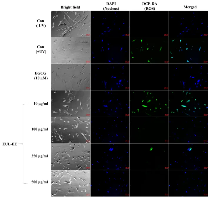
Oxidative stresses can be generated in connective tissues and in skin cells by photo-damage or inflammatory proc- esses. UV-induced ROS induces the activation of mitogen- activated protein kinase, activator protein 1, phosphor- ylation of c-Fos and c-Jun, and upregulation of MMP-1 that contribute for skin collagen degradation. To test the effects of EUL-EE on intracellular ROS generation induced by UVB exposure in CCD-986sk cells, the changes of ROS signal were observed qualitatively by fluorescence staining. EUL-EE was found to reduce UVB-induced ROS generation in CCD-986sk skin dermal cells (Fig. 5). Our data suggested that EUL-EE might effectively protect skin against UVB-induced oxida- tive damage. In addition, EGCG, a positive control, was known to block UV-induced increases of collagen degrada- tion and collagenase mRNA levels in fibroblast culture [10].
Taken together, EUL-EE was found to downregulate mel- anin production and to inhibit tyrosinase in α-MSH-stimu- lated melanocytes. Furthermore, EUL-EE significantly in- hibited collagenase activity, inhibited ROS generation, and increased collagen production in UVB-exposed CCD-986sk fibroblast. These results collectively show EUL-EE has poten- tial use as cosmeceutical and/or functional food ingredient
Fig. 4. Anti-wrinkle effects of EUL-EE in UVB-irradiated CCD- 986sk human dermal cells (15 mJ/cm2). (A) The effects on CCD-986sk cell viability without UVB irradiation, (B) cell viability within UVB irradiation, (C) collagen pro- duction, and (D) the release of MMP-1 into the culture media. Values with different superscript letters are sig- nificantly different (p<0.05) by Duncan’s multiple com- parison test (n=3).
Fig. 5. Inhibitory effects of EUL-EE on ROS generation in UVB-irradiated CCD-986sk human dermal cells. The cells were treated with EUL-EE after UVB-irradiation (15 mJ/cm2) and stained using DAPI (nuclear staining, blue signal) and DCF-DA ROS indicators (intracellular oxygen free radical, green signal).
with skin-whitening and anti-wrinkle activities for the first report.
Acknowledgements
This research was funded by Korea Institute of Planning and Evaluation for Technology in Food, Agriculture, Fores- try and Fisheries grant number 115009-3, and National Research Foundation of Korea grant number 2015RICIA2A 01055125.
References
1. An, B. J., Lee, C. E., Son, J. H., Lee, J. Y., Choi, G. H. and Park, T. S. 2005. Antioxidant, anticancer and tyrosinase in- hibition activities of extracts from Rhododendron mucronula- tum T. J. Kor. Soc. Appl. Biol. Chem. 48, 280-284.
2. Fagot, D., Asselineau, D. and Bernerd, F. 2002. Direct role of human dermal fibroblasts and indirect participation of epidermal keratinocytes in MMP-1 production after UV-B irradiation. Arch. Dermatol. Res. 293, 576-583.
3. Fordham, I. M. 2001. Fruit of autumn olive: A rich source of lycopene. Hortscience 36, 1136-1137.
4. Herrling, T., Jung, K. and Fuchs, J. 2006. Measurements of
UV-generated free radicals/reactive oxygen species (ROS) in skin. Spectrochim. Acta A Mol. Biomol. Spectrosc. 63, 840-84 5.
5. Khan, S. and Yeole, P. G. 2005. 26-protective effects of toma- to in coronary heart diseases: a nutraceutical perspective.
Health Administrator. XX, 104-108.
6. Khattak, K. F. 2012. Free radical scavenging activity, phy- tochemical composition and nutrient analysis of Elaeagnus umbellata berry. J. Med. Plants Res. 6, 5196-5203.
7. Kim, K. S., Kim, J. A., Eom, S. Y., Lee, S. H., Min, K. R.
and Kim, Y. 2006. Inhibitory effect of piperlonguminine on melanin production in melanoma B16 cell line by down- regulation of tyrosinase expression. Pigment Cell Res. 19, 90-98.
8. Kim, M. J., Lim, J. S. and Yang, S. A. 2016. Component anal- ysis and anti-proliferative effects of ethanol extracts of fruits, leaves, and stems from Elaeagnus umbellata in HepG2 cells.
J. Kor. Soc. Food Sci. Nutr. 45, 828-834.
9. Kim, S. Y., Kim, D. S., Kwon, S. B., Park, E. S., Huh, C.
H., Youn, S. W., Kim, S. W. and Park, K. C. 2005. Protective effects of EGCG on UVB-induced damage in living skin equivalents. Arch. Pharm. Res. 28, 784-790.
10. Kim, Y. J. and Uyama, H. 2005. Tyrosinase inhibitors from natural and synthetic sources: structure, inhibition mecha- nism and perspective for the future. Cell Mol. Life Sci. 62, 1707-1723.
11. Kohl, E., Steinbauer, J., Landthaler, M. and Szeimies, R. M.
2011. Skin ageing. J. Eur. Acad. Dermatol. Venereol. 25, 873- 884.
12. Langton, A. K., Sherratt, M. J., Griffiths, C. E. M. and Watson, R. E. B. 2010. A new wrinkle on old skin: the role of elastic fibres in skin ageing. Int. J. Cosmet. Sci. 32, 330-339.
13. Lim, H. and Kim, H. P. 2007. Inhibition of mammalian colla- genase, matrix metalloproteinase-1, by naturally-occurring flavonoids. Planta Med. 73, 1267-1274.
14. Lim, Y. J., Lee, E. H., Kang, T. H., Ha, S. K., Oh, M. S., Kim, S. M., Yoon, T. J., Kang, C., Park, J. H. and Kim, S.
Y. 2009. Inhibitory effects of arbutin on melanin biosynthesis of α-melanocyte stimulating hormone-induced hyperpig- mentation in cultured brownish guinea pig skin tissues.
Arch. Pharm. Res. 32, 367-373.
15. Mukherjee, P. K., Maity, N., Nema, N. K. and Sarkar, B.
K. 2011. Bioactive compounds from natural resources against skin aging. Phytomedicine 19, 64-73.
16. Palumbo, A., D’lschia, M., Misuraca, G. and Prota, G. 1991.
Mechanism of inhibition of melanogenesis by hydroquinone.
Biochim. Biophys. Acta 1073, 85-90.
17. Rho, H. S., Ghimeray, A. K., Yoo, D. S., Ahn, S. M., Kwon, S. S., Lee, K. H., Cho, D. H. and Cho, J. Y. 2011. Kaempferol and kaempferol rhamnosides with depigmenting and an- ti-inflammatory properties. Molecules 16, 3338-3344.
18. Sabir, M. S., Ahmad, D. S., Hussain, I. M. and Tahir, K. M.
2007. Antibacterial activity of Elaeagnus umbellata (Thunb.) a medicinal plant from Pakistan. Saudi. Med. J. 28, 259-263.
19. Sahni, S., Hannan, M. T., Blumberg, J., Cupples, L. A., Kiel, D. P. and Tucker, K. L. 2009. Protective effect of total car- otenoid and lycopene intake on the risk of hip fracture: a 17-year follow-up from the framingham osteoporosis study.
J. Bone Miner Res. 24, 1086-1094.
20. Sinha, R. P. and Häder, D. P. 2002. UV-induced DNA dam- age and repair: a review. Photochem. Photobiol. Sci. 1, 225-236.
21. Su, T. R., Lin, J. J., Tsai, C. C., Huang, T. K., Yang, Z. Y., Wu, M. O., Zheng, Y. Q., Su, C. C. and Wu, Y. J. 2013. Inhibi- tion of melanogenesis by gallic acid: Possible involvement of the PI3K/Akt, MEK/ERK and Wnt/β-catenin signaling pathways in B16F10 cells. Int. J. Mol. Sci. 14, 20443-20458.
22. Svobodová, A., Psotová, J. and Walterová, D. 2003. Natural phenolics in the prevention of UV-induced skin damage.
A review. Biomed. Pap. Med. Fac. Univ. Palacky Olomouc.
Czech. Repub. 147, 137-145.
23. Tsuji, N., Moriwaki, S., Suzuki, Y., Takema, Y. and Imokawa, G. 2001. The role of elastases secreted by fibroblasts in wrin- kle formation: Implication through selective inhibition of elastase activity. Photochem. Photobiol. 74, 283-290.
24. Wang, L., Liu, S., Manson, J. E., Gaziano, M., Buring, J. E.
and Sesso, H. D. 2006. The consumption of lycopene and tomato-based food products is not associated with the risk of type 2 diabetes in women. Nutr. Epidermiol. 136, 620-625.
25. Yilmaz, Y. and Toledo, R. T. 2004. Major Flavonoids in grape seeds and skins: antioxidant capacity of catechin, epicate- chin, and gallic acid. J. Agric. Food Chem. 52, 255-260.
26. Zillich, O. V., Schweiggert-Weisz, U., Eisner, P. and Kerscher, M. 2015. Polyphenols as active ingredients for cosmetic products. Int. J. Cosmet. Sci. 37, 455-464.
초록:보리수나무 잎 에탄올 추출물이 α-MSH 유도 B16-F0 세포의 멜라닌 생성 및 UVB 유도성 CCD- 986sk 세포 손상에 미치는 효과
박세호1,2․지광환2․양선아3*
(1계명대학교 자연과학연구소, 2금오공과대학교 응용화학과, 3계명대학교 식품가공학과)
본 연구는 보리수나무 잎 에탄올추출물의 피부와 관련된 생리활성을 보고하였다. 선행 연구에서 보리수나무 잎 에탄올 추출물의 정량 분석을 통해 갈산이 중요한 페놀 화합물임을 확인하였고 HepG2 간암 세포의 증식에 대한 억제 효과를 보고한 바 있다. 본 연구에서는 α-멜라닌 세포 자극 호르몬으로 유도된 B16-F0 세포에서의 멜라 닌 생성 및 타이로시나제 활성에 대한 보리수나무 잎 에탄올 추출물의 억제 효과를 측정하여 보리수나무 잎 에탄 올 추출물이 피부 미백에 미치는 영향을 평가하였다. 또한 UVB가 조사된 CCD-986sk 세포를 사용하고, type I procollagen과 metalloproteinase-1 (MMP-1) 방출을 측정하여 보리수나무 잎 에탄올 추출물의 주름 개선 효과를 조사하였다. 보리수나무 잎 에탄올 추출물이 처리되지 않은 B16-F0 세포와 비교하였을 때 세포 내 멜라닌 생성을 유의적으로 감소시켰다는 것을 나타내었다(100 μg/ml에서 33.0% 억제). α-멜라닌 세포 자극 호르몬으로 유도된 B16-F0 세포에서의 타이로시나제 활성은 보리수나무 잎 에탄올 추출물에 의해 감소되었다(100 μg/ml에서 47.8%
억제). 또한, 보리수나무 잎 에탄올 추출물을 처리하였을 때 UVB 조사 대조군에 비해 CCD-986sk 세포에서 용량 의존적으로 type I procollagen (250 μg/ml에서 1.74배)의 생산을 증가시켰다. 또한, 보리수나무 잎 에탄올 추출물 은 10-500 μg/ml의 농도에서 UVB가 조사된 CCD-986sk 세포의 MMP-1 방출을 억제했다. 이상의 결과로부터, 보 리수나무 잎 에탄올추출물이 피부미백 및 주름 생성 억제 활성을 나타내는 피부 건강에 유용한 식용 소재임을 확인하였다.